
Article Figures & Data
Figures
- Fig 1.
Sagittal diagram shows the craniofacial region and the relationship of the prechordal plate to the ventral notochord. Also shown is some of the signaling that originates in the prechordal plate to influence the optic field division and some of the signaling for normal midfacial development. (Modified from Fig 3, Francis-West PH, Robson L, Evans DJ. Craniofacial development: the tissue and molecular interactions that control development of the head. Adv Anat Embryol Cell Biol 2003;169:III-VI, 1–138. With permisison from Springer Science+Business Media.)
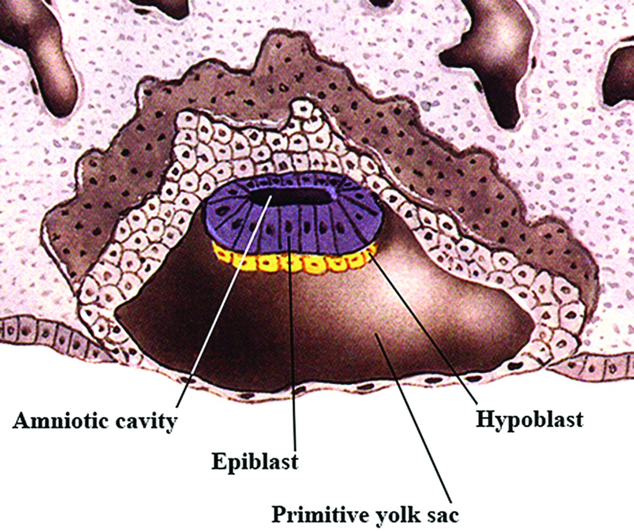
- Fig 2.
Sagittal drawing of a late 2-week-old embryo showing the embryonic disk with an epiblast layer and a the hypoblast below it. This is the bilaminar embryo just before gastrulation. (Modified with permission from Netter Illustration from www.netterimages.com. Elsevier Inc. © All rights reserved.16,17)
- Fig 3.
A, Oblique view from above of an early 3-week embryo shows the appearance of the primitive streak. Within the center of the primitive streak, a groove develops. Modified with permission from Cochard17 and Netter et al.18 B, Oblique view from above of an embryo a few days older than that in Fig 3A shows the development of the primitive node and the primitive pit at the ventral margin of the primitive streak. (Modified with permission from Netter Illustration from www.netterimages.com. Elsevier Inc. © All rights reserved.16,17)
- Fig 4.
Oblique view from above of an embryo slightly older than that in Fig 3B showing the relative relationship of the neural plate, prechordal plate and notochord, primitive streak, and primitive node. Also shown is the mesoderm spreading out under the epiblast. (Modified with permission from Netter Illustration from www.netterimages.com. Elsevier Inc. © All rights reserved.16,17)
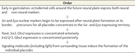
- Fig 5.
A, Location of the preplacodal territory and (B) the region around the anterior neural plate. The optic field develops to eventually be divided by signals from the prechordal plate. C, The placodes have separated from the preplacodal territory. (Modified with permission from Netter Illustration from www.netterimages.com. Elsevier Inc. © All rights reserved.16,17)
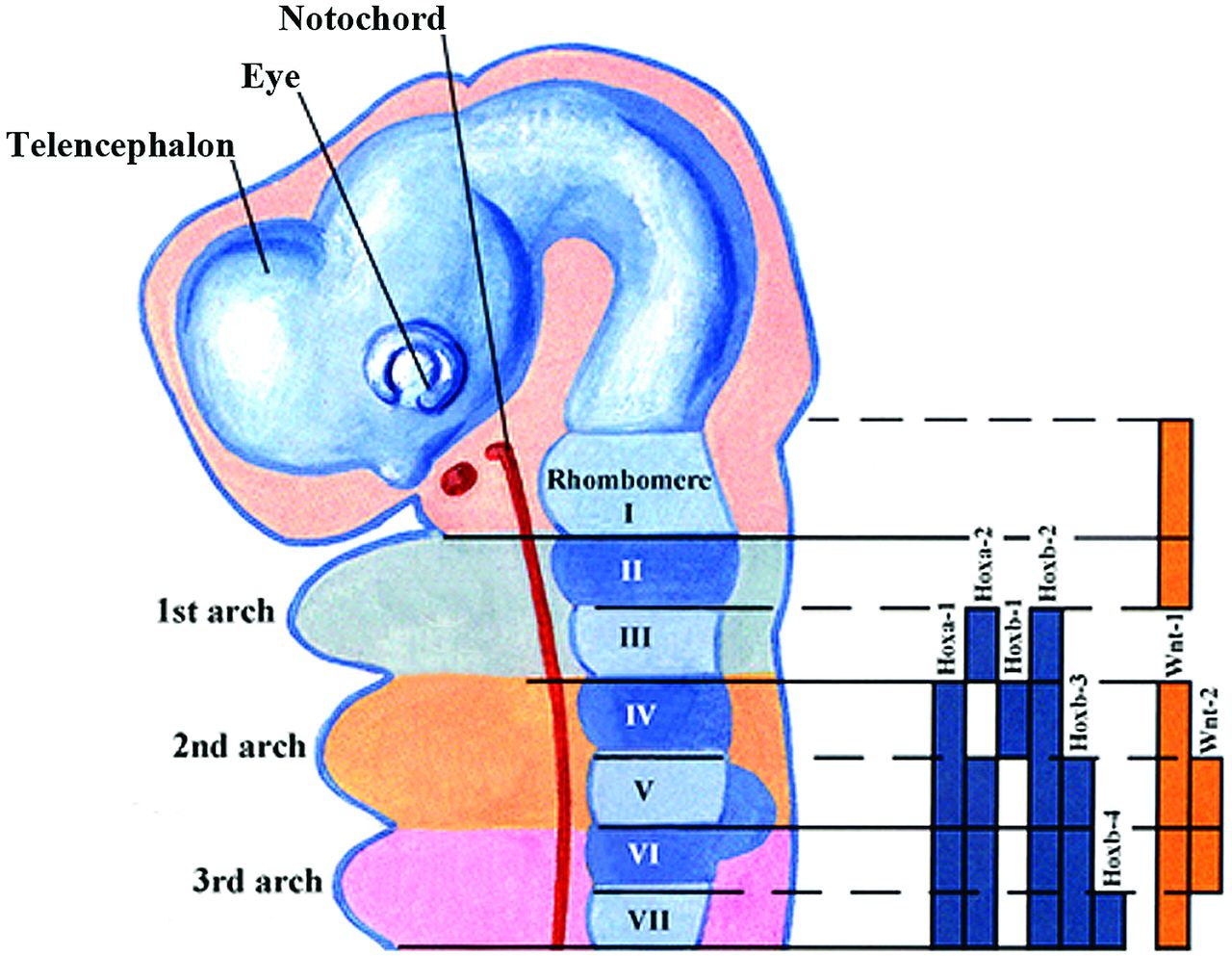
- Fig 6.
Sagittal drawing illustrating the Hox gene family signaling in the hindbrain and the pharyngeal arches. The solid bars represent the Hox gene expression in the neural crest cells and the neural tube. The downregulation of the Hoxa-2 gene in the first arch of neural crest cells is necessary for normal first arch development. (Modified with permission from Netter Illustration from www.netterimages.com. Elsevier Inc. © All rights reserved.16,17)
- Fig 7.
Serial coronal drawings showing the development of the palate and the genes associated with the stages of this development. A, The lateral palatal shelves emerge. B, The palatal shelves have elevated. C, The shelves have met in the midline, the medial edge epithelium dissolves, and the shelves eventually fuse.


Tables
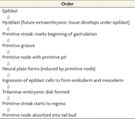
 Table 1:
Summary of the order of development of the trilaminar embryonic disk
Placode Inducer Families FGF PDGF RA Shh TGFβ family Wnta Adenohypophysis Shh Nodal, BMP4 Lens FGFR BMP4, BMP7 Olfactory FGF Otic FGFR Wnt FGF Epibranchial FGFR (VII, IX, X) FGF Trigeminal FGF PDGF Wnt PDGFR Note:—RA indicates retinoic acid; PDGFR, platelet-derived growth factor receptor; FGFR, fibroblast growth factor receptor; PDGF, platelet-derived growth factor.
↵a Modified from Table 2 in McCabe KL, Bronner-Fraser M. Molecular and tissue interactions governing induction of cranial ectodermal placodes. Dev Biol 2009;332:189–95, Elsevier Inc. © All rights reserved.3
Factor Some Observed Abnormalities RA Excess leads to fusion of 1st and 2nd branchial arches and acoustic-facial ganglia, small jaws, cleft palate, deformed pinna (Treacher-Collins syndrome); RA controls Shh and Fgf8 levels Fgf Controls outgrowth of facial primordia and migration of neural (Fgf) crest cells to facial processes; a decrease in FBGFR1 leads to midline clefting and Kallmann syndrome, small face and skull, achondroplasia, Crouzon syndrome, Apert syndrome TGF TGFβ required for fusion of lateral palatal processes; a decrease leads to defects in maxillary and mandibular development BMP A decrease leads to short frontal and nasal bones and small pterygoid processes, short stature, ear defects, odontogenic patterning defects, slower neural tube closure, small branchial arches, loss of incisor teeth Shh protein A decrease leads to holoprosencephaly, hypotelorism; an increase leads to a wide forehead, frontonasal dysplasia, Gorlin syndrome, Grieg cephalopolysyndactyly, Smith-Lemli-Opitz syndrome Wnts A decrease leads to loss of teeth, truncation of jaw, mesencephalic nucleus, and trigeminal nerve ET-1 A decrease leads to aplasia of 1st and 2nd arches, defects in maxilla and cleft palate, malformations of middle and external ear; 22q11.2 deletion syndrome (CATCH22 syndrome) Jagged 1 and 2 A decrease leads to Alagille syndrome, failure of palatal shelves to elevate, and fusion of shelves with tongue Platelet-derived growth factors A decrease leads to loss of some facial bones Homeobox-containing genes A decrease leads to primitive facial morphology, cleft palate, short maxilla and mandible, loss of maxillary molar teeth, ankyloglossia Note:—RA indicates retinoic acid; FGFR, fibroblast growth factor receptor; PDGFR, platelet-derived growth factor receptor.
Genes Associated Conditions Collagen genes, COL II and XI Otospondylomegaepiphyseal dysplasia, achondrogenesis type II, Stickler syndrome types I-III Diastrophic dysplasia sulfate transporter Diastrophic dysplasia FGFR2 Apert syndrome Homeobox MSX1 Cleft palate and hypodontia TGFβ R1 or TGFβR2 Aortic aneurysm, arterial tortuosity, hypertelorism, cleft palate, bifid uvula, craniosynostosis T-Box 1 (BX1) DiGeorge/Velo cardiofacial syndrome T-Box 22 (TBX22) X-linked cleft palate and ankyloglossia TCOF1 Treacher Collins syndrome TWIST Saethre-Chotzen syndrome Note:—FGFR2 indicates fibroblast growth factor receptor 2.
↵a Other genes and proteins associated with normal palatal formation include the following: Fgf-Shh signaling, Tbx22, BMPs, Jagged-2, Pax-9, serotonin, hyaluronan, PVRL1, TGF-β3, TGF-α, EGF, Lhx-8, Msx-1, and GABA (Fig 1). Table modified from Rice DP. Craniofacial anomalies: from development to molecular pathogenesis. Curr Mol Med 2005;5:699–722, Table 2, © Bentham Science Publishers.16
Gene Condition DHCR7 Smith-Lemli-Opitz syndrome EFNB1 Craniofrontonasal syndrome FGFR1 Kallmann syndrome IRF6 van der Woude syndrome OFD1 Oral-facial-digital syndrome type I MID1 Opitz syndrome MSX1 Cleft lip/palate with hypodontia PVRL1 Margarita Island ectodermal dysplasia (part of cleft lip/palate-ectodermal dysplasia syndrome) TP73 L (p63) Ectrodactyly, ectodermal dysplasia, and cleft lip/palate, ankyloblepharon-ectodermal dysplasia-clefting syndrome SIX3 HPE2 TGIF HPE4 PTCH1 HPE7 GLI2 HPE-like features Note:—HPE indicates holoprosencephaly.
↵a Table modified from Rice DP. Craniofacial anomalies: from development to molecular pathogenesis. Curr Mol Med 2005;5:699–722, Table 1, © Bentham Science Publishers.16


