Advertisement

Main menu

  • Home
  • Content
    • Current Issue
    • Accepted Manuscripts
    • Article Preview
    • Past Issue Archive
    • Video Articles
    • AJNR Case Collection
    • Case of the Week Archive
    • Case of the Month Archive
    • Classic Case Archive
  • Special Collections
    • AJNR Awards
    • Low-Field MRI
    • Alzheimer Disease
    • ASNR Foundation Special Collection
    • Photon-Counting CT
    • View All
  • Multimedia
    • AJNR Podcasts
    • AJNR SCANtastic
    • Trainee Corner
    • MRI Safety Corner
    • Imaging Protocols
  • For Authors
    • Submit a Manuscript
    • Submit a Video Article
    • Submit an eLetter to the Editor/Response
    • Manuscript Submission Guidelines
    • Statistical Tips
    • Fast Publishing of Accepted Manuscripts
    • Graphical Abstract Preparation
    • Imaging Protocol Submission
    • Author Policies
  • About Us
    • About AJNR
    • Editorial Board
    • Editorial Board Alumni
  • More
    • Become a Reviewer/Academy of Reviewers
    • Subscribers
    • Permissions
    • Alerts
    • Feedback
    • Advertisers
    • ASNR Home

User menu

  • Alerts
  • Log in

Search

  • Advanced search
American Journal of Neuroradiology

American Journal of Neuroradiology

ASHNR American Society of Functional Neuroradiology ASHNR American Society of Pediatric Neuroradiology ASSR
  • Alerts
  • Log in

Advanced Search

  • Home
  • Content
    • Current Issue
    • Accepted Manuscripts
    • Article Preview
    • Past Issue Archive
    • Video Articles
    • AJNR Case Collection
    • Case of the Week Archive
    • Case of the Month Archive
    • Classic Case Archive
  • Special Collections
    • AJNR Awards
    • Low-Field MRI
    • Alzheimer Disease
    • ASNR Foundation Special Collection
    • Photon-Counting CT
    • View All
  • Multimedia
    • AJNR Podcasts
    • AJNR SCANtastic
    • Trainee Corner
    • MRI Safety Corner
    • Imaging Protocols
  • For Authors
    • Submit a Manuscript
    • Submit a Video Article
    • Submit an eLetter to the Editor/Response
    • Manuscript Submission Guidelines
    • Statistical Tips
    • Fast Publishing of Accepted Manuscripts
    • Graphical Abstract Preparation
    • Imaging Protocol Submission
    • Author Policies
  • About Us
    • About AJNR
    • Editorial Board
    • Editorial Board Alumni
  • More
    • Become a Reviewer/Academy of Reviewers
    • Subscribers
    • Permissions
    • Alerts
    • Feedback
    • Advertisers
    • ASNR Home
  • Follow AJNR on Twitter
  • Visit AJNR on Facebook
  • Follow AJNR on Instagram
  • Join AJNR on LinkedIn
  • RSS Feeds

AJNR Awards, New Junior Editors, and more. Read the latest AJNR updates

Review ArticleReview Articles
Open Access

Review of Transcranial Doppler Ultrasound to Detect Microemboli during Orthopedic Surgery

B.S. Silbert, L.A. Evered, D.A. Scott, S. Rahardja, R.P. Gerraty and P.F. Choong
American Journal of Neuroradiology October 2014, 35 (10) 1858-1863; DOI: https://doi.org/10.3174/ajnr.A3688
B.S. Silbert
aFrom the Department of Anaesthesia (B.S.S., L.A.E., D.A.S., S.R.), Centre for Anaesthesia and Cognitive Function, St. Vincent's Hospital, Melbourne, Australia
bDepartment of Anaesthesia (B.S.S., L.A.E., D.A.S.), Perioperative and Pain Medicine Unit, Melbourne Medical School
  • Find this author on Google Scholar
  • Find this author on PubMed
  • Search for this author on this site
L.A. Evered
aFrom the Department of Anaesthesia (B.S.S., L.A.E., D.A.S., S.R.), Centre for Anaesthesia and Cognitive Function, St. Vincent's Hospital, Melbourne, Australia
bDepartment of Anaesthesia (B.S.S., L.A.E., D.A.S.), Perioperative and Pain Medicine Unit, Melbourne Medical School
  • Find this author on Google Scholar
  • Find this author on PubMed
  • Search for this author on this site
D.A. Scott
aFrom the Department of Anaesthesia (B.S.S., L.A.E., D.A.S., S.R.), Centre for Anaesthesia and Cognitive Function, St. Vincent's Hospital, Melbourne, Australia
bDepartment of Anaesthesia (B.S.S., L.A.E., D.A.S.), Perioperative and Pain Medicine Unit, Melbourne Medical School
  • Find this author on Google Scholar
  • Find this author on PubMed
  • Search for this author on this site
S. Rahardja
aFrom the Department of Anaesthesia (B.S.S., L.A.E., D.A.S., S.R.), Centre for Anaesthesia and Cognitive Function, St. Vincent's Hospital, Melbourne, Australia
  • Find this author on Google Scholar
  • Find this author on PubMed
  • Search for this author on this site
R.P. Gerraty
dEpworth Healthcare (R.P.G.), Richmond, Victoria, Australia
eDepartment of Medicine (R.P.G.), Epworth HealthCare, Monash University, Melbourne, Australia.
  • Find this author on Google Scholar
  • Find this author on PubMed
  • Search for this author on this site
P.F. Choong
cDepartment of Surgery (P.F.C.), University of Melbourne, Melbourne, Australia
  • Find this author on Google Scholar
  • Find this author on PubMed
  • Search for this author on this site
  • Article
  • Figures & Data
  • Supplemental
  • Info & Metrics
  • Responses
  • References
  • PDF
Loading

Abstract

SUMMARY: Transcranial Doppler ultrasonography has been used to detect microemboli in the middle cerebral artery during orthopedic surgery. We conducted a comprehensive systematic literature review of transcranial Doppler ultrasonography in orthopedic surgery to evaluate its status in this setting. Fourteen studies were selected for qualitative analysis. The highest number of patients studied was 45; emboli were detected in all studies, occurring in 20%–100% of patients. Most embolic counts were below 10, but some high counts were noted. No study reported all the technical parameters of the transcranial Doppler ultrasonography. All studies assessed neurologic status, and 6 studies evaluated cognitive function postoperatively. No study identified an association between postoperative cognitive function and embolic count. Six studies sought the presence of right-to-left shunts.

ABBREVIATIONS:

HITS
high-intensity transient signals
PFO
patent foramen ovale
TCD
transcranial Doppler ultrasonography
TEE
transesophageal echocardiography

Transcranial Doppler uses sonography to study cerebral blood flow velocity and can be used to detect microembolic material in the cerebral circulation. In orthopedic surgery, sonography was originally introduced to detect fat embolism in the venous system after long bone fracture1⇓–3 and subsequently to detect microemboli in the middle cerebral artery.4 In general, microemboli that enter the venous circulation are largely filtered out by the pulmonary circulation but, in some cases, may cross into the systemic circulation by either pulmonary shunts or a right-to-left shunt within the heart.

In addition to fat, it is believed that other solid particles such as thrombus, tissue, and cement may pass into the circulation as microemboli during orthopedic surgery. Gas microemboli have also been noted during orthopedic surgery,5 though the exact source is open to conjecture. Air may enter blood vessels in the surgical field, originate in the intravenous solutions, or arise from agitation during surgical manipulation.

Transcranial Doppler ultrasonography (TCD) has been used in orthopedic surgery to count these microemboli that are detected as high-intensity transient signals in (HITS) the MCA. In addition to counting the number of microemboli in the cerebral circulation, investigators have also sought to identify which specific surgical manipulations lead to microemboli entering the cerebral circulation and thus potentially placing the patient at risk.6,7

Large emboli in the arterial circulation that reach the brain may cause overt neurologic symptoms (ie, stroke). Smaller microemboli may cause more subtle neurologic damage manifesting as postoperative cognitive dysfunction.8 Several studies have sought to identify whether neurologic or cognitive change is related to the number of HITS (microemboli) detected in the MCA.

The use of TCD requires technical expertise and accurate interpretation of the signal. Differences in criteria used for signal identification originally caused confusion and led a Consensus Committee in 1995 to recommend the criteria for identification of HITS (Table 1).9 The Consensus Committee suggested that investigators specify ultrasonic frequency, dynamic range, gain settings, and identification criteria for classifying HITS. A subsequent International Consensus Group recommended that specific parameters of the ultrasonic device be reported (Table 2) in addition to the detection criteria and also stated that automatic emboli detection did not have the required sensitivity and specificity for clinical use.10

View this table:
  • View inline
  • View popup
Table 1:

Criteria for identification of high-intensity transient signals

View this table:
  • View inline
  • View popup
Table 2:

Recommended parameters to be reported for transcranial Doppler ultrasound

We systematically reviewed studies that used TCD intraoperatively in orthopedic surgery to detect cerebral emboli with the following specific aims:

  • 1)  To identify whether the investigators have documented the features of the TCD device and detection criteria (as recommended)

  • 2)  To determine the number of emboli released during orthopedic surgery.

We also recorded whether the studies also sought to identify right-to-left shunts as an explanation for the cerebral emboli count or measure neurologic or cognitive change as a result of the cerebral emboli count.

Materials and Methods

Eligibility Criteria

Eligibility criteria for article selection were any publication from 1965 through 2012 that used TCD during orthopedic surgery to detect HITS in the MCA. The publications must have been in the English language and contain surgical reports of intraoperative patient data of microemboli in the cerebral arteries in >1 case (ie, single case reports were excluded, but case series were included).

Study Identification

Electronic searches of MEDLINE and EMBASE were performed with results limited to the English language from 1965 to August 2012 inclusive. Three broad categories were combined to find appropriate articles for review. The categories were Doppler transcranial ultrasonography, orthopedic surgery, and embolism. The specific search strategies used in each data base are listed below.

Full-text articles were retrieved for any citations that were considered potentially relevant. Additional articles were sought by searching bibliographies of previous articles and review articles. Relevant journal and conference proceedings were also hand-searched. Inclusion criteria were English language, prospective, intraoperative, orthopedic surgery, and use of TCD. Exclusion criteria were the use of TCD preoperatively or postoperatively, Doppler used other than transcranially, and nonorthopedic surgery. Reports of single cases were excluded, but case series consisting of multiple cases were included.

MEDLINE (Searched on www.EBSCOhost.com)

A combination of MeSH terms and keywords were used as follows: [{(MH “Ultrasonography, Doppler” OR MH “Blood Flow Velocity” OR “blood flow velocity” OR “Doppler”) AND “transcranial”} OR MH “Ultrasonography, Doppler, Transcranial” OR “high-intensity transient signals”] AND [MH “Orthopedic Procedures +” OR “orthopedic*” OR “orthopedic*” OR “arthroplasty” OR “fracture*” OR MH “Musculoskeletal Diseases+/SU”] AND [MH “Intracranial Embolism” OR MH “Embolism” OR MH “Embolism, Fat+” OR “embol*” or “microembol*” or “micro-embol*”].

Twenty-nine results were selected from the MEDLINE search.

www.EMBASE.com

A combination of EMTREE terms and keywords were used as follows: [(“Doppler echography”/de OR “Doppler” OR “high-intensity transient signals” OR “blood flow velocity” OR “blood flow velocity”/de) AND “transcranial”] AND [“orthopedic surgery”/exp OR “orthopedic*” OR “orthopedic*” OR “arthroplasty” OR “fracture*”] AND [“embolism”/de OR “fat embolism”/de OR “embol*” OR “microembol*” OR “microembolism”].

Thirty results were selected from the EMBASE search.

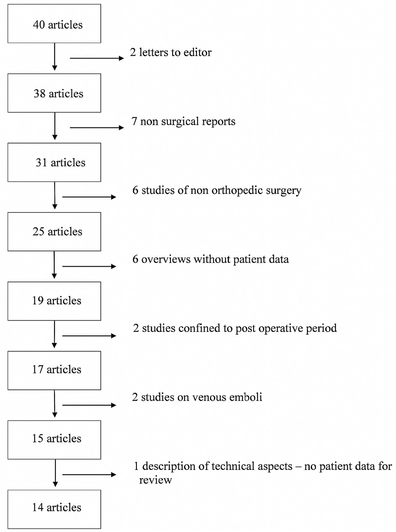
Fifty-nine articles were identified; screening removed 19 duplicates, leaving 40 eligible articles for review by 2 independent researchers. Fourteen articles satisfied the selection criteria (vide supra) for both researchers and were included for qualitative synthesis. Both independent researchers selected these 14 articles, so there was no need for an adjudication process. The reasons for eliminating articles are shown in Fig 1.

Fig 1.
  • Download figure
  • Open in new tab
  • Download powerpoint
Fig 1.

Reasons for exclusion of articles after selection.

Of the 2 articles by Rodriguez et al in 2001,11,12 the first describes 13 adolescents and the second describes 4 adolescents who appear to be a subset of the first article but have been included because they additionally underwent transesophageal echocardiography (TEE). Similarly, the 12 patients included by Patel et al13 were also described in the subsequent article as part of a larger study7; both articles have been included because cognitive decline was sought in the larger study but not in the former study.

Although the primary search related to the use of TCD to detect microemboli in the cerebral circulation, we also noted whether the studies additionally sought the presence of a patent foramen ovale (PFO) or assessed the neurologic or cognitive changes postoperatively.

Results

We found 14 publications that satisfied the inclusion criteria (Table 3). Ten of these publications additionally investigated the presence of PFO by using echocardiography or TCD after intravenous injection of agitated saline. All 14 publications made some comment on postoperative neurologic status, ranging from the presence or absence of neurologic complications to more subtle cognitive changes.

View this table:
  • View inline
  • View popup
Table 3:

Studies selected for review

On-line Tables 1 and 2 contain a description of each study included and whether the 14 specifications of the TCD used to detect HITS as recommended by the Consensus Committee10 were documented. Ideally, each of the 14 points should be made available in accordance with the consensus article. However, as evident from On-line Tables 1 and 3, none of the studies documented all 14 requirements.

For each study, On-line Table 3 shows the type of surgery, the number of patients, the criteria for emboli detection, the method of counting HITS, and monitoring of either unilateral or bilateral MCAs. Ten studies conformed to the recommended detection criteria, 3 studies raised the threshold amplitude >3 dB higher than background flow, and 1 study reduced the duration to <100 ms. On-line Table 4 lists the emboli count for each study and whether a PFO was sought and neurologic or cognitive change was identified. Emboli were detected in all 14 studies and ranged from 20% to 100% of patients (Fig 2). Most counts were low (<10), but high counts were present in some patients.

Fig 2.
  • Download figure
  • Open in new tab
  • Download powerpoint
Fig 2.

The percentage of patients with emboli detected in each of the 14 studies listed in Table 3.

On-line Table 4 also lists whether a right-to-left shunt was sought. The passage of microemboli from the venous into the arterial circulation can be confirmed by 2 methods either before or after surgery. Air microbubbles in an intravenous injection of agitated saline may pass into the arterial circulation via either cardiac or pulmonary communications and can be detected by TCD in the MCA as HITS. The early appearance of microemboli (eg, within the first 6 heart beats after injection14) suggests a cardiac communication (eg, functional or anatomic PFO) rather than a pulmonary communication (arteriovenous shunt). In the 2 studies by Patel et al,7,13 the diagnostic time window for microemboli was 25 seconds. Riding et al15 used a time window of 12 cardiac cycles, which is still arbitrary and open to challenge. Koch et al8 used contrast agents with microbubbles, which do not survive pulmonary passage.16

The second method involves the use of echocardiography (either TEE or transthoracic echocardiography) to identify right-to-left intracardiac shunts. This is also achieved by an intravenous injection of agitated saline. Echocardiography is then used to track these bubbles as they pass into the left atrium or ventricle as opposed to detecting them in the MCA. Unlike the detection of a right-to-left shunt by using TCD, which may not clearly distinguish cardiac from pulmonary communications, TEE definitively identifies cardiac communications. On-line Table 4 suggests that when TEE identifies a PFO, emboli counts may be high, but pulmonary communications also account for the appearance of HITS. No definitive conclusions can be drawn from these studies on the size and number of microemboli originating from cardiac or pulmonary communications.

Finally, On-line Table 4 lists whether the presence of neurologic or cognitive change was sought and whether any such change was related to the number of emboli.

The 14 articles spanned the 13 years from 1999 to 2010. Five studies were on hip replacements, 3 on knee replacements, 2 on both operations, 2 on fractured long bones, and 2 on scoliosis corrections in adolescents. Among the adult studies, the number of patients in each study ranged from 5 to 45. Two of the articles5,17 did not conform to the consensus criteria for detection of microemboli.9 Despite the use of automatic HITS counting, 13 articles reviewed the HITS count off-line. One article neither conformed to the consensus criteria nor stated how the emboli count was undertaken.18

Echocardiography was used to detect PFO in 6 studies (transthoracic echocardiography in 1 study, transthoracic echocardiography and TEE together in 1 study, and TEE in the remaining 4 studies). TCD was used to detect arteriovenous shunts in 3 studies and both TEE and TCD in 1 study.19 In general, higher counts were found in the presence of arteriovenous shunts.

All studies commented on the presence or absence of neurologic changes postoperatively. This appears to be assessed as part of routine follow-up, though 1 study specifically undertook neurologic examinations.20 Six studies measured cognitive function, and 1 study measured quality of life postoperatively as a proxy for cognition.13 No study found an association between neurologic or cognitive change and embolic count.

Discussion

Articles that described the use of TCD to detect microemboli during orthopedic surgery were sought and reviewed for the accuracy of reporting and the number of microemboli counted. Additionally, we recorded whether the detection of right-to-left shunts was sought and whether any neurologic or postoperative cognitive changes were noted. Fourteen articles were included for qualitative synthesis, and the largest number of patients in a single study was 45. The data for this important topic are thus limited, and this review highlights the need for larger and better reported studies in this area.

There are limitations on the quality of the reports. Few investigators explicitly stated all the features of the sonographic device used, particularly the 14 parameters recommended by the consensus statement.10 Settings that were consistently included were the insonated artery (MCA), insonation depth (40–60 mm), and sonographic frequency (2 MHz). The specific device, sample volume, threshold, and recording time were all inconsistent and poorly reported.

Once data have been recorded, it is common practice to use an independent observer to count the HITS. The criteria for classification of HITS should be documented to allow comparisons of studies. The Consensus Committee of the Ninth International Cerebral Hemodynamic Symposium addressed this concern in 1995 and recommended the criteria for classification.21 The results in On-line Tables 1 and 2 show that all studies stated the criteria for emboli detection, though 4 articles adjusted the threshold or duration from the recommended criteria. Additionally, while 12 of 14 articles included the guidelines used to count microemboli, the studies by Edmonds et al17 and Kalairajah et al18 did not specify how microemboli were counted. The validity of the results of these articles is of concern because the reliability of the TCD detection remains operator-dependent. Without independent analysis and strict classification guidelines, recorded data run the risk of including TCD signals which, in fact, are not real microemboli. Rodriguez et al11 noted that diathermy created interference that could easily be misinterpreted as HITS (especially by automatic detection), but this was not mentioned by other authors.

This review of the literature confirms that microemboli are detectable in the MCA during orthopedic surgery for the specific procedures identified. The embolic count may be absent but is more commonly present in small numbers (<10) or high numbers (>100), the highest reported being 550 for the unifrequency probe.6 A multifrequency probe detected 464 solid and 479 gas emboli in 1 patient,5 but counting was automatic with off-line verification by the same operator. The count distribution appears bimodal, and there is evidence that the high counts are associated with PFO when this has been sought by TEE (On-line Table 4). There is no apparent relationship between the type of surgery and the embolic count. Patient factors obviously play a role in addition to surgical factors. Rather than a specific surgical operation being implicated, any operation involving bone and intramedullary instrumentation is most likely to lead to embolic events.22

Of the 6 studies that tested for cognitive change, 1 did not explicitly describe the cognitive tests used, simply referring to a mental test score without describing the tests.18 The other 5 studies used a battery of neuropsychological tests at various time intervals before and after surgery. The definition of cognitive change is controversial.23,24 One study calculated the incidence of cognitive change by using the 20% rule (a decrease in cognitive score of 20% in 20% of the tests)25 and showed that the number of emboli was not different in patients with and without cognitive change.8 Three studies used group analyses to calculate the mean change in cognitive scores and showed that there was no difference in cognitive change between patients with and without detectable HITS.6,7,22 Only 1 study converted a cognitive score to a standardized score by using normative means and SDs19 but still found no association between the incidence of postoperative cognitive dysfunction and HITS count. Despite the use of highly differing methods of analysis, no study has found any association between HITS and neurologic or cognitive change.

This lack of association between embolic count and neuropsychological change brings into question the concept that microemboli released in orthopedic surgery may lead to cognitive changes. Even the patient with the highest embolic count (>500) was discharged with no detectable cognitive dysfunction.6

The early concern regarding microembolism to the brain during cardiac surgery being a cause of postoperative cognitive dysfunction26,27 has not been confirmed in more recent studies28 and systematic reviews.29,30 Liu et al31 reported a median embolic count of 430 (range, 155–2088) in patients undergoing cardiac surgery with cardiopulmonary bypass against a median embolic count of 2 (range, 0–66) in off-pump surgery but could show no difference in cognitive outcome. While the number of microemboli seen during cardiac surgery is much greater than that during orthopedic surgery, these are more likely to be predominantly gaseous. Cardiac surgical postoperative cognitive dysfunction has more recently been considered to be multifactorial.32,33

Several studies have used MR imaging and diffusion-weighted imaging after cardiac surgery to identify the relationship of new cerebral lesions to quantitative microemboli counts.34,35 None of the studies in this review used MR imaging or diffusion-weighted imaging to explore the incidence of cerebral lesions in relation to microemboli count.

Conclusions

Studies using TCD to detect cerebral microemboli in orthopedic surgery have used small numbers of patients. Many of these studies have not disclosed the full details of the TCD machine specifications, though most have used the recommended detection criteria. TCD often detects microemboli in the MCA during orthopedic surgery, in which the counts are frequently low but may be high in approximately 20% cases. High counts appear to be associated with right-to-left shunts when verified by agitated saline detected in the MCA by using TCD or when visualized by using TEE or transthoracic echocardiography. There is no evidence that these microemboli are associated with cognitive change after surgery. A study with high patient numbers, accurate reporting of TCD machine specifications, and criteria for emboli detection, together with comprehensive cognitive testing and analysis, is required to definitively confirm these conclusions.

Footnotes

  • Disclosures: Peter F. Choong—UNRELATED: Consultancy: Depuy, Zimmer, Comments: for being part of a surgeon consultation team involved in design of surgical instruments (Depuy), Grants/Grants Pending: National Health and Medical Research Council,* Australian Research Council,* Australian Orthopaedic Association Research Foundation,* Comments: peer-reviewed grant projects, Royalties: Zimmer, Comments: royalties for being part of design team of tumor endoprosthesis. *Money paid to the institution.

Indicates open access to non-subscribers at www.ajnr.org

REFERENCES

  1. 1.↵
    1. Gurd AR
    . Fat embolism: an aid to diagnosis. J Bone Joint Surg Br 1970;52:732–37
    PubMed
  2. 2.↵
    1. Kelly GL,
    2. Dodi G,
    3. Eiseman B
    . Ultrasound detection of fat emboli. Surg Forum 1972;23:459–61
    PubMed
  3. 3.↵
    1. Herndon JH,
    2. Bechtol CO,
    3. Crickenberger DP
    . Use of ultrasound to detect fat emboli during total hip replacement. Acta Orthop Scand 1975;46:108–18
    CrossRefPubMed
  4. 4.↵
    1. Forteza AM,
    2. Koch S,
    3. Romano JG, et al
    . Transcranial Doppler detection of fat emboli. Stroke 1999;30:2687–91
    Abstract/FREE Full Text
  5. 5.↵
    1. Barak M,
    2. Kabha M,
    3. Norman D, et al
    . Cerebral microemboli during hip fracture fixation: a prospective study. Anesthe Analg 2008;107:221–25
    CrossRef
  6. 6.↵
    1. Gray AC,
    2. Torrens L,
    3. Howie CR, et al
    . Cognitive function and cerebral emboli after primary hip arthroplasty. Hip Int 2008;18:40–45
    PubMed
  7. 7.↵
    1. Patel RV,
    2. Stygall J,
    3. Harrington J, et al
    . Cerebral microembolization during primary total hip arthroplasty and neuropsychologic outcome: a pilot study. Clin Orthop Relat Res 2010;468:1621–29
    CrossRefPubMed
  8. 8.↵
    1. Koch S,
    2. Forteza A,
    3. Lavernia C, et al
    . Cerebral fat microembolism and cognitive decline after hip and knee replacement. Stroke 2007;38:1079–81
    Abstract/FREE Full Text
  9. 9.↵
    Basic identification criteria of Doppler microembolic signals: Consensus Committee of the Ninth International Cerebral Hemodynamic Symposium. Stroke 1995;26:1123
    FREE Full Text
  10. 10.↵
    1. Ringelstein EB,
    2. Droste DW,
    3. Babikian VL, et al
    . Consensus on microembolus detection by TCD: International Consensus Group on Microembolus Detection. Stroke 1998;29:725–29
    Abstract/FREE Full Text
  11. 11.↵
    1. Rodriguez RA,
    2. Letts M,
    3. Jarvis J, et al
    . Cerebral microembolization during pediatric scoliosis surgery: a transcranial Doppler study. J Pediatr Orthop 2001;21:532–36
    CrossRefPubMed
  12. 12.↵
    1. Rodriguez RA,
    2. Sinclair B,
    3. Weatherdon D, et al
    . Patent foramen ovale and brain microembolization during scoliosis surgery in adolescents. Spine (Phila Pa 1976) 2001;26:1719–21
    CrossRef
  13. 13.↵
    1. Patel R,
    2. Stygall J,
    3. Harrington J, et al
    . Intra-operative cerebral microembolisation during primary hybrid total hip arthroplasty compared with primary hip resurfacing. Acta Orthop Belg 2009;75:671–77
    PubMed
  14. 14.↵
    1. Klötzsch C,
    2. Janssen G,
    3. Berlit P
    . Transesophageal echocardiography and contrast-TCD in the detection of a patent foramen ovale: experiences with 111 patients. Neurology 1994;44:1603–06
    CrossRef
  15. 15.↵
    1. Riding G,
    2. Daly K,
    3. Hutchinson S, et al
    . Paradoxical cerebral embolization: an explanation for fat embolism syndrome. J Bone Joint Surg Br 2004;86:95–98
    PubMed
  16. 16.↵
    1. Droste DW,
    2. Lakemeier S,
    3. Wichter T, et al
    . Optimizing the technique of contrast transcranial Doppler ultrasound in the detection of right-to-left shunts. Stroke 2002;33:2211–16
    Abstract/FREE Full Text
  17. 17.↵
    1. Edmonds CR,
    2. Barbut DM,
    3. Hager D, et al
    . Intraoperative cerebral arterial embolization during total hip arthroplasty. Anesthesiology 2000;93:315–18
    CrossRefPubMed
  18. 18.↵
    1. Kalairajah Y,
    2. Cossey AJ,
    3. Verrall GM, et al
    . Are systemic emboli reduced in computer-assisted knee surgery? A prospective, randomised, clinical trial. J Bone Joint Surg Br 2006;88:198–202
    CrossRefPubMed
  19. 19.↵
    1. Rodriguez RA,
    2. Tellier A,
    3. Grabowski J, et al
    . Cognitive dysfunction after total knee arthroplasty: effects of intraoperative cerebral embolization and postoperative complications. J Arthroplasty 2005;20:763–71
    CrossRefPubMed
  20. 20.↵
    1. Sulek CA,
    2. Davies LK,
    3. Enneking FK, et al
    . Cerebral microembolism diagnosed by transcranial Doppler during total knee arthroplasty: correlation with transesophageal echocardiography. Anesthesiology 1999;91:672
    CrossRefPubMed
  21. 21.↵
    1. Ringelstein EB
    . Skepticism toward carotid ultrasonography: a virtue, an attitude, or fanaticism? Stroke 1995;26:1743–46
    FREE Full Text
  22. 22.↵
    1. Gray AC,
    2. Torrens L,
    3. Christie J, et al
    . Cerebral emboli and cognitive function after intramedullary fracture fixation. Injury 2009;40:742–45
    CrossRefPubMed
  23. 23.↵
    1. Rasmussen LS,
    2. Larsen K,
    3. Houx P, et al
    . The assessment of postoperative cognitive function. Acta Anaesthesiol Scand 2001;45:275–89
    CrossRefPubMed
  24. 24.↵
    1. Lewis M,
    2. Maruff P,
    3. Silbert B
    . Statistical and conceptual issues in defining post-operative cognitive dysfunction. Neurosci Biobehav Rev 2004;28:433–40
    CrossRefPubMed
  25. 25.↵
    1. Lewis MS,
    2. Maruff P,
    3. Silbert BS, et al
    . The sensitivity and specificity of three common statistical rules for the classification of post-operative cognitive dysfunction following coronary artery bypass graft surgery. Acta Anaesthesiol Scand 2006;50:50–57
    CrossRefPubMed
  26. 26.↵
    1. Fearn SJ,
    2. Pole R,
    3. Wesnes K, et al
    . Cerebral injury during cardiopulmonary bypass: emboli impair memory. J Thoracic Cardiovasc Surg 2001;121:1150–60
    CrossRefPubMed
  27. 27.↵
    1. Braekken SK,
    2. Reinvang I,
    3. Russell D, et al
    . Association between intraoperative cerebral microembolic signals and postoperative neuropsychological deficit: comparison between patients with cardiac valve replacement and patients with coronary artery bypass grafting. J Neurol Neurosurg Psychiatry 1998;65:573–76
    Abstract/FREE Full Text
  28. 28.↵
    1. Stroobant N,
    2. Van Nooten G,
    3. Van Belleghem Y, et al
    . Relation between neurocognitive impairment, embolic load, and cerebrovascular reactivity following on- and off-pump coronary artery bypass grafting. Chest 2005;127:1967–76
    CrossRefPubMed
  29. 29.↵
    1. Martin KK,
    2. Wigginton JB,
    3. Babikian VL, et al
    . Intraoperative cerebral high-intensity transient signals and postoperative cognitive function: a systematic review. Am J Surg 2009;197:55–63
    CrossRefPubMed
  30. 30.↵
    1. Kruis RW,
    2. Vlasveld FA,
    3. Van Dijk D
    . The (un)importance of cerebral microemboli. Semin Cardiothorac Vasc Anesth 2010;14:111–18
    Abstract/FREE Full Text
  31. 31.↵
    1. Liu YH,
    2. Wang DX,
    3. Li LH, et al
    . The effects of cardiopulmonary bypass on the number of cerebral microemboli and the incidence of cognitive dysfunction after coronary artery bypass graft surgery. Anesth Analg 2009;109:1013–22
    CrossRefPubMed
  32. 32.↵
    1. Selnes OA,
    2. McKhann GM
    . Neurocognitive complications after coronary artery bypass surgery. Ann Neurol 2005;57:615–21
    CrossRefPubMed
  33. 33.↵
    1. Grocott HP,
    2. Homi HM,
    3. Puskas F
    . Cognitive dysfunction after cardiac surgery: revisiting etiology. Semin Cardiothorac Vasc Anesth 2005;9:123–29
    Abstract/FREE Full Text
  34. 34.↵
    1. Restrepo L,
    2. Wityk RJ,
    3. Grega MA, et al
    . Diffusion- and perfusion-weighted magnetic resonance imaging of the brain before and after coronary artery bypass grafting surgery. Stroke 2002;33:2909–15
    Abstract/FREE Full Text
  35. 35.↵
    1. Knipp SC,
    2. Matatko N,
    3. Wilhelm H, et al
    . Evaluation of brain injury after coronary artery bypass grafting: a prospective study using neuropsychological assessment and diffusion-weighted magnetic resonance imaging. Eur J Cardiothorac Surg 2004;25:791–800
    Abstract/FREE Full Text
  • © 2014 by American Journal of Neuroradiology
View Abstract
PreviousNext
Back to top

In this issue

American Journal of Neuroradiology: 35 (10)
American Journal of Neuroradiology
Vol. 35, Issue 10
1 Oct 2014
  • Table of Contents
  • Index by author
  • Complete Issue (PDF)
Advertisement
Print
Download PDF
Email Article

Thank you for your interest in spreading the word on American Journal of Neuroradiology.

NOTE: We only request your email address so that the person you are recommending the page to knows that you wanted them to see it, and that it is not junk mail. We do not capture any email address.

Enter multiple addresses on separate lines or separate them with commas.
Review of Transcranial Doppler Ultrasound to Detect Microemboli during Orthopedic Surgery
(Your Name) has sent you a message from American Journal of Neuroradiology
(Your Name) thought you would like to see the American Journal of Neuroradiology web site.
CAPTCHA
This question is for testing whether or not you are a human visitor and to prevent automated spam submissions.
Cite this article
B.S. Silbert, L.A. Evered, D.A. Scott, S. Rahardja, R.P. Gerraty, P.F. Choong
Review of Transcranial Doppler Ultrasound to Detect Microemboli during Orthopedic Surgery
American Journal of Neuroradiology Oct 2014, 35 (10) 1858-1863; DOI: 10.3174/ajnr.A3688

Citation Manager Formats

  • BibTeX
  • Bookends
  • EasyBib
  • EndNote (tagged)
  • EndNote 8 (xml)
  • Medlars
  • Mendeley
  • Papers
  • RefWorks Tagged
  • Ref Manager
  • RIS
  • Zotero
0 Responses
Respond to this article
Share
Bookmark this article
Review of Transcranial Doppler Ultrasound to Detect Microemboli during Orthopedic Surgery
B.S. Silbert, L.A. Evered, D.A. Scott, S. Rahardja, R.P. Gerraty, P.F. Choong
American Journal of Neuroradiology Oct 2014, 35 (10) 1858-1863; DOI: 10.3174/ajnr.A3688
del.icio.us logo Twitter logo Facebook logo Mendeley logo
  • Tweet Widget
  • Facebook Like
  • Google Plus One
Purchase

Jump to section

  • Article
    • Abstract
    • ABBREVIATIONS:
    • Materials and Methods
    • Results
    • Discussion
    • Conclusions
    • Footnotes
    • REFERENCES
  • Figures & Data
  • Supplemental
  • Info & Metrics
  • Responses
  • References
  • PDF

Related Articles

  • PubMed
  • Google Scholar

Cited By...

  • No citing articles found.
  • Crossref (14)
  • Google Scholar

This article has been cited by the following articles in journals that are participating in Crossref Cited-by Linking.

  • Point-of-Care Ultrasound in Neurology – Report of the EAN SPN/ESNCH/ERcNsono Neuro-POCUS Working Group
    Jurgita Valaikiene, Felix Schlachetzki, Elsa Azevedo, Manfred Kaps, Piergiorgio Lochner, Aristeidis H. Katsanos, Uwe Walter, Claudio Baracchini, Eva Bartels, David Školoudík
    Ultraschall in der Medizin - European Journal of Ultrasound 2022 43 04
  • Intraoperative monitoring of the central and peripheral nervous systems: a narrative review
    Christopher Ryalino, Marko M. Sahinovic, Gea Drost, Anthony R. Absalom
    British Journal of Anaesthesia 2024 132 2
  • Fat Embolism Syndrome: A Case Report and Review Literature
    Nattaphol Uransilp, Sombat Muengtaweepongsa, Nuttawut Chanalithichai, Nattapol Tammachote
    Case Reports in Medicine 2018 2018
  • Detection and differentiation of cerebral microemboli in patients undergoing major orthopaedic surgery using transcranial Doppler ultrasound
    C Kietaibl, A Engel, I Horvat Menih, M Huepfl, G Erdoes, B Kubista, R Ullrich, R Windhager, K Markstaller, K.U. Klein
    British Journal of Anaesthesia 2017 118 3
  • Paradoxical Cerebral Fat Embolism in Revision Hip Surgery
    Nicolás S. Piuzzi, Gerardo Zanotti, Fernando M. Comba, Martin A. Buttaro, Francisco Piccaluga
    Case Reports in Orthopedics 2014 2014
  • ASNM and ASN joint guidelines for transcranial Doppler ultrasonic monitoring: An update
    Alexander Y. Razumovsky, Faisal R. Jahangiri, Jeffrey Balzer, Andrei V. Alexandrov
    Journal of Neuroimaging 2022 32 5
  • Femoral Nailing in a Porcine Model Causes Bone Marrow Emboli in the Lungs and Systemic Emboli in the Heart and Brain
    Steinar Kristiansen, Anders Hagen Jarmund, Jonas Hilmo, Tom Eirik Mollnes, Martin Leth-Olsen, Siri Ann Nyrnes, Bent Aksel Nilsen, Renathe Henriksen Grønli, Bjørn Ove Faldaas, Benjamin Storm, Arild Espenes, Erik Waage Nielsen
    JBJS Open Access 2024 9 1
  • Oxidative stress and mitochondrial dysfunction contributes to postoperative cognitive dysfunction in elderly rats dependent on NLRP3 activation
    Sandra Bonfante, Martins Back Netto, Aloir Neri de Oliveira Junior, Khiany Mathias, Richard Simon Machado, Larissa Joaquim, Taina Cidreira, Marina Goulart da Silva, Guilherme Cabreira Daros, Lucinéia Gainski Danielski, Fernanda Gava, Isabela da Silva Lemos, Rafaela Tezza Matiola, Emily Córneo, Josiane Somariva Prophiro, Rafael Mariano de Bitencourt, Carlos Henrique Rocha Catalão, Jaqueline da Silva Generoso, Emílio Luiz Streck, Felipe Dal-Pizzol, Tatiana Barichello, Fabricia Petronilho
    Metabolic Brain Disease 2024 40 1
  • Robotic Assisted Transcranial Doppler Monitoring in Acute Neurovascular Care: A Feasibility and Safety Study
    Alvise Fattorello Salimbeni, Caterina Kulyk, Francesco Favruzzo, Ludovica De Rosa, Federica Viaro, Alessio Pieroni, Stefano Mozzetta, Milan R. Vosko, Claudio Baracchini
    Neurocritical Care 2025 42 2
  • Applications of Ultrasonography in Limb Lengthening and Reconstruction
    Alana M. Munger, Michael Amick, David B. Frumberg
    Journal of Limb Lengthening & Reconstruction 2023 9 1

More in this TOC Section

  • An Atlas of Neonatal Neurovascular Imaging Anatomy as Depicted with Microvascular Imaging: The Intracranial Arteries
  • An Atlas of Neonatal Neurovascular Imaging Anatomy as Depicted with Microvascular Imaging: The Intracranial Veins
  • Clinical Translation of Hyperpolarized 13C Metabolic Probes for Glioma Imaging
Show more Review articles

Similar Articles

Advertisement

Indexed Content

  • Current Issue
  • Accepted Manuscripts
  • Article Preview
  • Past Issues
  • Editorials
  • Editor's Choice
  • Fellows' Journal Club
  • Letters to the Editor
  • Video Articles

Cases

  • Case Collection
  • Archive - Case of the Week
  • Archive - Case of the Month
  • Archive - Classic Case

More from AJNR

  • Trainee Corner
  • Imaging Protocols
  • MRI Safety Corner
  • Book Reviews

Multimedia

  • AJNR Podcasts
  • AJNR Scantastics

Resources

  • Turnaround Time
  • Submit a Manuscript
  • Submit a Video Article
  • Submit an eLetter to the Editor/Response
  • Manuscript Submission Guidelines
  • Statistical Tips
  • Fast Publishing of Accepted Manuscripts
  • Graphical Abstract Preparation
  • Imaging Protocol Submission
  • Evidence-Based Medicine Level Guide
  • Publishing Checklists
  • Author Policies
  • Become a Reviewer/Academy of Reviewers
  • News and Updates

About Us

  • About AJNR
  • Editorial Board
  • Editorial Board Alumni
  • Alerts
  • Permissions
  • Not an AJNR Subscriber? Join Now
  • Advertise with Us
  • Librarian Resources
  • Feedback
  • Terms and Conditions
  • AJNR Editorial Board Alumni

American Society of Neuroradiology

  • Not an ASNR Member? Join Now

© 2025 by the American Society of Neuroradiology All rights, including for text and data mining, AI training, and similar technologies, are reserved.
Print ISSN: 0195-6108 Online ISSN: 1936-959X

Powered by HighWire