
Article Figures & Data
Figures
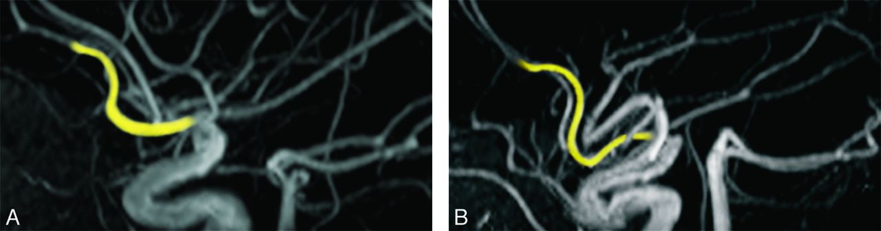
- Figure.
Anterior cerebral artery configuration in Williams syndrome. MRA from a normally developing person (A) and a participant with Williams syndrome (B) showing elongation of the postcommunicating segment of the anterior cerebral artery in the participant with Williams syndrome.

Tables
Participant Age (y) Pulse (bpm) Blood Pressure (mm Hg) Medical History Cardiovascular History Systolic Diastolic 1 18 84 130 90 Inguinal hernia Hypertension 2 18 Not available Hypercalcemia, joint limitation SVAS 3 18 72 90 70 Chronic otitis media, hip subluxation, kyphosis, constipation Ventricular septal defect, SVPS, prolonged Q-T interval 4 19 64 110 80 Constipation Negative 5 22 64 130 80 Chronic otitis media, scoliosis, rectal prolapse, constipation Mitral valve prolapse, hypertension 6 22 Not available Left conductive hearing loss, chronic otitis media, joint limitation SVAS, mitral valve prolapse 7 23 64 120 60 Inguinal hernia, umbilical hernia, joint impairment SVAS, SVPS, mitral valve prolapse 8 24 80 130 80 Chronic otitis media, inguinal hernia, joint impairment SVAS, peripheral pulmonary stenosis, hypertension 9 29 72 120 92 Hypercalcemia SVAS 10 30 76 120 70 Inguinal hernia SVAS, SVPS 11 33 68 165 100 Constipation, inguinal hernia SVAS, hypertension, renal artery stenosis 12 37 100 110 70 Diverticulitis, constipation SVAS, carotid bruit 13 44 62 120 91 Hypercalcemia, kyphosis, chronic abdominal pain, constipation SVAS 14 45 96 130 85 Diverticulitis No history of hypertension Medical Condition Study Wint et ala (n = 14) AAPb (n = 315) Cherniske et alc (n = 20) n % % % Cardiovascular 12 86 80 70 SVAS 9 64 75 65 SVPS 3 21 25 15 Peripheral pulmonary stenosis 1 7 50 – Mitral valve prolapse 3 21 − 15 Other 1 7 − 35 Hypertension 4 29 50 60 Gastrointestinal 11 79 − 75 Inguinal hernia 5 36 40 − Constipation 7 50 40 25 Diverticular disease 3 21 30 40 Rectal prolapse 1 7 15 − Neurologic Hyperreflexia 7 50 75 65 Gait disturbance 11 79 60 70 Ataxia 10 71 − − Skeletal 6 43 50 − Spinal 3 21 60 60 Peripheral 5 36 50 55 Chronic otitis 4 29 50 − Note:—This is a comparison of the prevalence of medical problems in this study's participants with those reported in 2 large studies of Williams syndrome. The number of participants with each condition is listed in the leftmost column.
↵a Present study.
↵b American Academy of Pediatrics, 2001.32
↵c Cherniske et al, 2004.33


