
Article Figures & Data
Figures
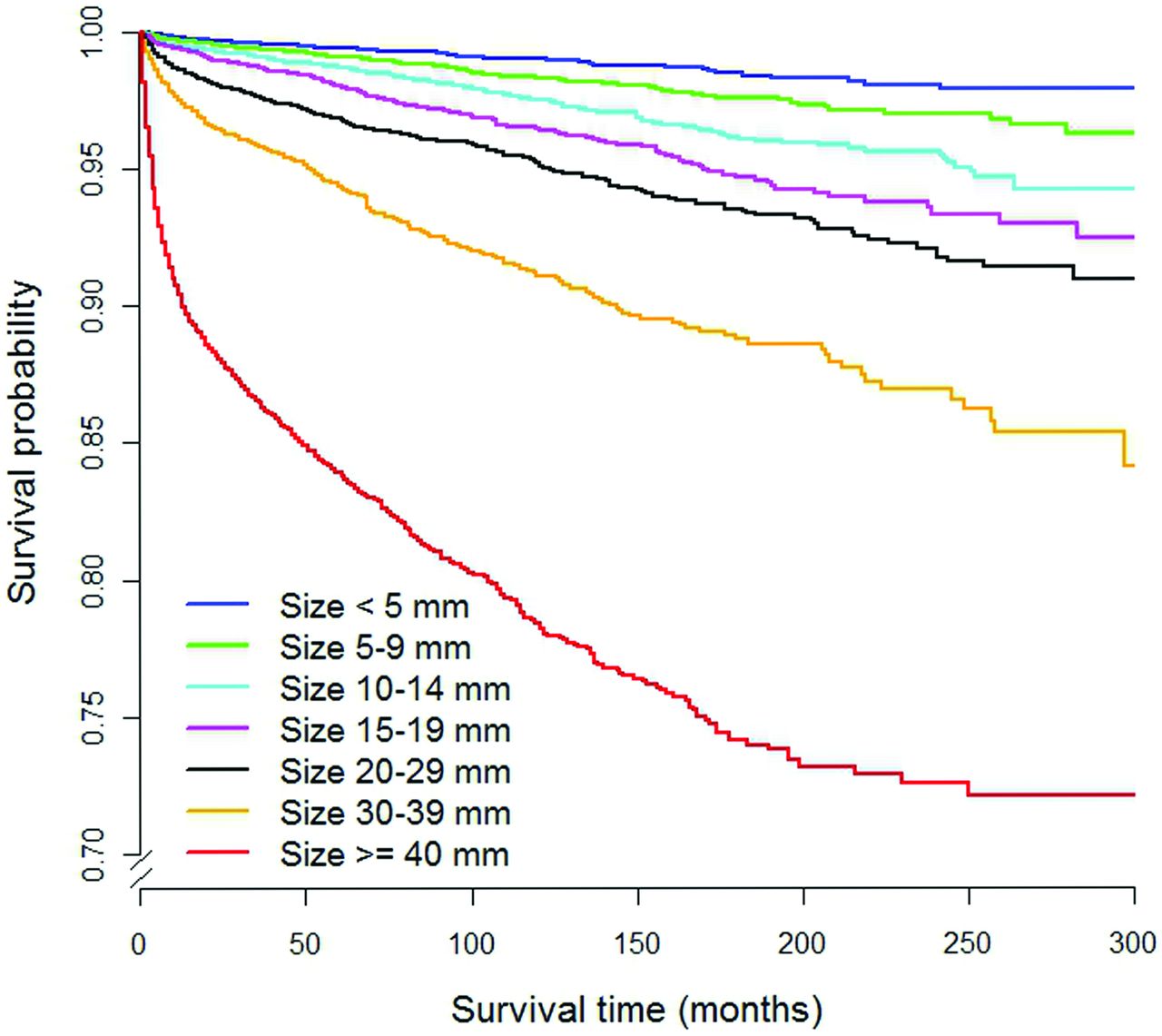
- Fig 1.
Cause-specific survival curves as a function of tumor size for thyroid cancers in the SEER data base. This represents method A of risk categorization.
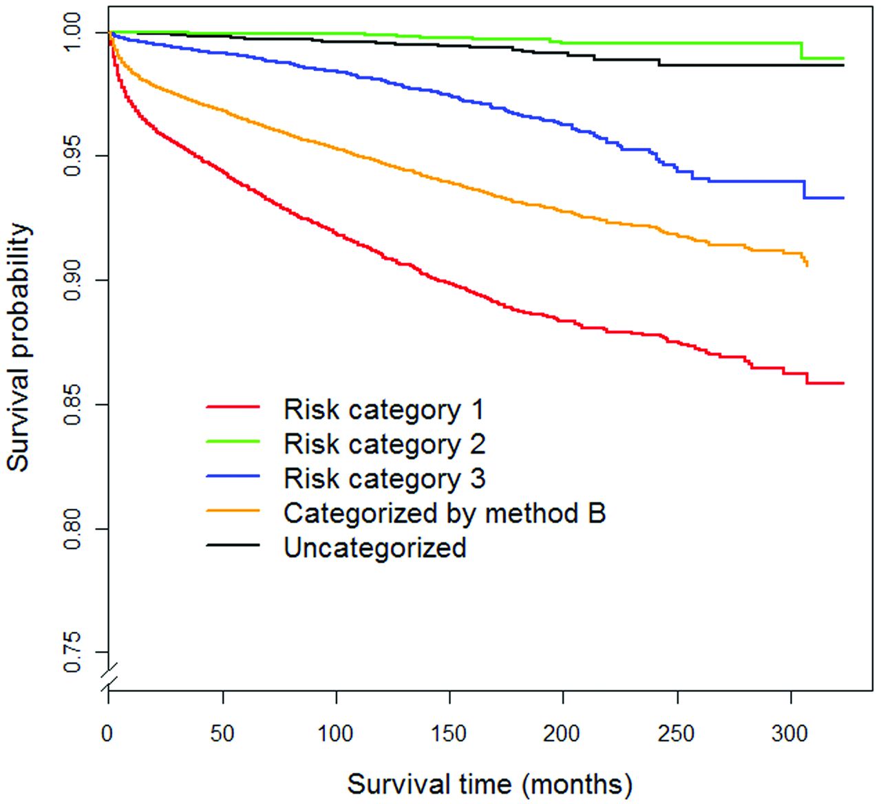
- Fig 2.
Cause-specific survival curves of thyroid cancers in SEER stratified by the 3-tiered system (method B).


Tables
- Table 1:
Survival of patients with thyroid carcinoma by patient demographics and tumor characteristics
Percentage Total 10-Year Relative Survival (%) 95% CI 10-Year Thyroid Cancer–Specific Survival (%) 95% CI Total 100% 97.4 97.0–97.7 95.7 95.5–95.9 Sex Male 23% 93.8 92.9–94.7 92.0 91.4–92.5 Female 77% 98.4 98.0–98.6 96.8 96.6–97.0 Age (yr) 0–19 2.2% 99.1 98.3–99.5 99.3 98.7–99.6 20–34 21% 99.3 98.9–99.5 99.7 99.5–99.8 35–49 36% 99.3 98.9–99.5 98.9 98.7–99.0 50–64 27% 96.2 95.3–96.9 94.6 94.1–95.1 ≥65 15% 90.3 88.1–92.1 81.0 79.9–82.0 SEER summary stage Localized or unstaged 61% 99.9 99.8–99.9 99.0 98.9–99.1 Regional 35% 95.6 95.0–96.2 94.4 94.0–94.8 Distant 4.1% 56.1 53.5–58.5 58.2 56.0–60.4 Tumor size (mm) <5 12% 99.9 99.7–99.9 99.4 99.0–99.6 5–10 14% 99.8 99.6–99.9 98.9 98.6–99.2 10–14 15% 99.8 99.5–99.9 99.0 98.7–99.2 15–19 13% 99.8 99.5–99.9 98.3 97.9–98.6 20–29 20% 99.4 98.3–99.8 97.0 96.7–97.4 30–39 11% 96.6 95.5–97.4 95.2 94.6–95.7 ≥40 14% 84.0 82.7–85.2 83.3 82.4–84.2 - Table 2:
Survival in patients with thyroid malignancy in the SEER data base using risk-categorization methods of size cutoff (method A) and the 3-tiered system (method B)a
Percentage Total 10-Year Relative Survival (%) 95% CI 10-Year Thyroid Cancer-Specific Survival (%) 95% CI Method A (size cutoff) ≥10 mm 74% 96.4 96.0–96.7 94.7 94.4–94.9 <10 mm 26% 99.8 99.7–99.9 99.1 98.9–99.3 ≥15 mm 59% 95.4 94.9–95.8 93.7 93.4–94.0 <15 mm 41% 99.8 99.8–99.9 99.1 98.9–99.2 ≥20 mm 46% 94.1 93.5–94.6 92.4 92.0–92.7 <20 mm 54% 99.9 99.8–99.9 98.9 98.7–99.0 Method B (3-tiered risk categories) Categories 1, 2, and 3 74% 96.0 95.6–96.3 94.6 94.3–94.8 Category 1 39% 91.5 90.9–92.1 90.7 90.3–91.1 Category 2 13% 99.7 99.2–99.9 99.9 99.7–99.9 Category 3 23% 99.8 99.7–99.9 98.0 97.7–98.3 Not categorized 26% 99.9 99.8–99.9 99.5 99.3–99.7 ↵a Nodules smaller than the size cutoff and in the “not categorized” group for method B would represent nodules that would not receive work-up if the methods were applied.
Categorization Method Patients Total Method A (size cutoff) ≥10 mm 57 43% <10 mm 76 57% ≥15 mm 22 17% <15 mm 111 83% ≥20 mm 12 9% <20 mm 121 91% Method B (3-tiered system) Category 1, 2, or 3 31 23% Category 1 2 2% Category 2 9 7% Category 3 20 15% Not categorized 102 77% Total 133 ↵a Incidental thyroid nodules from retrospective CT review were stratified by size cutoff (method A) and the 3-tiered system (method B). The nodules smaller than the size cutoff and in the “not categorized” group for method B would represent nodules that would not receive work-up if the methods were applied.


Jump to section
Related Articles
Cited By...
- Benign and Malignant Thyroid Incidentalomas Are Rare in Routine Clinical Practice: A Review of 97,908 Imaging Studies
- Radiology Reports for Incidental Thyroid Nodules on CT and MRI: High Variability across Subspecialties
- Imaging-Detected Incidental Thyroid Nodules that Undergo Surgery: A Single-Center Experience Over 1 Year
- High Variability in Radiologists' Reporting Practices for Incidental Thyroid Nodules Detected on CT and MRI
- An Exponential Growth in Incidence of Thyroid Cancer: Trends and Impact of CT Imaging