Advertisement

Main menu

  • Home
  • Content
    • Current Issue
    • Accepted Manuscripts
    • Article Preview
    • Past Issue Archive
    • Video Articles
    • AJNR Case Collection
    • Case of the Week Archive
    • Case of the Month Archive
    • Classic Case Archive
  • Special Collections
    • AJNR Awards
    • Low-Field MRI
    • Alzheimer Disease
    • ASNR Foundation Special Collection
    • Photon-Counting CT
    • View All
  • Multimedia
    • AJNR Podcasts
    • AJNR SCANtastic
    • Trainee Corner
    • MRI Safety Corner
    • Imaging Protocols
  • For Authors
    • Submit a Manuscript
    • Submit a Video Article
    • Submit an eLetter to the Editor/Response
    • Manuscript Submission Guidelines
    • Statistical Tips
    • Fast Publishing of Accepted Manuscripts
    • Graphical Abstract Preparation
    • Imaging Protocol Submission
    • Author Policies
  • About Us
    • About AJNR
    • Editorial Board
    • Editorial Board Alumni
  • More
    • Become a Reviewer/Academy of Reviewers
    • Subscribers
    • Permissions
    • Alerts
    • Feedback
    • Advertisers
    • ASNR Home

User menu

  • Alerts
  • Log in

Search

  • Advanced search
American Journal of Neuroradiology

American Journal of Neuroradiology

ASHNR American Society of Functional Neuroradiology ASHNR American Society of Pediatric Neuroradiology ASSR
  • Alerts
  • Log in

Advanced Search

  • Home
  • Content
    • Current Issue
    • Accepted Manuscripts
    • Article Preview
    • Past Issue Archive
    • Video Articles
    • AJNR Case Collection
    • Case of the Week Archive
    • Case of the Month Archive
    • Classic Case Archive
  • Special Collections
    • AJNR Awards
    • Low-Field MRI
    • Alzheimer Disease
    • ASNR Foundation Special Collection
    • Photon-Counting CT
    • View All
  • Multimedia
    • AJNR Podcasts
    • AJNR SCANtastic
    • Trainee Corner
    • MRI Safety Corner
    • Imaging Protocols
  • For Authors
    • Submit a Manuscript
    • Submit a Video Article
    • Submit an eLetter to the Editor/Response
    • Manuscript Submission Guidelines
    • Statistical Tips
    • Fast Publishing of Accepted Manuscripts
    • Graphical Abstract Preparation
    • Imaging Protocol Submission
    • Author Policies
  • About Us
    • About AJNR
    • Editorial Board
    • Editorial Board Alumni
  • More
    • Become a Reviewer/Academy of Reviewers
    • Subscribers
    • Permissions
    • Alerts
    • Feedback
    • Advertisers
    • ASNR Home
  • Follow AJNR on Twitter
  • Visit AJNR on Facebook
  • Follow AJNR on Instagram
  • Join AJNR on LinkedIn
  • RSS Feeds

AJNR Awards, New Junior Editors, and more. Read the latest AJNR updates

Research ArticleBrain
Open Access

Longitudinal Restriction Spectrum Imaging Is Resistant to Pseudoresponse in Patients with High-Grade Gliomas Treated with Bevacizumab

P.D. Kothari, N.S. White, N. Farid, R. Chung, J.M. Kuperman, H.M. Girard, A. Shankaranarayanan, S. Kesari, C.R. McDonald and A.M. Dale
American Journal of Neuroradiology September 2013, 34 (9) 1752-1757; DOI: https://doi.org/10.3174/ajnr.A3506
P.D. Kothari
aFrom the School of Medicine (P.D.K.)
  • Find this author on Google Scholar
  • Find this author on PubMed
  • Search for this author on this site
N.S. White
bDepartments of Radiology (N.S.W., N.F., R.C., J.M.K., A.M.D.)
eMultimodal Imaging Laboratory (N.S.W., N.F., J.M.K., H.M.G., C.R.M., A.M.D.)
  • Find this author on Google Scholar
  • Find this author on PubMed
  • Search for this author on this site
N. Farid
bDepartments of Radiology (N.S.W., N.F., R.C., J.M.K., A.M.D.)
eMultimodal Imaging Laboratory (N.S.W., N.F., J.M.K., H.M.G., C.R.M., A.M.D.)
  • Find this author on Google Scholar
  • Find this author on PubMed
  • Search for this author on this site
R. Chung
bDepartments of Radiology (N.S.W., N.F., R.C., J.M.K., A.M.D.)
  • Find this author on Google Scholar
  • Find this author on PubMed
  • Search for this author on this site
J.M. Kuperman
bDepartments of Radiology (N.S.W., N.F., R.C., J.M.K., A.M.D.)
eMultimodal Imaging Laboratory (N.S.W., N.F., J.M.K., H.M.G., C.R.M., A.M.D.)
  • Find this author on Google Scholar
  • Find this author on PubMed
  • Search for this author on this site
H.M. Girard
eMultimodal Imaging Laboratory (N.S.W., N.F., J.M.K., H.M.G., C.R.M., A.M.D.)
  • Find this author on Google Scholar
  • Find this author on PubMed
  • Search for this author on this site
A. Shankaranarayanan
gGE Healthcare (A.S.), Milwaukee, Wisconsin.
  • Find this author on Google Scholar
  • Find this author on PubMed
  • Search for this author on this site
S. Kesari
cNeurosciences (S.K., A.M.D.)
fTranslational Neuro-Oncology Laboratories (S.K.), Moores Cancer Center; University of California, San Diego, La Jolla, California
  • Find this author on Google Scholar
  • Find this author on PubMed
  • Search for this author on this site
C.R. McDonald
dPsychiatry (C.R.M., A.M.D.)
eMultimodal Imaging Laboratory (N.S.W., N.F., J.M.K., H.M.G., C.R.M., A.M.D.)
  • Find this author on Google Scholar
  • Find this author on PubMed
  • Search for this author on this site
A.M. Dale
bDepartments of Radiology (N.S.W., N.F., R.C., J.M.K., A.M.D.)
cNeurosciences (S.K., A.M.D.)
dPsychiatry (C.R.M., A.M.D.)
eMultimodal Imaging Laboratory (N.S.W., N.F., J.M.K., H.M.G., C.R.M., A.M.D.)
  • Find this author on Google Scholar
  • Find this author on PubMed
  • Search for this author on this site
  • Article
  • Figures & Data
  • Info & Metrics
  • Responses
  • References
  • PDF
Loading

Article Figures & Data

Figures

  • Tables
  • Fig 1.
    • Download figure
    • Open in new tab
    • Download powerpoint
    Fig 1.

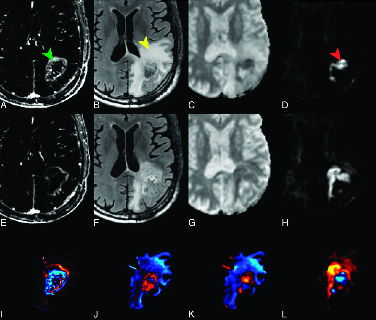
    A 67-year-old man with left parietal GBM status after resection and chemoradiation. Top row shows the T1 postcontrast–T1 precontrast (A), FLAIR (B), ADC (C), and RSI-CM (D) images before the start of bevacizumab, while the middle row shows the T1 postcontrast–T1 precontrast (E), FLAIR (F), ADC (G), and RSI-CM (H) images after the initiation of bevacizumab. Arrowheads indicate the contrast-enhancing region (green), the surrounding region of FLAIR-HI (yellow), and the region of RD on RSI-CMs (red). Although there is a decrease in contrast enhancement and surrounding FLAIR-HI after the initiation of bevacizumab, the region of RD increases and becomes more confluent; this change suggests worsening residual/recurrent tumor. Moreover, this increase in the region of RD is much more conspicuous on RSI-CMs compared with the ADC. The bottom row depicts these changes on “change maps” (change in T1 postcontrast–precontrast) (I), change in FLAIR (J), change in ADC (K), and change in the RSI-CMs (L), with red-yellow indicating an increase in signal intensity and blue-cyan indicating a decrease in signal intensity. Note that on the ADC change map (K), the area of increased RD is essentially masked by the decreased signal intensity within the region of surrounding FLAIR-HI.

  • Fig 2.
    • Download figure
    • Open in new tab
    • Download powerpoint
    Fig 2.

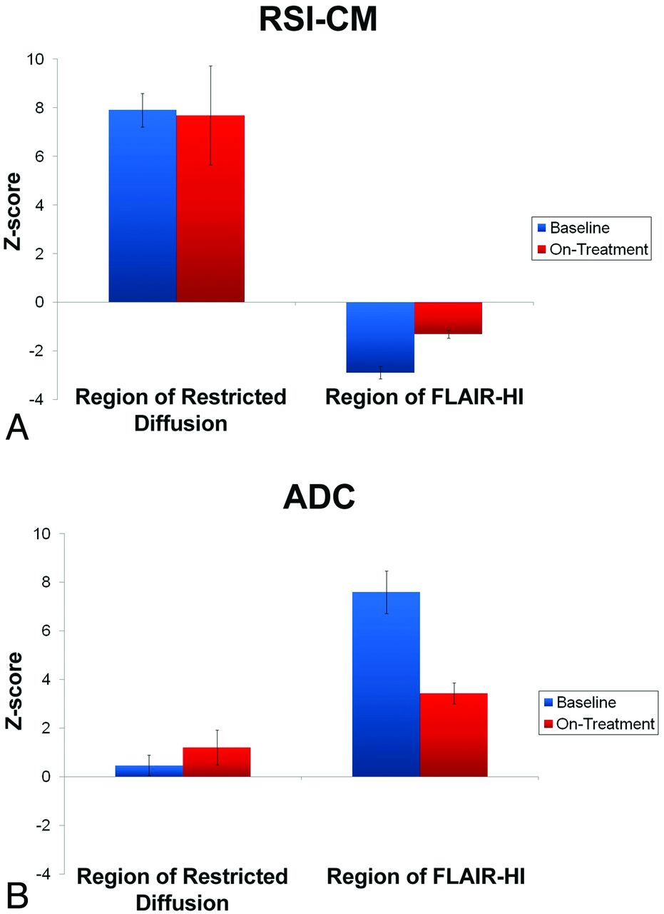
    Bar graphs depicting the mean normalized intensity values (z scores) of the RSI-CMs (A) and the ADC (B) in regions of RD and FLAIR-HI before and on treatment with bevacizumab. Error bars reflect the standard error.

  • Fig 3.
    • Download figure
    • Open in new tab
    • Download powerpoint
    Fig 3.

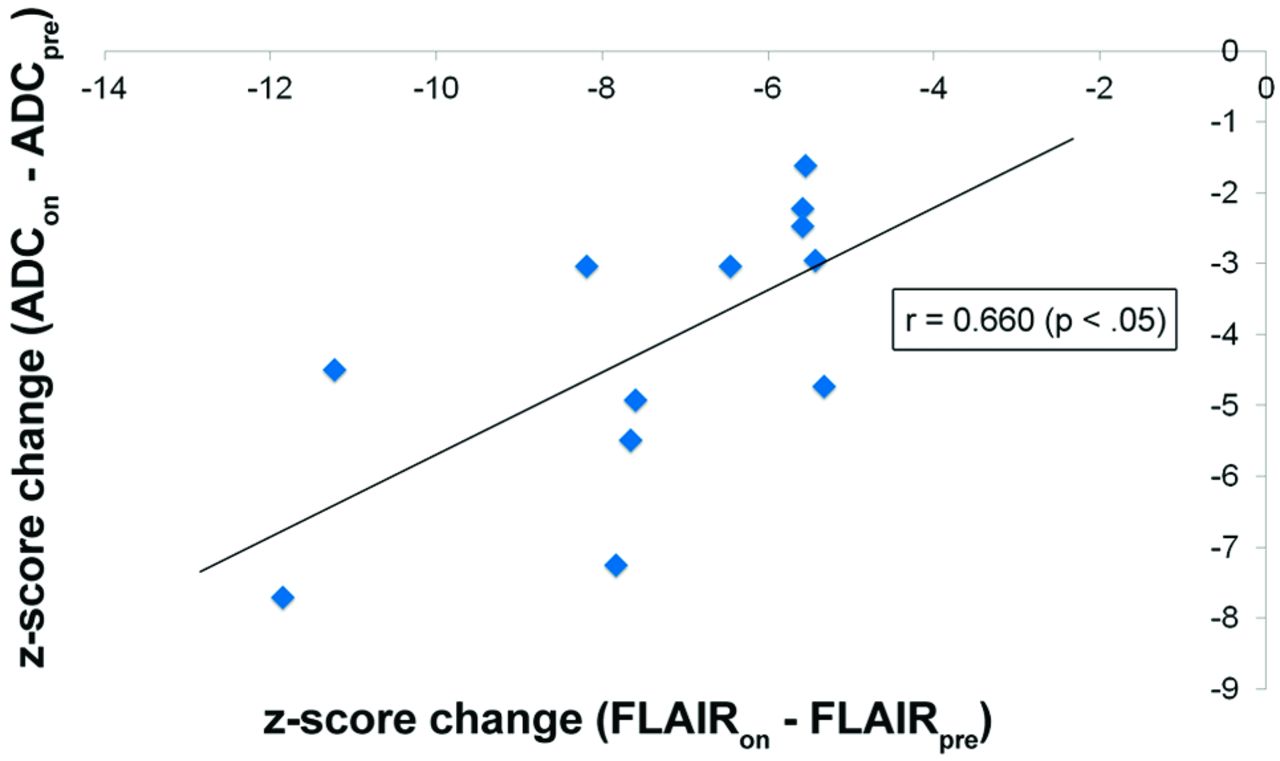
    Scatterplot of the relationship between change in ADC z scores (on treatment–pretreatment) and change in FLAIR-HI z scores (on treatment–pretreatment) within the regions of FLAIR-HI. Z scores represent intensity values normalized to NAWM.

Tables

  • Figures
  • Demographic and treatment-related characteristics of the patient sample

    PatientAge (yr)/SexPathologyInitial TherapySurgery Prior to Bevacizumab Scan (mo)aPre and Post-Bevacizumab Scans (day)bPost-Bevacizumab Pathologyc
    158/FGBM WHO IVXRT + TMZ; TMZ 5/28GTR, 1−31, +29–
    243/MAGN/WHO III/IVXRT + TMZ; TMZ 3/14GTR, 12−22, +16AA/WHO III/IV
    362/FGBM / WHO IVXRT + TMZ; TMZ 3/14STR, 1+−3, +112GBM/WHO IV
    449/MGBM/WHO IVXRT + TMZ; TMZ 3/14STR, 1−10, +22–
    557/FGBM/WHO IVXRT + TMZ; TMZ 5/28, TMZ 3/14STR, 6+−13, +49GBM/WHO IV
    663/FGBM/WHO IVXRT + TMZ; TMZ 5/28STR, 1−28, +46GBM/WHO IV + radiation effect
    766/MGBM/WHO IVXRT + TMZ; TMZ 5/28STR, 5−2, +34GBM/WHO IV + radiation necrosis
    856/MAOA/WHO IIIXRT + TMZ; TMZ 5/28RXN, 4+−15 days, +15–
    967/MGBM/WHO IVXRT + TMZ; TMZ 5/28GTR, 7−7, +29 days–
    1027/FAA/WHO IIIXRT + TMZBx, 2+−8, +16–
    1140/MGBM/WHO IVXRT + TMZ; TMZ 5/28STR, 5+−37, +35–
    1256/MGBM/WHO IVXRT + TMZ; TMZ 5/28STR, 7−49, +27–
    • Note:—AOA indicates anaplastic oligoastrocytoma; AA, anaplastic astrocytoma; XRT + TMZ, radiotherapy plus adjuvant temozolomide; TMZ 5/28, temozolomide for 5 days every 28 days; TMZ 3/14, temozolomide for 3 days every 14 days; GTR, gross total resection; STR, subtotal resection; RXN, craniotomy and resection; Bx, biopsy; AGN, anaplastic glioneural neoplasm; WHO = World Health Organization.

    • ↵a Months shown indicate the interval between the surgical event and the first scan obtained during bevacizumab treatment.

    • ↵b Interval in days between the scan prior to bevacizumab treatment (pre-bevacizumab scan), and initiation of bevacizumab therapy, followed by the interval in days between initiation of bevacizumab therapy and first scan during bevacizumab treatment (post-bevacizumab scan).

    • ↵c Pathology after bevacizumab therapy had started.

PreviousNext
Back to top

In this issue

American Journal of Neuroradiology: 34 (9)
American Journal of Neuroradiology
Vol. 34, Issue 9
1 Sep 2013
  • Table of Contents
  • Index by author
  • Complete Issue (PDF)
Advertisement
Print
Download PDF
Email Article

Thank you for your interest in spreading the word on American Journal of Neuroradiology.

NOTE: We only request your email address so that the person you are recommending the page to knows that you wanted them to see it, and that it is not junk mail. We do not capture any email address.

Enter multiple addresses on separate lines or separate them with commas.
Longitudinal Restriction Spectrum Imaging Is Resistant to Pseudoresponse in Patients with High-Grade Gliomas Treated with Bevacizumab
(Your Name) has sent you a message from American Journal of Neuroradiology
(Your Name) thought you would like to see the American Journal of Neuroradiology web site.
CAPTCHA
This question is for testing whether or not you are a human visitor and to prevent automated spam submissions.
Cite this article
P.D. Kothari, N.S. White, N. Farid, R. Chung, J.M. Kuperman, H.M. Girard, A. Shankaranarayanan, S. Kesari, C.R. McDonald, A.M. Dale
Longitudinal Restriction Spectrum Imaging Is Resistant to Pseudoresponse in Patients with High-Grade Gliomas Treated with Bevacizumab
American Journal of Neuroradiology Sep 2013, 34 (9) 1752-1757; DOI: 10.3174/ajnr.A3506

Citation Manager Formats

  • BibTeX
  • Bookends
  • EasyBib
  • EndNote (tagged)
  • EndNote 8 (xml)
  • Medlars
  • Mendeley
  • Papers
  • RefWorks Tagged
  • Ref Manager
  • RIS
  • Zotero
0 Responses
Respond to this article
Share
Bookmark this article
Longitudinal Restriction Spectrum Imaging Is Resistant to Pseudoresponse in Patients with High-Grade Gliomas Treated with Bevacizumab
P.D. Kothari, N.S. White, N. Farid, R. Chung, J.M. Kuperman, H.M. Girard, A. Shankaranarayanan, S. Kesari, C.R. McDonald, A.M. Dale
American Journal of Neuroradiology Sep 2013, 34 (9) 1752-1757; DOI: 10.3174/ajnr.A3506
del.icio.us logo Twitter logo Facebook logo Mendeley logo
  • Tweet Widget
  • Facebook Like
  • Google Plus One
Purchase

Jump to section

  • Article
    • Abstract
    • ABBREVIATIONS:
    • Materials and Methods
    • Results
    • Discussion
    • Conclusions
    • Acknowledgments
    • Footnotes
    • REFERENCES
  • Figures & Data
  • Info & Metrics
  • Responses
  • References
  • PDF

Related Articles

  • No related articles found.
  • PubMed
  • Google Scholar

Cited By...

  • Characterization of the Diffusion Signal of Breast Tissues using Multi-exponential Models
  • Edge Contrast of the FLAIR Hyperintense Region Predicts Survival in Patients with High-Grade Gliomas following Treatment with Bevacizumab
  • Lessons From Anti-Vascular Endothelial Growth Factor and Anti-Vascular Endothelial Growth Factor Receptor Trials in Patients With Glioblastoma
  • Diffusion-Weighted Imaging in Cancer: Physical Foundations and Applications of Restriction Spectrum Imaging
  • Advanced Magnetic Resonance Imaging of the Physical Processes in Human Glioblastoma
  • Pretreatment ADC Histogram Analysis Is a Predictive Imaging Biomarker for Bevacizumab Treatment but Not Chemotherapy in Recurrent Glioblastoma
  • Crossref (34)
  • Google Scholar

This article has been cited by the following articles in journals that are participating in Crossref Cited-by Linking.

  • Current Clinical Brain Tumor Imaging
    Javier E. Villanueva-Meyer, Marc C. Mabray, Soonmee Cha
    Neurosurgery 2017 81 3
  • Diffusion-Weighted Imaging in Cancer: Physical Foundations and Applications of Restriction Spectrum Imaging
    Nathan S. White, Carrie R. McDonald, Niky Farid, Josh Kuperman, David Karow, Natalie M. Schenker-Ahmed, Hauke Bartsch, Rebecca Rakow-Penner, Dominic Holland, Ahmed Shabaik, Atle Bjørnerud, Tuva Hope, Jona Hattangadi-Gluth, Michael Liss, J. Kellogg Parsons, Clark C. Chen, Steve Raman, Daniel Margolis, Robert E. Reiter, Leonard Marks, Santosh Kesari, Arno J. Mundt, Christopher J. Kane, Bob S. Carter, William G. Bradley, Anders M. Dale
    Cancer Research 2014 74 17
  • Lessons From Anti–Vascular Endothelial Growth Factor and Anti–Vascular Endothelial Growth Factor Receptor Trials in Patients With Glioblastoma
    Christine Lu-Emerson, Dan G. Duda, Kyrre E. Emblem, Jennie W. Taylor, Elizabeth R. Gerstner, Jay S. Loeffler, Tracy T. Batchelor, Rakesh K. Jain
    Journal of Clinical Oncology 2015 33 10
  • Advanced Magnetic Resonance Imaging of the Physical Processes in Human Glioblastoma
    Jayashree Kalpathy-Cramer, Elizabeth R. Gerstner, Kyrre E. Emblem, Ovidiu C. Andronesi, Bruce Rosen
    Cancer Research 2014 74 17
  • Dose-Dependent Cortical Thinning After Partial Brain Irradiation in High-Grade Glioma
    Roshan Karunamuni, Hauke Bartsch, Nathan S. White, Vitali Moiseenko, Ruben Carmona, Deborah C. Marshall, Tyler M. Seibert, Carrie R. McDonald, Nikdokht Farid, Anithapriya Krishnan, Joshua Kuperman, Loren Mell, James B. Brewer, Anders M. Dale, Jona A. Hattangadi-Gluth
    International Journal of Radiation Oncology*Biology*Physics 2016 94 2
  • New Directions in Anti-Angiogenic Therapy for Glioblastoma
    Nancy Wang, Rakesh K. Jain, Tracy T. Batchelor
    Neurotherapeutics 2017 14 2
  • Pretreatment ADC Histogram Analysis Is a Predictive Imaging Biomarker for Bevacizumab Treatment but Not Chemotherapy in Recurrent Glioblastoma
    B. M. Ellingson, S. Sahebjam, H. J. Kim, W. B. Pope, R. J. Harris, D. C. Woodworth, A. Lai, P. L. Nghiemphu, W. P. Mason, T. F. Cloughesy
    American Journal of Neuroradiology 2014 35 4
  • Assessment of Glioblastoma Response in the Era of Bevacizumab: Longstanding and Emergent Challenges in the Imaging Evaluation of Pseudoresponse
    Octavio D. Arevalo, Carolina Soto, Pejman Rabiei, Arash Kamali, Leomar Y. Ballester, Yoshua Esquenazi, Jay-Jiguang Zhu, Roy Francisco Riascos
    Frontiers in Neurology 2019 10
  • Multimodal imaging of gliomas in the context of evolving cellular and molecular therapies
    Olivier Keunen, Torfinn Taxt, Renate Grüner, Morten Lund-Johansen, Joerg-Christian Tonn, Tina Pavlin, Rolf Bjerkvig, Simone P. Niclou, Frits Thorsen
    Advanced Drug Delivery Reviews 2014 76
  • Distinct effects of nuclear volume fraction and cell diameter on high b‐value diffusion MRI contrast in tumors
    Nathan S. White, Anders M. Dale
    Magnetic Resonance in Medicine 2014 72 5

More in this TOC Section

  • White Matter Alterations in the Brains of Patients with Active, Remitted, and Cured Cushing Syndrome: A DTI Study
  • Qualitative and Quantitative Analysis of MR Imaging Findings in Patients with Middle Cerebral Artery Stroke Implanted with Mesenchymal Stem Cells
  • Fast Contrast-Enhanced 4D MRA and 4D Flow MRI Using Constrained Reconstruction (HYPRFlow): Potential Applications for Brain Arteriovenous Malformations
Show more Brain

Similar Articles

Advertisement

Indexed Content

  • Current Issue
  • Accepted Manuscripts
  • Article Preview
  • Past Issues
  • Editorials
  • Editor's Choice
  • Fellows' Journal Club
  • Letters to the Editor
  • Video Articles

Cases

  • Case Collection
  • Archive - Case of the Week
  • Archive - Case of the Month
  • Archive - Classic Case

More from AJNR

  • Trainee Corner
  • Imaging Protocols
  • MRI Safety Corner
  • Book Reviews

Multimedia

  • AJNR Podcasts
  • AJNR Scantastics

Resources

  • Turnaround Time
  • Submit a Manuscript
  • Submit a Video Article
  • Submit an eLetter to the Editor/Response
  • Manuscript Submission Guidelines
  • Statistical Tips
  • Fast Publishing of Accepted Manuscripts
  • Graphical Abstract Preparation
  • Imaging Protocol Submission
  • Evidence-Based Medicine Level Guide
  • Publishing Checklists
  • Author Policies
  • Become a Reviewer/Academy of Reviewers
  • News and Updates

About Us

  • About AJNR
  • Editorial Board
  • Editorial Board Alumni
  • Alerts
  • Permissions
  • Not an AJNR Subscriber? Join Now
  • Advertise with Us
  • Librarian Resources
  • Feedback
  • Terms and Conditions
  • AJNR Editorial Board Alumni

American Society of Neuroradiology

  • Not an ASNR Member? Join Now

© 2025 by the American Society of Neuroradiology All rights, including for text and data mining, AI training, and similar technologies, are reserved.
Print ISSN: 0195-6108 Online ISSN: 1936-959X

Powered by HighWire