
Article Figures & Data
Figures
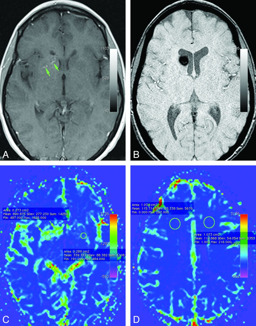
- Fig 1.
Axial contrast-enhanced T1-weighted (A) and susceptibility-weighted (B) images demonstrate a small developmental venous anomaly (arrows) in the right basal ganglia with a cavernous malformation in the right caudate head (B). Corresponding cerebral blood volume map shows a wide zone of higher cerebral blood volume (indicated by green on this color map) in the brain around the DVA. Note that the elevation of CBV is not restricted to the location of the individual venous channels of the DVA (arrows) but involves a wider confluent zone of brain around the draining vein. Note placement of regions of interest to get the objective parameters for quantification of perfusion around DVAs (C) and for brain with normal venous drainage (D).
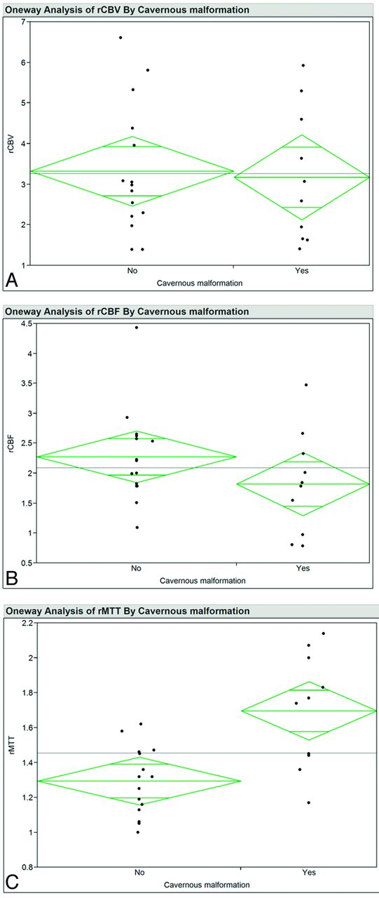
- Fig 2.
Ninety-five percent mean diamond plots for rCBV (A), rCBF (B), and rMTT (C) for patients with and without cavernous malformations. The horizontal line is the grand mean. The heights of the diamonds represent the 95% confidence intervals, and the widths of the diamonds are proportional to the sample sizes. If the overlap line of one diamond is closer to the mean of another diamond than is the overlap line of that diamond, there is no difference between the groups.
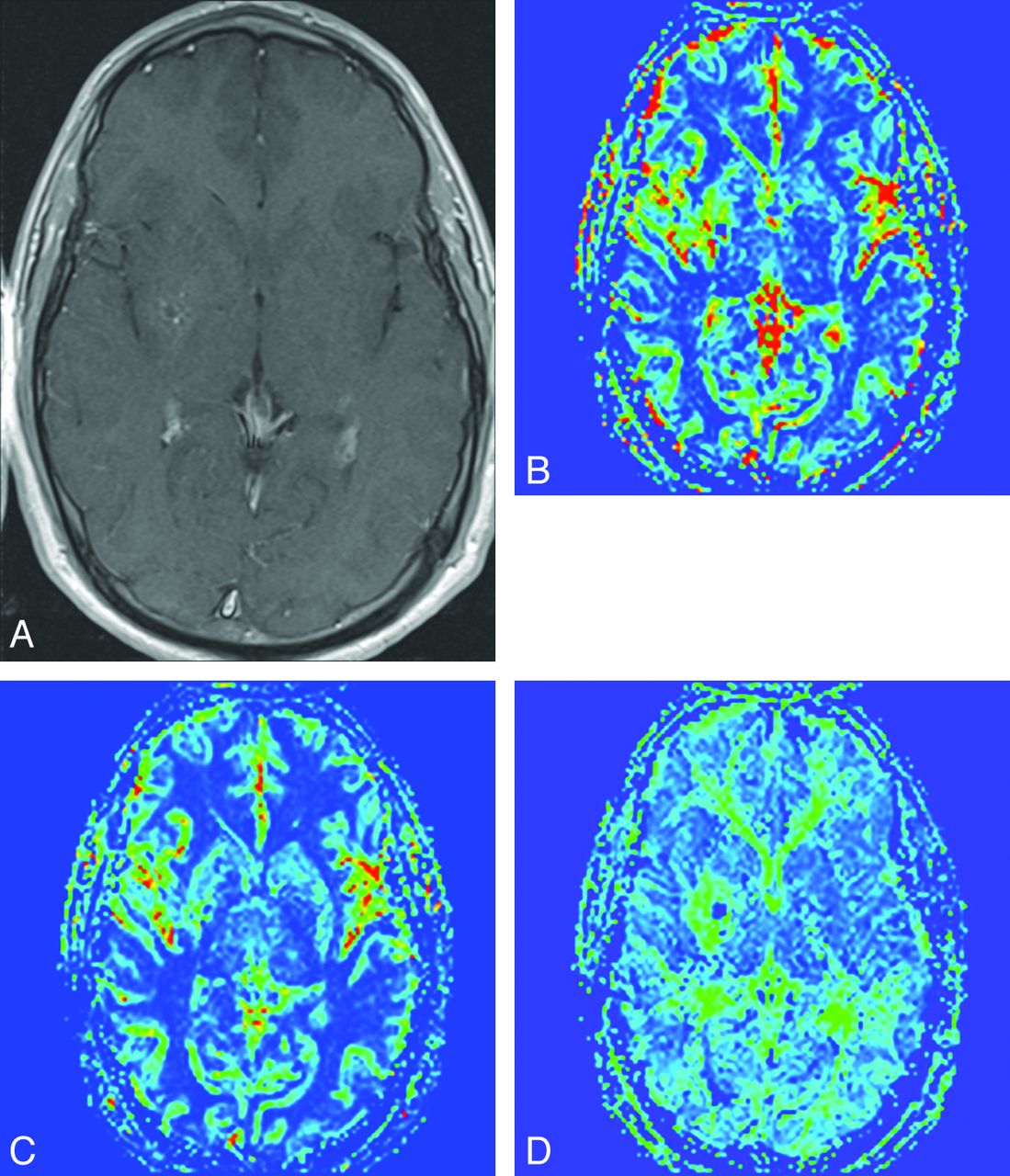
- Fig 3.
Axial contrast-enhanced T1-weighted image (A) demonstrates tributaries of the DVA in the right lentiform nucleus, seen as punctate enhancing foci. Corresponding rCBV (B), rCBF (C), and rMTT (D) maps demonstrate a zone of perfusion alteration around these tributaries incorporating otherwise normal-appearing brain tissue. Note that in this case, the alteration in rCBV and rMTT maps was more pronounced relative to the rCBF.


Tables
- Table 1:
Descriptive statistics for relative perfusion parameters for brain parenchyma drained by DVAs and for control regions with normal venous drainage
Perfusion Parametera Mean 95% CI Median Minimum Maximum P Valueb rCBV 3.26 2.61–3.91 2.98 1.39 6.61 .04 rCBF 2.09 1.75–2.43 2.00 0.79 4.43 .19 rMTT 1.46 1.32–1.59 1.44 1.00 2.14 .15 cCBV 1.00 0.98–1.02 0.99 0.89 1.06 .15 cCBF 1.01 0.98–1.04 1.00 0.94 1.27 <.01 cMTT 1.00 0.98–1.02 1.00 0.91 1.15 .11 ↵a rCBV, rCBF, and rMTT represent relative cerebral blood volume, relative cerebral blood flow, and relative mean transit time respectively, for brain tissue around the DVA. cCBV, cCBF, and cMTT represent corresponding control values as measured in brain tissue with normal venous drainage (ipsilateral to the DVA).
↵b P value for Shapiro-Wilk W-test for data distribution normality. A value < .05 indicates a non-normal distribution.
Perfusion Parametera CM Mean 95% CI Median Minimum Maximum P Valueb rCBV − 3.32 2.45–4.20 2.98 1.39 6.61 .16 + 3.17 2.01–4.34 2.83 1.41 5.92 .24 rCBF − 2.28 1.85–2.70 2.21 1.09 4.43 .07 + 1.82 1.20–2.43 1.81 0.79 3.47 .60 rMTT − 1.29 1.19–1.40 1.32 1.00 1.62 .67 + 1.70 1.46–1.93 1.75 1.17 2.14 .62 cCBV − 1.01 0.99–1.03 1.01 0.94 1.06 .22 + 0.98 0.95–1.02 0.98 0.89 1.05 .85 cCBF − 1.02 0.98–1.06 1.01 0.95 1.27 <.01 + 0.99 0.96–1.03 0.98 0.94 1.08 .33 cMTT − 1.00 0.97–1.03 1.00 0.91 1.15 .35 + 0.99 0.97–1.02 0.99 0.94 1.05 .99 Note:— +, present; −, absent.
↵a rCBV, rCBF, and rMTT represent relative cerebral blood volume, relative cerebral blood flow, and relative mean transit time respectively, as measured in brain tissue around the DVA. cCBV, cCBF, and cMTT represent corresponding control values as measured in brain tissue with normal venous drainage (ipsilateral to the DVA).
↵b P value for Shapiro-Wilk W-test for data distribution normality. A value < .05 indicates a non-normal distribution.


Jump to section
Related Articles
Cited By...
- Prevalence of Developmental Venous Anomalies in Association with Sporadic Cavernous Malformations on 7T MRI
- Symptomatic Developmental Venous Anomaly: State-of-the-Art Review on Genetics, Pathophysiology, and Imaging Approach to Diagnosis
- Cerebral cavernous malformations do not fall in the spectrum of PIK3CA-related overgrowth
- Neonatal Developmental Venous Anomalies: Clinicoradiologic Characterization and Follow-Up
- Cavernous malformations with DVA: Hold those knives
- Interaction of Developmental Venous Anomalies with Resting-State Functional MRI Measures
- The Central Vein: FLAIR Signal Abnormalities Associated with Developmental Venous Anomalies in Patients with Multiple Sclerosis
- A benchmark approach to hemorrhage risk management of cavernous malformations
- Increased Prevalence of Developmental Venous Anomalies in Children with Intracranial Neoplasms
- Brain Metabolic Abnormalities Associated with Developmental Venous Anomalies
- Diffusion and Perfusion MRI Findings of the Signal-Intensity Abnormalities of Brain Associated with Developmental Venous Anomaly
- Brain Parenchymal Signal Abnormalities Associated with Developmental Venous Anomalies in Children and Young Adults