Advertisement

Main menu

  • Home
  • Content
    • Current Issue
    • Accepted Manuscripts
    • Article Preview
    • Past Issue Archive
    • Video Articles
    • AJNR Case Collection
    • Case of the Week Archive
    • Case of the Month Archive
    • Classic Case Archive
  • Special Collections
    • AJNR Awards
    • Low-Field MRI
    • Alzheimer Disease
    • ASNR Foundation Special Collection
    • Photon-Counting CT
    • View All
  • Multimedia
    • AJNR Podcasts
    • AJNR SCANtastic
    • Trainee Corner
    • MRI Safety Corner
    • Imaging Protocols
  • For Authors
    • Submit a Manuscript
    • Submit a Video Article
    • Submit an eLetter to the Editor/Response
    • Manuscript Submission Guidelines
    • Statistical Tips
    • Fast Publishing of Accepted Manuscripts
    • Graphical Abstract Preparation
    • Imaging Protocol Submission
    • Author Policies
  • About Us
    • About AJNR
    • Editorial Board
    • Editorial Board Alumni
  • More
    • Become a Reviewer/Academy of Reviewers
    • Subscribers
    • Permissions
    • Alerts
    • Feedback
    • Advertisers
    • ASNR Home

User menu

  • Alerts
  • Log in

Search

  • Advanced search
American Journal of Neuroradiology

American Journal of Neuroradiology

ASHNR American Society of Functional Neuroradiology ASHNR American Society of Pediatric Neuroradiology ASSR
  • Alerts
  • Log in

Advanced Search

  • Home
  • Content
    • Current Issue
    • Accepted Manuscripts
    • Article Preview
    • Past Issue Archive
    • Video Articles
    • AJNR Case Collection
    • Case of the Week Archive
    • Case of the Month Archive
    • Classic Case Archive
  • Special Collections
    • AJNR Awards
    • Low-Field MRI
    • Alzheimer Disease
    • ASNR Foundation Special Collection
    • Photon-Counting CT
    • View All
  • Multimedia
    • AJNR Podcasts
    • AJNR SCANtastic
    • Trainee Corner
    • MRI Safety Corner
    • Imaging Protocols
  • For Authors
    • Submit a Manuscript
    • Submit a Video Article
    • Submit an eLetter to the Editor/Response
    • Manuscript Submission Guidelines
    • Statistical Tips
    • Fast Publishing of Accepted Manuscripts
    • Graphical Abstract Preparation
    • Imaging Protocol Submission
    • Author Policies
  • About Us
    • About AJNR
    • Editorial Board
    • Editorial Board Alumni
  • More
    • Become a Reviewer/Academy of Reviewers
    • Subscribers
    • Permissions
    • Alerts
    • Feedback
    • Advertisers
    • ASNR Home
  • Follow AJNR on Twitter
  • Visit AJNR on Facebook
  • Follow AJNR on Instagram
  • Join AJNR on LinkedIn
  • RSS Feeds

AJNR Awards, New Junior Editors, and more. Read the latest AJNR updates

Research ArticleHead & NeckF

Hyperintense Optic Nerve Heads on Diffusion-Weighted Imaging: A Potential Imaging Sign of Papilledema

R. Viets, M. Parsons, G. Van Stavern, C. Hildebolt and A. Sharma
American Journal of Neuroradiology July 2013, 34 (7) 1438-1442; DOI: https://doi.org/10.3174/ajnr.A3388
R. Viets
aFrom the Mallinckrodt Institute of Radiology (R.V., M.P., C.H., A.S.)
  • Find this author on Google Scholar
  • Find this author on PubMed
  • Search for this author on this site
M. Parsons
aFrom the Mallinckrodt Institute of Radiology (R.V., M.P., C.H., A.S.)
  • Find this author on Google Scholar
  • Find this author on PubMed
  • Search for this author on this site
G. Van Stavern
bDepartments of Ophthalmology and Neurology (G.V.S.), Washington University School of Medicine, St. Louis, Missouri.
  • Find this author on Google Scholar
  • Find this author on PubMed
  • Search for this author on this site
C. Hildebolt
aFrom the Mallinckrodt Institute of Radiology (R.V., M.P., C.H., A.S.)
  • Find this author on Google Scholar
  • Find this author on PubMed
  • Search for this author on this site
A. Sharma
aFrom the Mallinckrodt Institute of Radiology (R.V., M.P., C.H., A.S.)
  • Find this author on Google Scholar
  • Find this author on PubMed
  • Search for this author on this site
  • Article
  • Figures & Data
  • Supplemental
  • Info & Metrics
  • Responses
  • References
  • PDF
Loading

Article Figures & Data

Figures

  • Tables
  • Fig 1.
    • Download figure
    • Open in new tab
    • Download powerpoint
    Fig 1.

    Axial DWI at the level of the ON heads in a control participant (A) and in 2 patients with clinically proven papilledema (B and C) showing lack of any hyperintensity at the ON head in the control participant, with a signal at the ON heads less than that of the globe margins. In contrast, mild (B) and prominent (C) hyperintensity at the ON heads is observed in patients with papilledema, with signal intensity minimally and substantially higher compared with the globe margins.
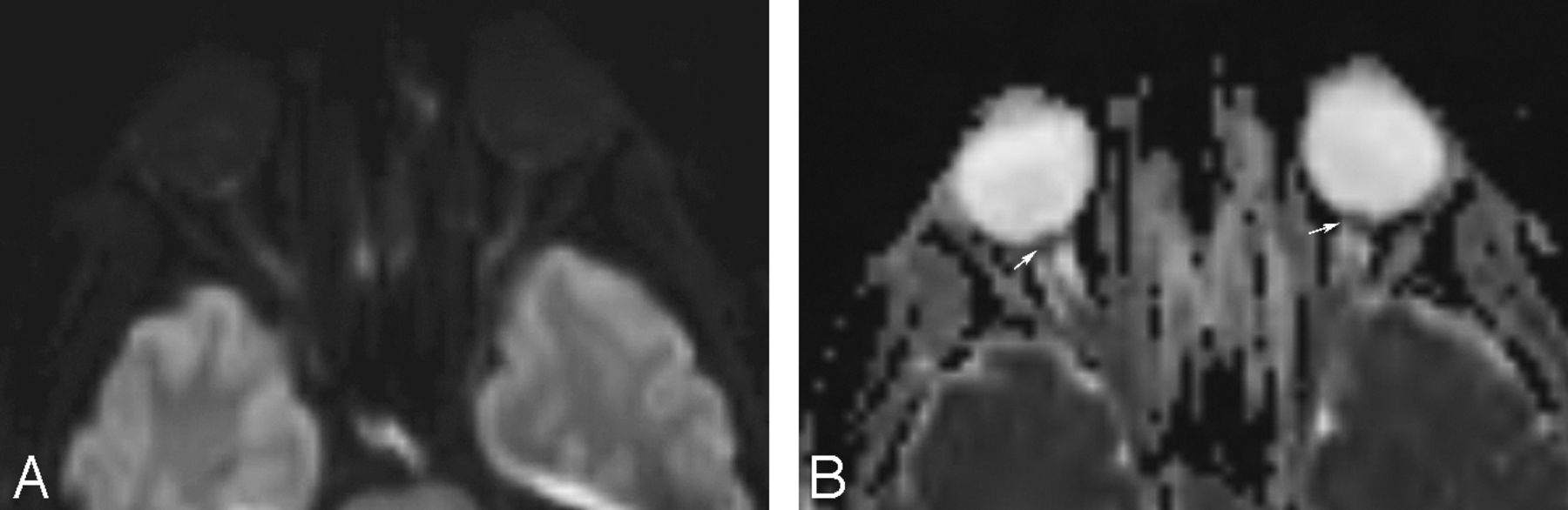
  • Fig 2.
    • Download figure
    • Open in new tab
    • Download powerpoint
    Fig 2.

    Axial diffusion-weighted image (A) and ADC map (B) in a patient with clinically proven papilledema showing prominent hyperintensity at both ON heads with a corresponding hypointense signal on the ADC map suggestive of restricted diffusion.

Tables

  • Figures
    • View popup
    Table 1:

    Presence of DWI hyperintensity at ON heads in patients with papilledema and control participants

    Control Participants (n = 20)Patients (n = 19)
    1 EyeBoth Eyes1/Both Eyes1 EyeBoth Eyes1/Both Eyes
    Reader 15.0% (1/20)0% (0/20)5.0% (1/20)26.3% (5/19)26.3% (5/19)52.6% (10/19)
    Reader 210.0% (2/20)0% (0/20)10.0% (2/20)36.8% (7/19)42.1% (8/19)78.9% (15/19)
    • View popup
    Table 2:

    Fisher exact test P values for difference between patients with papilledema and control participants

    1 EyeBoth Eyes1/Both Eyes
    Reader 1.091.020.001
    Reader 2.065.001<.001
    • View popup
    Table 3:

    Presence of DWI hyperintensity at ON heads based on total number of patient eyes

    Control Participants 40 Total EyesPatients 38 Total EyesP Value
    Reader 12.5% (1/40)39.5% (15/38)<.001
    Reader 25.0% (2/40)60.5% (23/38)<.001
    • View popup
    Table 4:

    Presence of DWI hyperintensity at ON heads in patients stratified by Frisen papilledema grade

    Grade II or HigherGrade III or HigherGrade IV or Higher
    Patients (n = 16)Total Eyes (n = 28)Patients (n = 11)Total Eyes (n = 19)Patients (n = 4)Total Eyes (n = 7)
    Reader 156.3% (9/16)42.9% (12/28)63.6% (7/11)57.9% (11/19)100.0% (4/4)85.7% (6/7)
    Reader 281.3% (13/16)64.3% (18/28)81.8% (9/11)78.9% (15/19)100.0% (4/4)85.7% (6/7)
PreviousNext
Back to top

In this issue

American Journal of Neuroradiology: 34 (7)
American Journal of Neuroradiology
Vol. 34, Issue 7
1 Jul 2013
  • Table of Contents
  • Index by author
Advertisement
Print
Download PDF
Email Article

Thank you for your interest in spreading the word on American Journal of Neuroradiology.

NOTE: We only request your email address so that the person you are recommending the page to knows that you wanted them to see it, and that it is not junk mail. We do not capture any email address.

Enter multiple addresses on separate lines or separate them with commas.
Hyperintense Optic Nerve Heads on Diffusion-Weighted Imaging: A Potential Imaging Sign of Papilledema
(Your Name) has sent you a message from American Journal of Neuroradiology
(Your Name) thought you would like to see the American Journal of Neuroradiology web site.
CAPTCHA
This question is for testing whether or not you are a human visitor and to prevent automated spam submissions.
Cite this article
R. Viets, M. Parsons, G. Van Stavern, C. Hildebolt, A. Sharma
Hyperintense Optic Nerve Heads on Diffusion-Weighted Imaging: A Potential Imaging Sign of Papilledema
American Journal of Neuroradiology Jul 2013, 34 (7) 1438-1442; DOI: 10.3174/ajnr.A3388

Citation Manager Formats

  • BibTeX
  • Bookends
  • EasyBib
  • EndNote (tagged)
  • EndNote 8 (xml)
  • Medlars
  • Mendeley
  • Papers
  • RefWorks Tagged
  • Ref Manager
  • RIS
  • Zotero
0 Responses
Respond to this article
Share
Bookmark this article
Hyperintense Optic Nerve Heads on Diffusion-Weighted Imaging: A Potential Imaging Sign of Papilledema
R. Viets, M. Parsons, G. Van Stavern, C. Hildebolt, A. Sharma
American Journal of Neuroradiology Jul 2013, 34 (7) 1438-1442; DOI: 10.3174/ajnr.A3388
del.icio.us logo Twitter logo Facebook logo Mendeley logo
  • Tweet Widget
  • Facebook Like
  • Google Plus One
Purchase

Jump to section

  • Article
    • Abstract
    • ABBREVIATION:
    • Materials and Methods
    • Results
    • Discussion
    • Conclusions
    • REFERENCES
  • Figures & Data
  • Supplemental
  • Info & Metrics
  • Responses
  • References
  • PDF

Related Articles

  • PubMed
  • Google Scholar

Cited By...

  • Contrast-Enhanced 3D-FLAIR Imaging of the Optic Nerve and Optic Nerve Head: Novel Neuroimaging Findings of Idiopathic Intracranial Hypertension
  • Crossref (26)
  • Google Scholar

This article has been cited by the following articles in journals that are participating in Crossref Cited-by Linking.

  • Clinical applications of diffusion weighted imaging in neuroradiology
    Marta Drake-Pérez, Jose Boto, Aikaterini Fitsiori, Karl Lovblad, Maria Isabel Vargas
    Insights into Imaging 2018 9 4
  • MRI findings as markers of idiopathic intracranial hypertension
    Arhem F. Barkatullah, Lakshmi Leishangthem, Heather E. Moss
    Current Opinion in Neurology 2021 34 1
  • Utility of MRI Diffusion Techniques in the Evaluation of Tumors of the Head and Neck
    José Martínez Barbero, Inmaculada Rodríquez Jiménez, Teodoro Martin Noguerol, Antonio Luna Alcalá
    Cancers 2013 5 3
  • Clinical applications of diffusion-weighted sequence in brain imaging: beyond stroke
    Siddhartha Gaddamanugu, Omid Shafaat, Houman Sotoudeh, Amir Hossein Sarrami, Ali Rezaei, Zahra Saadatpour, Aparna Singhal
    Neuroradiology 2022 64 1
  • Magnetic Resonance Imaging of Idiopathic Intracranial Hypertension: Before and After Treatment
    Hale Z. Batur Caglayan, Murat Ucar, Murat Hasanreisoglu, Bijen Nazliel, Nil Tokgoz
    Journal of Neuro-Ophthalmology 2019 39 3
  • Neuroimaging Features of Idiopathic Intracranial Hypertension Persist After Resolution of Papilloedema
    Randy O. Chang, Brigid K. Marshall, Noushin Yahyavi, Aseem Sharma, Julia Huecker, Mae O. Gordon, Collin McClelland, Gregory P. Van Stavern
    Neuro-Ophthalmology 2016 40 4
  • Correlation Between Papilledema Grade and Diffusion-Weighted Magnetic Resonance Imaging in Idiopathic Intracranial Hypertension
    David M. Salvay, Leena V. Padhye, Julie B. Huecker, Mae O. Gordon, Ryan Viets, Aseem Sharma, Gregory P. Van Stavern
    Journal of Neuro-Ophthalmology 2014 34 4
  • Contrast-Enhanced 3D-FLAIR Imaging of the Optic Nerve and Optic Nerve Head: Novel Neuroimaging Findings of Idiopathic Intracranial Hypertension
    E. Golden, R. Krivochenitser, N. Mathews, C. Longhurst, Y. Chen, J.-P.J. Yu, T.A. Kennedy
    American Journal of Neuroradiology 2019 40 2
  • Features of Idiopathic Intracranial Hypertension on MRI With MR Elastography: Prospective Comparison With Control Individuals and Assessment of Postintervention Changes
    Petrice M. Cogswell, Matthew C. Murphy, Ajay A. Madhavan, M. Tariq Bhatti, Jeremy K. Cutsforth-Gregory, Matthew L. Senjem, John Huston, John J. Chen
    American Journal of Roentgenology 2022 219 6
  • A Unique Radiologic Case of Optic Nerve Infarction in a Patient With Mucormycosis
    Alexandria L. Chaulk, Thai H. Do, Emilio P. Supsupin, Meenakshi B. Bhattacharjee, Karina Richani, Ore-Ofe O. Adesina
    Journal of Neuro-Ophthalmology 2021 41 3

More in this TOC Section

  • Chondrosarcoma vs Synovial Chondromatosis: Imaging
  • WHO Classification Update: Nasal&Skull Base Tumors
  • Peritumoral Signal in Vestibular Schwannomas
Show more Head & Neck

Similar Articles

Advertisement

Indexed Content

  • Current Issue
  • Accepted Manuscripts
  • Article Preview
  • Past Issues
  • Editorials
  • Editor's Choice
  • Fellows' Journal Club
  • Letters to the Editor
  • Video Articles

Cases

  • Case Collection
  • Archive - Case of the Week
  • Archive - Case of the Month
  • Archive - Classic Case

More from AJNR

  • Trainee Corner
  • Imaging Protocols
  • MRI Safety Corner
  • Book Reviews

Multimedia

  • AJNR Podcasts
  • AJNR Scantastics

Resources

  • Turnaround Time
  • Submit a Manuscript
  • Submit a Video Article
  • Submit an eLetter to the Editor/Response
  • Manuscript Submission Guidelines
  • Statistical Tips
  • Fast Publishing of Accepted Manuscripts
  • Graphical Abstract Preparation
  • Imaging Protocol Submission
  • Evidence-Based Medicine Level Guide
  • Publishing Checklists
  • Author Policies
  • Become a Reviewer/Academy of Reviewers
  • News and Updates

About Us

  • About AJNR
  • Editorial Board
  • Editorial Board Alumni
  • Alerts
  • Permissions
  • Not an AJNR Subscriber? Join Now
  • Advertise with Us
  • Librarian Resources
  • Feedback
  • Terms and Conditions
  • AJNR Editorial Board Alumni

American Society of Neuroradiology

  • Not an ASNR Member? Join Now

© 2025 by the American Society of Neuroradiology All rights, including for text and data mining, AI training, and similar technologies, are reserved.
Print ISSN: 0195-6108 Online ISSN: 1936-959X

Powered by HighWire