
Article Figures & Data
Figures
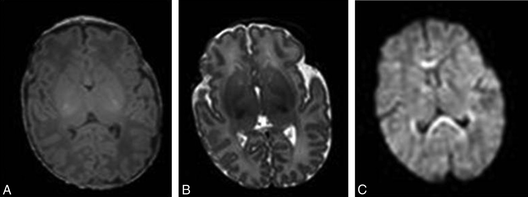
- Fig 1.
T1-weighted (A), T2-weighted (B), and diffusion-weighted (C) axial MR brain images of a 5-day-old full-term neonate without motion artifacts acquired at 1.5T.
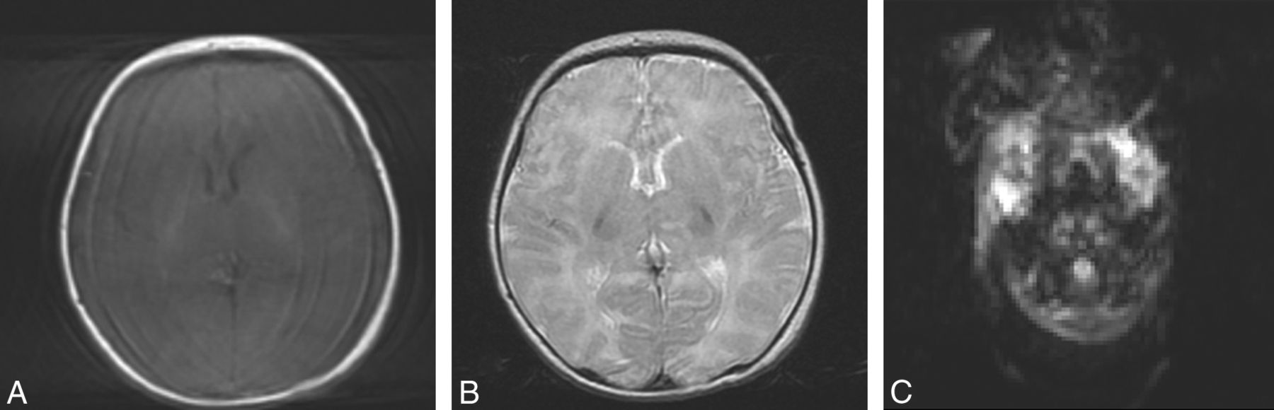
- Fig 2.
T1-weighted (A), T2-weighted (B), and diffusion-weighted (C) axial MR brain images of a 14-day-old full-term neonate acquired at 1.5T. Motion artifacts in the form of high-signal-intensity ghosts can be seen.
- Fig 3.
T1-weighted gradient-echo axial brain images at 1.5T (TR, 142 ms; TE, 6 ms; section thickness, 4 mm; scanning time, 16 seconds) of a 28-week-old fetus without motion artifacts after a successful breath-hold (A) and with motion artifacts after an incomplete breath-hold (B). Breathing artifacts appear in the form of high-signal ghosts in the operator-selected phase-encoding direction and severely degrade image quality.
- Fig 4.
Successive snapshots of a cine bFFE acquisition obtained at 1.5T (TR, 3.21 ms; TE, 1.59 ms; slab thickness, 30–40 mm; scanning duration, 30 seconds for 100 dynamic scans) of a 25-week-old fetus moving inside the uterus. The range and direction of movement of the fetal head (red star) and body and legs (dotted lines) at 6 (A), 12 (B), 18 (C), 24 (D), and 30 (E) seconds can be appreciated.
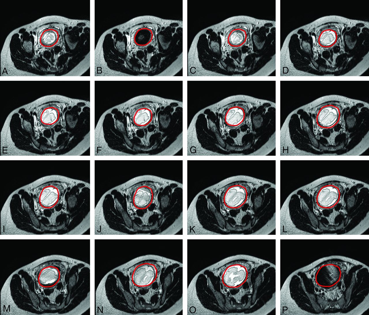
- Fig 5.
Successive axially planned sections of a single-shot fast-spin-echo acquisition at 1.5T (TR, 1000 ms; TE, 127 ms; section thickness, 4 mm; scanning duration, 26 seconds) of a 32-week-old fetus (E) with significant fetal motion occurring during data acquisition and resulting in blurring (C, J, M), contrast changes (P), and ultimately signal void when motion is extreme (B). Please note that though sections were planned in the axial plane, fetal movement resulted in plane transposition in the produced images (A–P) (fetal brain is circumscribed in red to distinguish it from neighboring maternal tissues).
- Fig 6.
Ringing artifacts (arrow) at the back of the brain of a 4-week-old full-term neonate on an axial maximum intensity projection of an optimized neonatal MR angiography protocol79 acquired at 3T. Ringing artifacts occur due to data undersampling, and should not be confused with motion-artifacts ghosts.
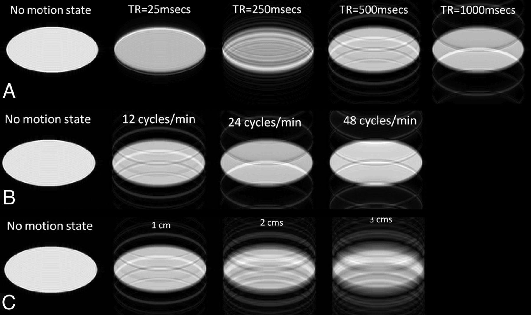
- Fig 7.
The effect of varying imaging-acquisition parameters such as TR (A) and motion characteristics (varying speed of motion, [B] and varying amplitude of motion [C]) on motion artifacts appearance, compared with the nonmotion status (first column). Note that the longer the TR (A) (range, 25–1000 ms) and the faster the motion (measured in cycles/minute; range, 12–48 cycles/min) (B), the farther apart the ghosts appear; also the bigger the amplitude of motion (measured in centimeters; range, 1–3 cm), the brighter is the ghost and the longer, its trace (C).
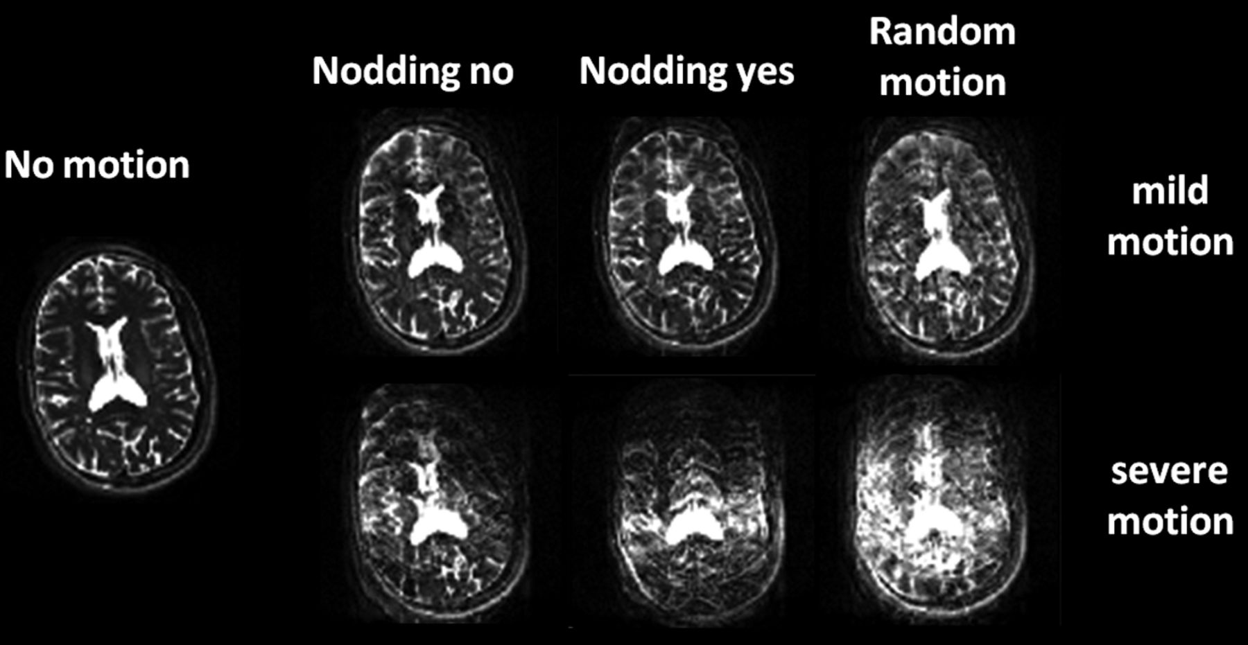
- Fig 8.
The effect of in-plane motion (side-to-side head nodding or “nodding no,” first column), through-plane motion (up and down head nodding or “nodding yes,” second column), random motion (combination of in-plane and through-plane motion, third column), and different motion intensities (top row: mild motion; bottom row: severe motion) on image quality of axial T2-weighted fast spin-echo acquisitions of a healthy adult volunteer. Through-plane severe patient motion is detrimental to image quality.
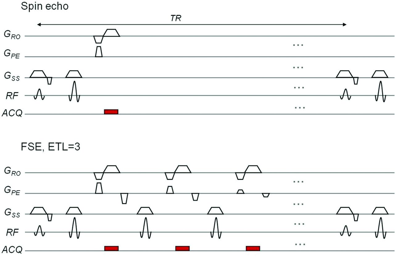
- Fig 9.
A sequence diagram for a spin-echo (top) and fast spin-echo sequence with an echo-train length or number of refocusing 180° pulses of 3 (bottom), which significantly decreases scanning time from 2-and-a-half minutes to 1 minute. ACQ indicates data acquisition.
- Fig 10.
A sequence diagram for gradient-echo (top) and EPI sequences with an EPI factor (or number of gradient reversals) of 12 (bottom), which significantly decreases scanning time from 1 minute to 6 seconds. ACQ indicates data acquisition.
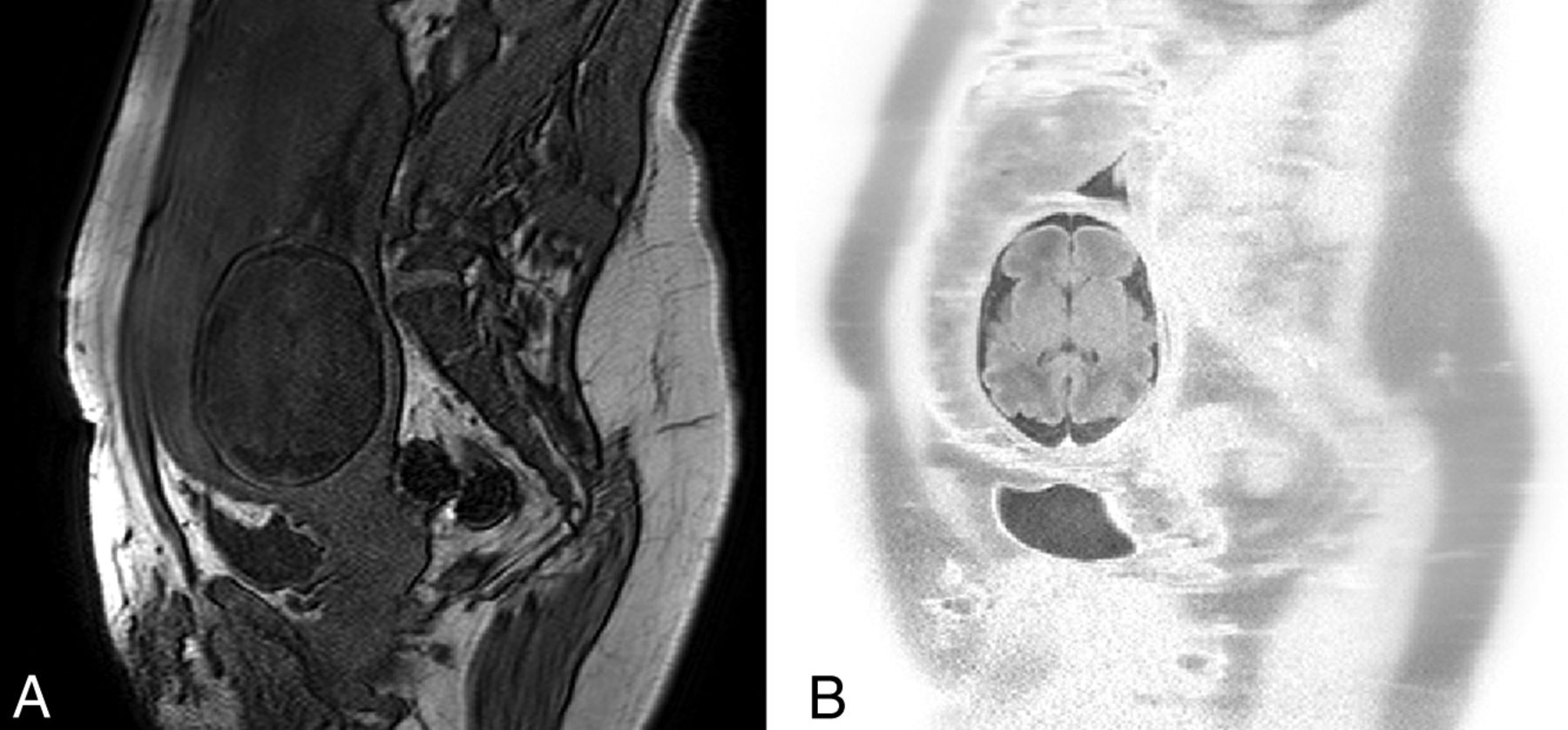
- Fig 11.
An axial T1-weighted gradient-echo breath-hold acquisition of the fetal brain (A) and a motion-resistant axial T1-weighted free-breathing SNAPIR (Snapshot Inversion Recovery) acquisition of the same fetal patient acquired at 34 weeks' gestation at 1.5T. (B) Sections were anatomically matched for comparison. Depiction of anatomic structures (cerebral cortex, ventricular system) is improved with the optimized SNAPIR acquisition compared with the breath-hold protocol.
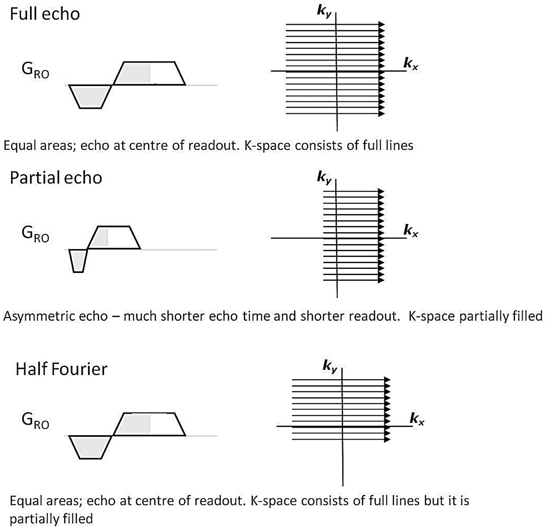
- Fig 12.
Readout gradient diagrams and k-space sampling strategies for different data-truncation techniques such as partial echo and half-Fourier compared with full-echo. ky indicates the y-axis of the k-space; kx, the x-axis of the k-space. With partial echo or half-Fourier, scanning time can be reduced.


In this issue


Jump to section
Related Articles
Cited By...
- Automated 3D reconstruction of the fetal thorax in the standard atlas space from motion-corrupted MRI stacks for 21-36 weeks GA range
- In utero diffusion tensor imaging of the fetal brain: a reproducibility study
- 'Feed and wrap' or sedate and immobilise for neonatal brain MRI?
- Choice of Diffusion Tensor Estimation Approach Affects Fiber Tractography of the Fornix in Preterm Brain