
Article Figures & Data
Figures
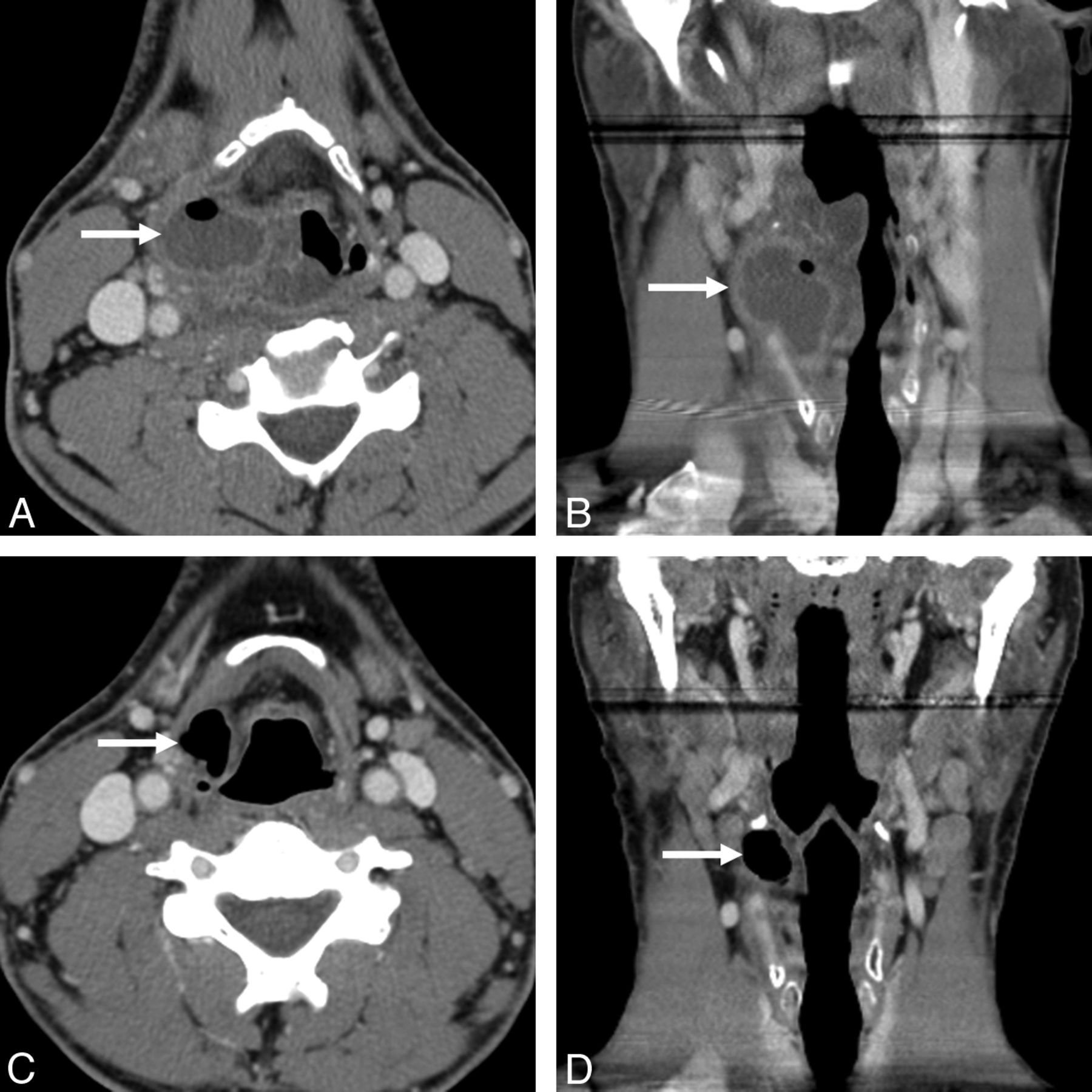
- Fig. 1.
Thirty-eight-year-old man with 1-week history of sore throat and productive cough and 3-day history of fever, dysphagia, odynophagia, and inability to tolerate liquids and solids. A and B, Contrast-enhanced axial (A) and coronal (B) neck CT images demonstrated a peripherally enhancing fluid collection with an air-fluid level in the right paraglottic space, bulging through the thyrohyoid membrane (arrows), consistent with a laryngopyocele. There was surrounding epiglottic and posterior pharyngeal wall edema with narrowing of the airway. C and D, Axial (C) and coronal (D) neck CT images performed 2 months after the acute presentation demonstrated interval resolution of the peripherally enhancing fluid collection and a residual mixed right laryngocele (arrows), protruding through the thyrohyoid membrane.
- Fig. 2.
Kopans needle and hookwire localization kit (upper) with hookwire tip at the needle tip (middle) and the hookwire deployed through the needle (lower). When the black marker on the wire (black arrows) is at the level of the needle hub (middle image), the tip of the wire is at the tip of the needle. The distal 1 cm of the wire has a curved hook (yellow arrows), which allows the wire to be advanced in the lumen of the introducer needle (middle image) and then to spring open when the needle is withdrawn >1 cm (lower image). The thicker component on the distal wire (open arrow in upper image) is just proximal to the hook and allows the surgeon to anticipate the depth to the tip.
- Fig. 3.
Hookwire placement technique. A, Preliminary noncontrast axial CT image was obtained for planning the needle approach (line in A) and measuring the distance between the skin-entry site and the desired needle-tip position at the level of the lateral laryngocele wall. The needle path should avoid major vascular structures: the carotid artery (red) and internal jugular vein (blue). B and C, Axial CT images during (B) and immediately following (C) the needle-hookwire placement demonstrate the tip of the Kopans needle (arrow in B) and the tip of the hookwire (open arrow in C) at the level of the lateral laryngocele wall. Note the wire protruding through the skin (arrowhead in C).


In this issue


Jump to section
Related Articles
Cited By...
- No citing articles found.