Advertisement

Main menu

  • Home
  • Content
    • Current Issue
    • Accepted Manuscripts
    • Article Preview
    • Past Issue Archive
    • Video Articles
    • AJNR Case Collection
    • Case of the Week Archive
    • Case of the Month Archive
    • Classic Case Archive
  • Special Collections
    • AJNR Awards
    • Low-Field MRI
    • Alzheimer Disease
    • ASNR Foundation Special Collection
    • Photon-Counting CT
    • View All
  • Multimedia
    • AJNR Podcasts
    • AJNR SCANtastic
    • Trainee Corner
    • MRI Safety Corner
    • Imaging Protocols
  • For Authors
    • Submit a Manuscript
    • Submit a Video Article
    • Submit an eLetter to the Editor/Response
    • Manuscript Submission Guidelines
    • Statistical Tips
    • Fast Publishing of Accepted Manuscripts
    • Graphical Abstract Preparation
    • Imaging Protocol Submission
    • Author Policies
  • About Us
    • About AJNR
    • Editorial Board
    • Editorial Board Alumni
  • More
    • Become a Reviewer/Academy of Reviewers
    • Subscribers
    • Permissions
    • Alerts
    • Feedback
    • Advertisers
    • ASNR Home

User menu

  • Alerts
  • Log in

Search

  • Advanced search
American Journal of Neuroradiology

American Journal of Neuroradiology

ASHNR American Society of Functional Neuroradiology ASHNR American Society of Pediatric Neuroradiology ASSR
  • Alerts
  • Log in

Advanced Search

  • Home
  • Content
    • Current Issue
    • Accepted Manuscripts
    • Article Preview
    • Past Issue Archive
    • Video Articles
    • AJNR Case Collection
    • Case of the Week Archive
    • Case of the Month Archive
    • Classic Case Archive
  • Special Collections
    • AJNR Awards
    • Low-Field MRI
    • Alzheimer Disease
    • ASNR Foundation Special Collection
    • Photon-Counting CT
    • View All
  • Multimedia
    • AJNR Podcasts
    • AJNR SCANtastic
    • Trainee Corner
    • MRI Safety Corner
    • Imaging Protocols
  • For Authors
    • Submit a Manuscript
    • Submit a Video Article
    • Submit an eLetter to the Editor/Response
    • Manuscript Submission Guidelines
    • Statistical Tips
    • Fast Publishing of Accepted Manuscripts
    • Graphical Abstract Preparation
    • Imaging Protocol Submission
    • Author Policies
  • About Us
    • About AJNR
    • Editorial Board
    • Editorial Board Alumni
  • More
    • Become a Reviewer/Academy of Reviewers
    • Subscribers
    • Permissions
    • Alerts
    • Feedback
    • Advertisers
    • ASNR Home
  • Follow AJNR on Twitter
  • Visit AJNR on Facebook
  • Follow AJNR on Instagram
  • Join AJNR on LinkedIn
  • RSS Feeds

AJNR Awards, New Junior Editors, and more. Read the latest AJNR updates

Research ArticleHead & Neck

Ocular Volumetry Using Fast High-Resolution MRI during Visual Fixation

K. Tanitame, T. Sone, T. Miyoshi, N. Tanitame, K. Otani, Y. Akiyama, M. Takasu, S. Date, Y. Kiuchi and K. Awai
American Journal of Neuroradiology April 2013, 34 (4) 870-876; DOI: https://doi.org/10.3174/ajnr.A3305
K. Tanitame
aFrom the Departments of Diagnostic Radiology (K.T., S.D., M.T., K.A.)
  • Find this author on Google Scholar
  • Find this author on PubMed
  • Search for this author on this site
T. Sone
bOphthalmology and Visual Science (T.S., Y.K.), Graduate School of Biomedical Sciences
  • Find this author on Google Scholar
  • Find this author on PubMed
  • Search for this author on this site
T. Miyoshi
dDepartment of Clinical Radiology (T.M., Y.A.), Hiroshima University Hospital, Hiroshima, Japan
  • Find this author on Google Scholar
  • Find this author on PubMed
  • Search for this author on this site
N. Tanitame
eDepartment of Radiology (N.T.), Hiroshima City Asa Hospital, Hiroshima, Japan.
  • Find this author on Google Scholar
  • Find this author on PubMed
  • Search for this author on this site
K. Otani
cDepartment of Environmetrics and Biometrics Research Institute for Radiation Biology and Medicine (K.O.), Hiroshima University, Hiroshima, Japan
  • Find this author on Google Scholar
  • Find this author on PubMed
  • Search for this author on this site
Y. Akiyama
dDepartment of Clinical Radiology (T.M., Y.A.), Hiroshima University Hospital, Hiroshima, Japan
  • Find this author on Google Scholar
  • Find this author on PubMed
  • Search for this author on this site
M. Takasu
aFrom the Departments of Diagnostic Radiology (K.T., S.D., M.T., K.A.)
  • Find this author on Google Scholar
  • Find this author on PubMed
  • Search for this author on this site
S. Date
aFrom the Departments of Diagnostic Radiology (K.T., S.D., M.T., K.A.)
  • Find this author on Google Scholar
  • Find this author on PubMed
  • Search for this author on this site
Y. Kiuchi
bOphthalmology and Visual Science (T.S., Y.K.), Graduate School of Biomedical Sciences
  • Find this author on Google Scholar
  • Find this author on PubMed
  • Search for this author on this site
K. Awai
aFrom the Departments of Diagnostic Radiology (K.T., S.D., M.T., K.A.)
  • Find this author on Google Scholar
  • Find this author on PubMed
  • Search for this author on this site
  • Article
  • Figures & Data
  • Info & Metrics
  • Responses
  • References
  • PDF
Loading

Article Figures & Data

Figures

  • Tables
  • Fig. 1.
    • Download figure
    • Open in new tab
    • Download powerpoint
    Fig. 1.

    A, Retinal detachment phantom for evaluating the accuracy of ocular MR volumetry. This phantom consisted of a cut 38-mm-diameter table tennis ball and a rubber film. The film was placed on the cut end of the ball, a small hole was made in the posterior pole of the ball, and a tube was connected to the hole. To have the phantom simulate several types of retinal detachment, we injected different volumes of a 0.5-mmol/L Gd-DTPA solution from a small syringe via the tube connected to the hole. Representative half-Fourier single-shot RARE (B) and enlarged (×1.6) FSPGR (C) images of the retinal detachment phantom injected with 2.00 mL of a solution containing Gd-DTPA are shown.

  • Fig. 2.
    • Download figure
    • Open in new tab
    • Download powerpoint
    Fig. 2.

    Sequential contiguous half-Fourier single-shot RARE images of the right eye of a female volunteer. Two radiologists manually measured the areas of the anterior chamber and the whole eyeball (enclosed with a gray and a white freehand line in the centered figure, respectively) on each image and calculated their volumes by multiplying the sum of the areas by each section thickness.

  • Fig. 3.
    • Download figure
    • Open in new tab
    • Download powerpoint
    Fig. 3.

    Imaging and intraobserver reproducibility on ocular MR volumetry. The right eyes of 15 volunteers were scanned twice in 1 week. Bland-Altman analysis shows the comparison of the volumes of the anterior chamber or whole eyeball between the first and second scans. Data are shown for each of the 2 observers. The central line (mean) indicates the bias, and the outer broken lines (mean ± 2 SDs) indicate the limits of agreement.
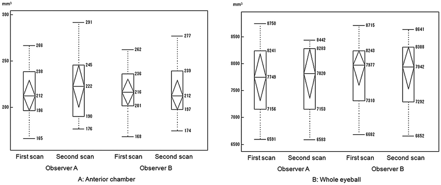
  • Fig. 4.
    • Download figure
    • Open in new tab
    • Download powerpoint
    Fig. 4.

    Boxplots showing volumetric data measured by each of the 2 observers in the first and second scans (A, anterior chamber. B, whole eyeball). Although no interobserver difference for the measured anterior chamber volumes was evident, volumes of the whole eyeball measured by observer B were higher than those measured by observer A.

  • Fig. 5.
    • Download figure
    • Open in new tab
    • Download powerpoint
    Fig. 5.

    Half-Fourier single-shot RARE images in the follow-up of a 7-year-old girl with Sturge-Weber syndrome. In the first (A) and the second (B) scans, contiguous half-Fourier single-shot RARE images show the convex-shaped relatively low-signal-intensity subretinal fluid and relatively high-signal-intensity choroidal hemangioma in the left eye. The measured volumes of the whole eyeball, the tumor, and the subretinal fluid in the first scan were 6.55 × 103 mm3, 1.32 × 103 mm3, and 0.61 × 103 mm3, respectively. The corresponding volumes in the second scan were 6.50 × 103 mm3, 1.33 × 103 mm3, and 0.34 × 103 mm3, respectively. Although no notable change in the volumes of the whole eyeball and the tumor were found, the volume of subretinal fluid decreased by 44% after 11 weeks.

  • Fig. 6.
    • Download figure
    • Open in new tab
    • Download powerpoint
    Fig. 6.

    Ocular MR images of a 40-year-old woman with spindle-shaped choroidal hemangioma (arrows) with exudative subretinal fluid (arrowheads). Axial (A) and sagittal (B) half-Fourier single-shot RARE images show an intermediate-signal-intensity tumor and a low-signal-intensity fluid in the posterior wall of her right eye. Axial fat-saturated FSPGR image (C) shows a low-signal-intensity tumor in the posterior wall of the eye. Axial (D) and sagittal (E) Gd-enhanced fat-saturated FSPGR images show the homogeneous-enhanced tumor and nonenhanced subretinal fluid.

Tables

  • Figures
  • Results of the volumetric phantom studya

    Infusion Volume (×103 mm3)Measured Volume (×103 mm3) (Error Rate)
    Observer 1Observer 2
    FSPGRRAREFSPGRRARE
    1.001.17 (0.170)1.08 (0.080)1.13 (0.130)1.07 (0.070)
    1.501.64 (0.093)1.61 (0.067)1.59 (0.060)1.57 (0.047)
    2.002.14 (0.070)2.02 (0.010)2.16 (0.080)2.09 (0.045)
    2.502.74 (0.080)2.60 (0.040)2.73 (0.092)2.47 (−0.012)
    3.003.15 (0.050)3.06 (0.020)3.16 (0.053)3.08 (0.027)
    3.503.74 (0.069)3.54 (0.011)3.69 (0.054)3.44 (0.017)
    4.004.10 (0.025)4.05 (0.013)4.17 (0.043)3.98 (−0.025)
    • ↵a Volumes ranging from 1.00 to 4.00 mL were injected into the subretinal space. For volumetry using FSPGR on the phantom with 1.00 mL of subretinal fluid, the measurement error rates of each of the 2 observers were >0.10. For other phantom volumetry using FSPGR and all phantom volumetry using half-Fourier single-shot RARE, measurement error rates were <0.10. For both observers, all error rates in the measurement of subretinal volumes were smaller on half-Fourier single-shot RARE images than on FSPGR images.

PreviousNext
Back to top

In this issue

American Journal of Neuroradiology: 34 (4)
American Journal of Neuroradiology
Vol. 34, Issue 4
1 Apr 2013
  • Table of Contents
  • Index by author
Advertisement
Print
Download PDF
Email Article

Thank you for your interest in spreading the word on American Journal of Neuroradiology.

NOTE: We only request your email address so that the person you are recommending the page to knows that you wanted them to see it, and that it is not junk mail. We do not capture any email address.

Enter multiple addresses on separate lines or separate them with commas.
Ocular Volumetry Using Fast High-Resolution MRI during Visual Fixation
(Your Name) has sent you a message from American Journal of Neuroradiology
(Your Name) thought you would like to see the American Journal of Neuroradiology web site.
CAPTCHA
This question is for testing whether or not you are a human visitor and to prevent automated spam submissions.
Cite this article
K. Tanitame, T. Sone, T. Miyoshi, N. Tanitame, K. Otani, Y. Akiyama, M. Takasu, S. Date, Y. Kiuchi, K. Awai
Ocular Volumetry Using Fast High-Resolution MRI during Visual Fixation
American Journal of Neuroradiology Apr 2013, 34 (4) 870-876; DOI: 10.3174/ajnr.A3305

Citation Manager Formats

  • BibTeX
  • Bookends
  • EasyBib
  • EndNote (tagged)
  • EndNote 8 (xml)
  • Medlars
  • Mendeley
  • Papers
  • RefWorks Tagged
  • Ref Manager
  • RIS
  • Zotero
0 Responses
Respond to this article
Share
Bookmark this article
Ocular Volumetry Using Fast High-Resolution MRI during Visual Fixation
K. Tanitame, T. Sone, T. Miyoshi, N. Tanitame, K. Otani, Y. Akiyama, M. Takasu, S. Date, Y. Kiuchi, K. Awai
American Journal of Neuroradiology Apr 2013, 34 (4) 870-876; DOI: 10.3174/ajnr.A3305
del.icio.us logo Twitter logo Facebook logo Mendeley logo
  • Tweet Widget
  • Facebook Like
  • Google Plus One
Purchase

Jump to section

  • Article
    • Abstract
    • ABBREVIATIONS:
    • Materials and Methods
    • Results
    • Discussion
    • Conclusions
    • Footnotes
    • References
  • Figures & Data
  • Info & Metrics
  • Responses
  • References
  • PDF

Related Articles

  • No related articles found.
  • PubMed
  • Google Scholar

Cited By...

  • No citing articles found.
  • Crossref (5)
  • Google Scholar

This article has been cited by the following articles in journals that are participating in Crossref Cited-by Linking.

  • Quantitative analysis with volume rendering of pathological myopic eyes by high-resolution three-dimensional magnetic resonance imaging
    Jinqiong Zhou, Ying Tu, Qinghua Chen, Wenbin Wei
    Medicine 2020 99 42
  • Magnetic resonance imaging of the normal canine eye using a T1‐weighted volumetric acquisition
    Katherine Edith Leonora Manchip, Philip George Sansom, David Donaldson, Chris Warren‐Smith
    Veterinary Record 2021 189 8
  • Manual of Normative Measurements in Head and Neck Imaging
    Mathew B. Macey, Juan E. Small, Daniel Thomas Ginat
    2021
  • Ocular biometry through fully refocused steady-state magnetic resonance imaging sequence: reliability and agreement with the IOLMaster® 500
    Lorenzo Ismael Perez-Sanchez, Julia Gutierrez-Vazquez, Maria Satrustegui-Lapetra, Francisco Ferreira-Manuel, Juan Jose Arevalo-Manso, Juan Jesus Gomez-Herrera, Juan Jose Criado-Alvarez
    International Ophthalmology 2021 41 5
  • Precise size evaluation of extraocular muscles using fat-suppressed fast T1-weighted gradient-recalled echo imaging and multiple gaze fixation targets
    Yuji Takahashi, Keizo Tanitame, Kazushi Yokomachi, Yuji Akiyama, Yoko Kaichi, Kazuo Awai
    Japanese Journal of Radiology 2013 31 12

More in this TOC Section

  • Chondrosarcoma vs Synovial Chondromatosis: Imaging
  • WHO Classification Update: Nasal&Skull Base Tumors
  • Peritumoral Signal in Vestibular Schwannomas
Show more Head & Neck

Similar Articles

Advertisement

Indexed Content

  • Current Issue
  • Accepted Manuscripts
  • Article Preview
  • Past Issues
  • Editorials
  • Editor's Choice
  • Fellows' Journal Club
  • Letters to the Editor
  • Video Articles

Cases

  • Case Collection
  • Archive - Case of the Week
  • Archive - Case of the Month
  • Archive - Classic Case

More from AJNR

  • Trainee Corner
  • Imaging Protocols
  • MRI Safety Corner

Multimedia

  • AJNR Podcasts
  • AJNR Scantastics

Resources

  • Turnaround Time
  • Submit a Manuscript
  • Submit a Video Article
  • Submit an eLetter to the Editor/Response
  • Manuscript Submission Guidelines
  • Statistical Tips
  • Fast Publishing of Accepted Manuscripts
  • Graphical Abstract Preparation
  • Imaging Protocol Submission
  • Evidence-Based Medicine Level Guide
  • Publishing Checklists
  • Author Policies
  • Become a Reviewer/Academy of Reviewers
  • News and Updates

About Us

  • About AJNR
  • Editorial Board
  • Editorial Board Alumni
  • Alerts
  • Permissions
  • Not an AJNR Subscriber? Join Now
  • Advertise with Us
  • Librarian Resources
  • Feedback
  • Terms and Conditions
  • AJNR Editorial Board Alumni

American Society of Neuroradiology

  • Not an ASNR Member? Join Now

© 2025 by the American Society of Neuroradiology All rights, including for text and data mining, AI training, and similar technologies, are reserved.
Print ISSN: 0195-6108 Online ISSN: 1936-959X

Powered by HighWire