Advertisement

Main menu

  • Home
  • Content
    • Current Issue
    • Accepted Manuscripts
    • Article Preview
    • Past Issue Archive
    • Video Articles
    • AJNR Case Collection
    • Case of the Week Archive
    • Case of the Month Archive
    • Classic Case Archive
  • Special Collections
    • Advancing NeuroMRI with High-Relaxivity Contrast Agents
    • AJNR Awards
    • Low-Field MRI
    • Alzheimer Disease
    • ASNR Foundation Special Collection
    • Photon-Counting CT
    • View All
  • Multimedia
    • AJNR Podcasts
    • AJNR SCANtastic
    • Trainee Corner
    • MRI Safety Corner
    • Imaging Protocols
  • For Authors
    • Submit a Manuscript
    • Submit a Video Article
    • Submit an eLetter to the Editor/Response
    • Manuscript Submission Guidelines
    • Statistical Tips
    • Fast Publishing of Accepted Manuscripts
    • Graphical Abstract Preparation
    • Imaging Protocol Submission
    • Author Policies
  • About Us
    • About AJNR
    • Editorial Board
    • Editorial Board Alumni
  • More
    • Become a Reviewer/Academy of Reviewers
    • Subscribers
    • Permissions
    • Alerts
    • Feedback
    • Advertisers
    • ASNR Home

User menu

  • Alerts
  • Log in

Search

  • Advanced search
American Journal of Neuroradiology

American Journal of Neuroradiology

ASHNR American Society of Functional Neuroradiology ASHNR American Society of Pediatric Neuroradiology ASSR
  • Alerts
  • Log in

Advanced Search

  • Home
  • Content
    • Current Issue
    • Accepted Manuscripts
    • Article Preview
    • Past Issue Archive
    • Video Articles
    • AJNR Case Collection
    • Case of the Week Archive
    • Case of the Month Archive
    • Classic Case Archive
  • Special Collections
    • Advancing NeuroMRI with High-Relaxivity Contrast Agents
    • AJNR Awards
    • Low-Field MRI
    • Alzheimer Disease
    • ASNR Foundation Special Collection
    • Photon-Counting CT
    • View All
  • Multimedia
    • AJNR Podcasts
    • AJNR SCANtastic
    • Trainee Corner
    • MRI Safety Corner
    • Imaging Protocols
  • For Authors
    • Submit a Manuscript
    • Submit a Video Article
    • Submit an eLetter to the Editor/Response
    • Manuscript Submission Guidelines
    • Statistical Tips
    • Fast Publishing of Accepted Manuscripts
    • Graphical Abstract Preparation
    • Imaging Protocol Submission
    • Author Policies
  • About Us
    • About AJNR
    • Editorial Board
    • Editorial Board Alumni
  • More
    • Become a Reviewer/Academy of Reviewers
    • Subscribers
    • Permissions
    • Alerts
    • Feedback
    • Advertisers
    • ASNR Home
  • Follow AJNR on Twitter
  • Visit AJNR on Facebook
  • Follow AJNR on Instagram
  • Join AJNR on LinkedIn
  • RSS Feeds

AJNR Awards, New Junior Editors, and more. Read the latest AJNR updates


Improved Turnaround Times | Median time to first decision: 12 days

Research ArticleHead and Neck Imaging

Prediction of Nodal Metastasis in Head and Neck Cancer Using a 3T MRI ADC Map

M.-C. Lee, H.-Y. Tsai, K.-S. Chuang, C.-K. Liu and M.-K. Chen
American Journal of Neuroradiology April 2013, 34 (4) 864-869; DOI: https://doi.org/10.3174/ajnr.A3281
M.-C. Lee
aFrom the Biomedical Engineering and Environmental Sciences Department (M.-C.L., K.-S.C.), National Tsing Hua University, Hsinchu, Taiwan
bDepartment of Medical Imaging (M.-C.L., C.-K.L.)
  • Find this author on Google Scholar
  • Find this author on PubMed
  • Search for this author on this site
H.-Y. Tsai
dDepartment of Medical Imaging and Radiological Sciences (H.-Y.T.), Chang Gung University, Taoyuan, Taiwan.
  • Find this author on Google Scholar
  • Find this author on PubMed
  • Search for this author on this site
K.-S. Chuang
aFrom the Biomedical Engineering and Environmental Sciences Department (M.-C.L., K.-S.C.), National Tsing Hua University, Hsinchu, Taiwan
  • Find this author on Google Scholar
  • Find this author on PubMed
  • Search for this author on this site
C.-K. Liu
bDepartment of Medical Imaging (M.-C.L., C.-K.L.)
  • Find this author on Google Scholar
  • Find this author on PubMed
  • Search for this author on this site
M.-K. Chen
cSuperintendent's Office (M.-K.C.), Changhua Christian Hospital, Changhua, Taiwan
  • Find this author on Google Scholar
  • Find this author on PubMed
  • Search for this author on this site
  • Article
  • Figures & Data
  • Supplemental
  • Info & Metrics
  • Responses
  • References
  • PDF
Loading

Article Figures & Data

Figures

  • Tables
  • Fig. 1.
    • Download figure
    • Open in new tab
    • Download powerpoint
    Fig. 1.

    Box-and-whisker plot presenting the scatterplot of mean apparent diffusion coefficients in all lymph nodes and subcentimeter and supracentimeter lymph nodes.

  • Fig. 2.
    • Download figure
    • Open in new tab
    • Download powerpoint
    Fig. 2.

    ROC curves were created for all lymph nodes and subcentimeter and supracentimeter lymph nodes. The cutoff ADC values were 0.851 × 10⁻³ mm2/s, 0.884 × 10⁻³ mm2/s, and 0.851 × 10⁻³ mm2/s for all lymph nodes and subcentimeter and supracentimeter lymph nodes and were used to differentiate malignant from benign lymph nodes. The best results obtained were sensitivities of 91.3%, 100%, and 100%, specificities of 91.1%, 80.4%, and 91.8%, respectively. The areas under the curve were 0.97, 0.96, and 0.99.
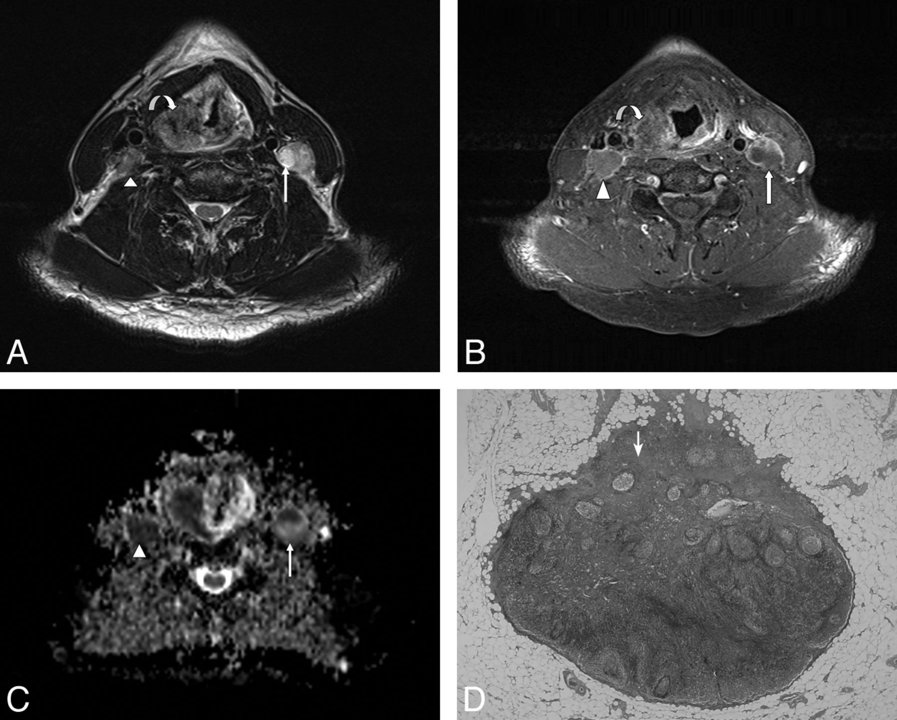
  • Fig. 3.
    • Download figure
    • Open in new tab
    • Download powerpoint
    Fig. 3.

    A 60-year-old male patient with pathologically proved squamous cell carcinoma of the hypopharynx. A, The axial T2-weighted MR image shows an infiltrative neoplasm in the right pyriform apex of the hypopharynx (curved arrow) and enlarged lymph nodes in the bilateral level III, showing heterogeneous signal intensity (arrow and arrowhead). B, The axial postgadolinium fat-suppressed T1-weighted FSE image reveals mild peripheral enhancement in the right pyriform apex tumor (curved arrow) and heterogeneous enhancement in bilateral level III lymph nodes (arrow and arrowhead). C, The ADC value within the right level III lymph node measured 0.712 × 10⁻³ mm2/s (arrowhead), and the left level III lymph node measured 0.659 × 10⁻³ mm2/s (arrow). D, Corresponding H&E-stained histopathologic slide shows intranodal tumor cell metastasis (arrow) (original magnification, ×20).

  • Fig. 4.
    • Download figure
    • Open in new tab
    • Download powerpoint
    Fig. 4.

    A 59-year-old male patient with pathologically proved squamous cell carcinoma of the hypopharynx and metastatic lymphadenopathy of the right level V lymph nodes. A, The axial T2-weighted MR image shows a subcentimeter lymph node with high signal intensity at right level V (arrow). B, The axial postgadolinium fat-suppressed T1-weighted image reveals moderate enhancement in the right pyriform apex tumor (arrowhead) and heterogeneous enhancement in the right level V lymph nodes (arrow). C, The ADC value was 0.811 × 10⁻³ mm2/s (arrow) in the right level V lymph node.

  • Fig. 5.
    • Download figure
    • Open in new tab
    • Download powerpoint
    Fig. 5.

    A 28-year-old female patient with pathologically proved squamous cell carcinoma of the right tongue body with right level I lymph node metastasis. A, The axial T2-weighted MR image shows no necrotic change of the right level I lymph node (arrow). B, The axial T1-weighted MR image shows normal configuration and size of the right level I lymph node with fatty content in its hilar region (arrow). C, The axial postgadolinium fat-suppressed T1-weighted FSE image reveals homogeneous enhancement of the right level I lymph node (arrow). D, The ADC value within the right level I lymph node measured 0.839 × 10⁻³ mm2/s (arrow).

Tables

  • Figures
    • View popup
    Table 1:

    Tumor location, clinical tumor stages, and nodal stages according to TSE MR imaging, histopathology, and DWI

    Patient No.Primary Tumor LocationClinical Tumor StageNodal Stage
    TSEHistopathologyDWI
    1BuccalT1N0N0N0
    2Oropharynx (palate)T2N2bN2bN0
    3BuccalT3N2bN1N1
    4TongueT1NxN0N0
    5Mouth floorT1N0N0N0
    6TongueT2N0N0N0
    7TongueT4aN2bN2cN2c
    8TongueT1N1N0N1
    9TongueT2N0N0N0
    10Hypopharynx and epiglottisT2N1N0N1
    11TongueT1N0N0N0
    12PalateT4aN2bN0N0
    13TongueT1N0N0N0
    14TongueT4aN2bN0N0
    15TongueT2N0N0N0
    16TongueT3N2cN0N2c
    17TongueT2N0N0N0
    18TongueT4aN2bN2bN2b
    19TongueT2N0N2bN2b
    20Hypopharynx and pyriform sinusT4aN1N2bN1
    21RetromolarT4bN2bN2bN2b
    22Hypopharynx and pyriform sinusT2N2cN2cN2c
    • View popup
    Table 2:

    3T ADC values in malignant neck lymph nodes

    Patient No.Age (yr)SexLesionLymph NodeaLN Size (cm)ADC (×10−3 mm2/s)a
    1844MTongue cancerLN30.840.715 ± 0.126
    LN61.080.635 ± 0.143
    LN130.630.460 ± 0.165
    LN140.780.735 ± 0.067
    LN160.600.678 ± 0.193
    2059MHypopharynx cancerLN10.560.811 ± 0.069
    744MTongue cancerLN41.440.753 ± 0.089
    LN50.890.884 ± 0.207
    LN130.360.723 ± 0.162
    LN150.740.437 ± 0.221
    LN190.440.666 ± 0.270
    2260MHypopharynx cancerLN11.040.655 ± 0.090
    LN21.360.731 ± 0.082
    LN31.350.751 ± 0.127
    LN41.580.659 ± 0.073
    LN51.820.712 ± 0.154
    LN61.720.598 ± 0.028
    2150MOral cancerLN20.850.676 ± 0.139
    LN70.500.816 ± 0.094
    351MBuccal cancerLN140.990.559 ± 0.043
    1928FTongue cancerLN20.550.839 ± 0.057
    LN60.670.875 ± 0.090
    LN71.840.851 ± 0.109
    • Note:—LN indicates lymph node.

    • ↵a Mean ± SD.

    • View popup
    Table 3:

    ADC value based on supracentimeter and subcentimeter lymph node size

    LN(a) groupLN Diameter (cm)B/MADC Cutoff Value (×10−3 mm2/s)ADC Value (×10−3mm2/s)a Benign MalignantSensitivity (%)Specificity (%)PAUCb
    BenignMalignant
    All0.27–2.89146/230.8511.086 ± 0.222 (0.578–1.932)0.705 ± 0.118 (0.437–0.884)91.391.1<.00010.97 (0.93–0.99)
    Subcentimeter0.27–0.9997/140.8841.076 ± 0.238 (0.578–1.932)0.705 ± 0.141 (0.437–0.884)10080.4<.00010.95 (0.90–0.98)
    Supracentimeter1.00–2.8949/90.8511.105 ± 0.188 (0.771–1.494)0.705 ± 0.077 (0.598–0.851)10091.8<.00010.97 (0.92–0.99)
    • Note:—LN indicates lymph node; B/M, benign/malignant.

    • ↵a Mean ± SD. Parentheses show range of ADC values.

    • ↵b Parentheses show 95% confidential intervals.

PreviousNext
Back to top

In this issue

American Journal of Neuroradiology: 34 (4)
American Journal of Neuroradiology
Vol. 34, Issue 4
1 Apr 2013
  • Table of Contents
  • Index by author
Advertisement
Print
Download PDF
Email Article

Thank you for your interest in spreading the word on American Journal of Neuroradiology.

NOTE: We only request your email address so that the person you are recommending the page to knows that you wanted them to see it, and that it is not junk mail. We do not capture any email address.

Enter multiple addresses on separate lines or separate them with commas.
Prediction of Nodal Metastasis in Head and Neck Cancer Using a 3T MRI ADC Map
(Your Name) has sent you a message from American Journal of Neuroradiology
(Your Name) thought you would like to see the American Journal of Neuroradiology web site.
CAPTCHA
This question is for testing whether or not you are a human visitor and to prevent automated spam submissions.
Cite this article
M.-C. Lee, H.-Y. Tsai, K.-S. Chuang, C.-K. Liu, M.-K. Chen
Prediction of Nodal Metastasis in Head and Neck Cancer Using a 3T MRI ADC Map
American Journal of Neuroradiology Apr 2013, 34 (4) 864-869; DOI: 10.3174/ajnr.A3281

Citation Manager Formats

  • BibTeX
  • Bookends
  • EasyBib
  • EndNote (tagged)
  • EndNote 8 (xml)
  • Medlars
  • Mendeley
  • Papers
  • RefWorks Tagged
  • Ref Manager
  • RIS
  • Zotero
0 Responses
Respond to this article
Share
Bookmark this article
Prediction of Nodal Metastasis in Head and Neck Cancer Using a 3T MRI ADC Map
M.-C. Lee, H.-Y. Tsai, K.-S. Chuang, C.-K. Liu, M.-K. Chen
American Journal of Neuroradiology Apr 2013, 34 (4) 864-869; DOI: 10.3174/ajnr.A3281
del.icio.us logo Twitter logo Facebook logo Mendeley logo
  • Tweet Widget
  • Facebook Like
  • Google Plus One
Purchase

Jump to section

  • Article
    • Abstract
    • ABBREVIATIONS:
    • Materials and Methods
    • Results
    • Discussion
    • Conclusions
    • Footnotes
    • References
  • Figures & Data
  • Supplemental
  • Info & Metrics
  • Responses
  • References
  • PDF

Related Articles

  • PubMed
  • Google Scholar

Cited By...

  • ADC for Differentiation between Posttreatment Changes and Recurrence in Head and Neck Cancer: A Systematic Review and Meta-analysis
  • The Diagnostic Value of Diffusion-Weighted Imaging in Differentiating Metastatic Lymph Nodes of Head and Neck Squamous Cell Carcinoma: A Systematic Review and Meta-Analysis
  • Crossref
  • Google Scholar

This article has not yet been cited by articles in journals that are participating in Crossref Cited-by Linking.

More in this TOC Section

  • Diagnostic Performance of US in Neck Node NIRADS 2
  • Hydrops Herniation into the Semicircular Canals
  • DIVr of NPC Predicted Outcome
Show more HEAD AND NECK IMAGING

Similar Articles

Advertisement

Indexed Content

  • Current Issue
  • Accepted Manuscripts
  • Article Preview
  • Past Issues
  • Editorials
  • Editor's Choice
  • Fellows' Journal Club
  • Letters to the Editor
  • Video Articles

Cases

  • Case Collection
  • Archive - Case of the Week
  • Archive - Case of the Month
  • Archive - Classic Case

More from AJNR

  • Trainee Corner
  • Imaging Protocols
  • MRI Safety Corner
  • Book Reviews

Multimedia

  • AJNR Podcasts
  • AJNR Scantastics

Resources

  • Turnaround Time
  • Submit a Manuscript
  • Submit a Video Article
  • Submit an eLetter to the Editor/Response
  • Manuscript Submission Guidelines
  • Statistical Tips
  • Fast Publishing of Accepted Manuscripts
  • Graphical Abstract Preparation
  • Imaging Protocol Submission
  • Evidence-Based Medicine Level Guide
  • Publishing Checklists
  • Author Policies
  • Become a Reviewer/Academy of Reviewers
  • News and Updates

About Us

  • About AJNR
  • Editorial Board
  • Editorial Board Alumni
  • Alerts
  • Permissions
  • Not an AJNR Subscriber? Join Now
  • Advertise with Us
  • Librarian Resources
  • Feedback
  • Terms and Conditions
  • AJNR Editorial Board Alumni

American Society of Neuroradiology

  • Not an ASNR Member? Join Now

© 2025 by the American Society of Neuroradiology All rights, including for text and data mining, AI training, and similar technologies, are reserved.
Print ISSN: 0195-6108 Online ISSN: 1936-959X

Powered by HighWire