
Article Figures & Data
Figures
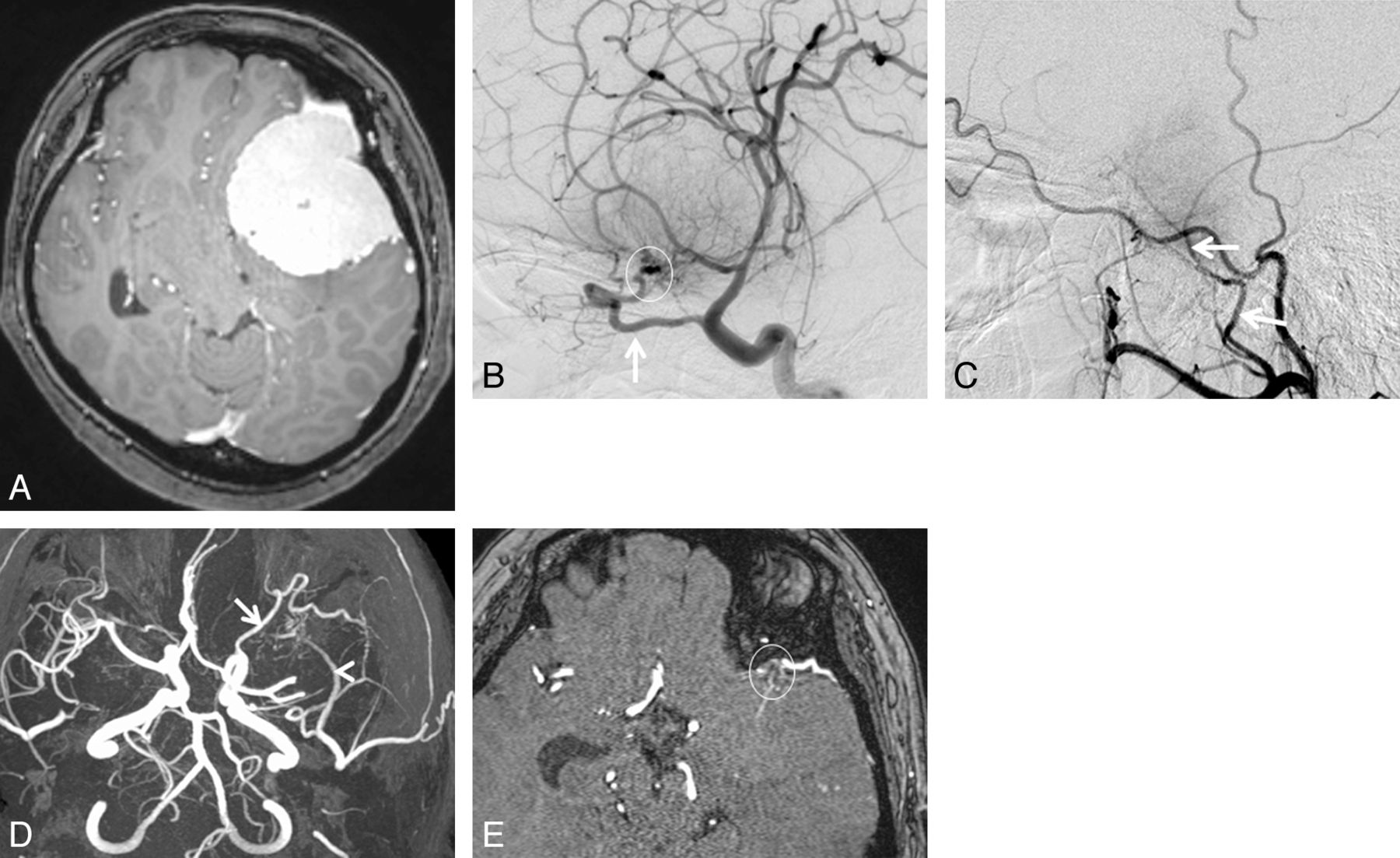
- Fig. 1.
A 43-year-old woman with sphenoid ridge meningioma. A, Axial contrast-enhanced 3D turbo field echo image showing a large enhanced mass in the left anterior to middle cranial fossa regions. B, DSA (lateral projection from the left internal carotid artery) reveals a tumor fed primarily by the recurrent meningeal artery of the ophthalmic artery (arrow). Based on the surgical findings, the dural attachment was the sphenoid ridge (circle). C, DSA (lateral projection from the left external carotid artery) shows a tumor fed partially by the middle meningeal artery (arrows). The right middle meningeal artery was judged to be the secondary feeder. D, 3D TOF MRA (axial projection) depicts dilated branches from the left ophthalmic (arrow) and middle meningeal arteries (arrowhead). E, This axial source 3D TOF MRA image shows tumor-feeding branches from the left ophthalmic artery at the left sphenoid ridge (circle). Both readers judged that the ophthalmic artery was the primary feeder and that the dural attachment was the left sphenoid ridge.
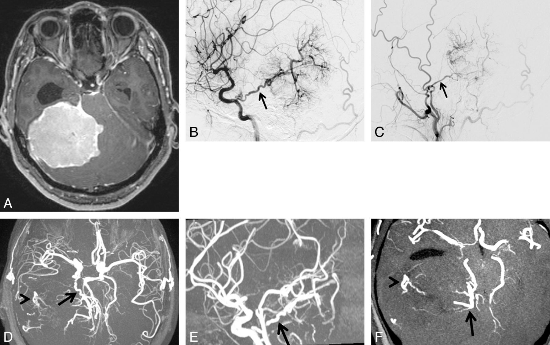
- Fig. 2.
A 49-year-old woman with tentorial meningioma. A, Axial contrast-enhanced 3D turbo field echo image showing a large enhanced mass in the supra- to infratentorial regions. B, DSA (lateral projection from the right internal carotid artery) reveals a tumor fed primarily by the tentorial artery of the meningohypophyseal trunk (arrow). Based on the surgical findings, the dural attachment was the tentorium cerebelli. C, DSA (lateral projection from the right external carotid artery) shows a tumor fed partially by the middle meningeal artery (arrow). The right middle meningeal artery was judged to be the secondary feeder. D, 3D TOF MRA (axial projection) depicts dilated branches from the right tentorial- (arrow) and middle meningeal arteries (arrowhead). E, 3D TOF MRA (sagittal projection) shows the dilated tentorial artery (arrow) from the meningohypophyseal trunk of the internal carotid artery. F, This axial partial MIP MRA shows tumor-feeding branches from the tentorial artery at the medial portion of the tumor (arrow) and from the middle meningeal artery at the lateral portion of the tumor (arrowhead). Both readers judged that the tentorial artery of the meningohypophyseal trunk was the primary feeder and that the dural attachment was the tentorium cerebelli.
- Fig. 3.
A 49-year-old man with cerebellopontine angle meningioma. A, Axial contrast-enhanced 3D turbo field echo image showing an enhanced mass in the cerebellopontine angle region. B, DSA (lateral projection from the right vertebral artery) depicts a tumor fed primarily by the anterior inferior cerebellar artery (arrow). C, DSA (lateral projection from the right external carotid artery) shows a tumor fed partially by the ascending pharyngeal artery (arrow). The ascending pharyngeal artery was judged to be the secondary feeder. Based on the surgical findings, the dural attachment was the cerebellopontine angle. On the 3D TOF MRA image (anteroposterior projection) (D) and sagittal partial MIP-MRA image (E), the right ascending pharyngeal (arrow) and anterior inferior cerebellar artery (arrowheads) are well visualized. F, Axial partial MIP-MRA image showing tumor-feeding branches from the right anterior inferior cerebellar artery (arrowhead). G, Axial source MRA image showing tumor-feeding jugular branches from the right ascending pharyngeal artery (arrow). One reader judged that the anterior inferior cerebellar artery was the primary feeder and that the ascending pharyngeal artery was the secondary feeder. The other reader came to the opposite conclusion. Both readers concluded that the dural attachment was the cerebellopontine angle.


Tables
Case No. Age Sex Clinical Manifestation Sizea (mm) Primary Feeders Secondary Feeders Location of Dural Attachmentb 1 48 F Hemiparesis 77 MMA – Cerebral convexity 2 65 F Headache 54 MCA MMA Cerebral convexity 3 75 F Headache 59 MMA OphA Cerebral convexity 4 67 F Speech dist. 48 MMA MCA Cerebral convexity 5 68 F Gait dist. 31 MMA ACA Falx 6 67 F Speech dist. 67 MMA OA Parasagittal 7 77 F Hemiparesis 40 MMA ACA Parasagittal 8 59 F Visual dist. 65 ILT AMA Sphenoid ridge 9 66 M Headache 54 OphA MCA Sphenoid ridge 10 55 F Visual dist. 73 OphA MHT Sphenoid ridge 11 47 F Hemiparesis 46 OphA MHT Sphenoid ridge 12 43 F Visual dist. 62 OphA MMA Sphenoid ridge 13 62 F Visual dist. 50 AMA MHT Anterior clinoidal 14 71 F Visual dist. 50 ILT MHT Central skull base 15 42 M Headache 73 MHT MMA Middle cranial fossa 16 49 F Headache 91 MHT MMA Tentorial 17 50 F Trigeminal n. 21 MHT – Petroclival 18 59 F Gait dist. 41 ILT – Petroclival 19 44 F Headache 43 OA – T-S sinus junction 20 49 M Headache 24 AICA APhA Cerebellopontine angle 21 44 F Gait dist. 54 OA MMA Cerebellar convexity Note:—F indicates female; M, male; Speech dist., speech disturbance; Visual dist., visual disturbance; Trigeminal n., trigeminal neuralgia; Gait dist., gait disturbance; MMA, middle meningeal artery; AMA, accessory meningeal artery; APhA, ascending pharyngeal artery; OA, occipital artery; OphA, ophthalmic artery; ILT, inferolateral trunk; MHT, meningohypophyseal trunk; MCA, middle cerebral artery; ACA, anterior cerebral artery; AICA, anterior inferior cerebellar artery; T-S sinus junction, transverse-sigmoid sinus junction; –, None.
↵a Maximum diameter of the tumor.
↵b Final diagnosis of dural attachment was determined by surgical findings. When total tumor resection was not obtained, DSA findings were also used for determining the dural attachment of meningiomas.
- Table 2:
Interobserver and intermodality agreement for the identification of primary feeders
MRA Interobserver Agreementa MRAb DSA Intermodality Agreementc Reader 1 Reader 2 MMA 6 6 6 6 AMA 1 1 1 1 APhA 0 1 1 0 OA 3 2 2 2 OphA 4 3 4 4 ILT 3 3 18 (86%) 3 3 20 (95%) MHT 2 4 κ = 0.83 3 3 κ = 0.94 ACA 0 0 [0.66–1.00] 0 0 [0.84–1.00] MCA 1 1 1 1 PCA 0 0 0 0 SCA 0 0 0 0 AICA 1 0 0 1 PICA 0 0 0 0 Other 0 0 0 0 Note:—Data are number of meningiomas. Data in parentheses are the percentage of times that results that were concordant, and data in brackets are 95% confidence intervals. MMA indicates middle meningeal artery; AMA, accessory meningeal artery; APhA, ascending pharyngeal artery; OA, occipital artery; OphA, ophthalmic artery; ILT, inferolateral trunk; MHT, meningohypophyseal trunk; MCA, middle cerebral artery; ACA, anterior cerebral artery; PCA, posterior cerebral artery; SCA, superior cerebellar artery; AICA, anterior inferior cerebellar artery; PICA, posterior inferior cerebellar artery.
↵a Agreement of MRA between Reader 1 and Reader 2.
↵b Consensus reading at MRA of Reader 1 and Reader 2.
↵c Agreement between the consensus reading of MRA of Reader 1 and Reader 2 and DSA.
- Table 3:
Interobserver and intermodality agreement for the identification of secondary feeders
MRA Interobserver Agreementa MRAb DSA Intermodality Agreementc Reader 1 Reader 2 MMA 3 2 3 5 AMA 0 0 0 1 APhA 1 0 0 1 OA 0 0 0 1 OphA 1 2 1 1 ILT 0 0 15 (71%) 0 0 16 (76%) MHT 5 3 κ = 0.58 5 4 κ = 0.72 ACA 2 2 [0.34–0.82] 2 2 [0.51–0.93] MCA 2 4 2 2 PCA 0 0 0 0 SCA 0 0 0 0 AICA 0 1 1 0 PICA 0 0 0 0 None 7 7 7 4 Other 0 0 0 0 Note:—Data are number of meningiomas. Data in parentheses are the percentage of times that results that were concordant, and data in brackets are 95% confidence intervals. MMA indicates middle meningeal artery; AMA, accessory meningeal artery; APhA, ascending pharyngeal artery; OA, occipital artery; OphA, ophthalmic artery; ILT, inferolateral trunk; MHT, meningohypophyseal trunk; MCA, middle cerebral artery; ACA, anterior cerebral artery; PCA, posterior cerebral artery; SCA, superior cerebellar artery; AICA, anterior inferior cerebellar artery; PICA, posterior inferior cerebellar artery.
↵a Agreement of MRA between Reader 1 and Reader 2.
↵b Consensus reading of MRA of Reader 1 and Reader 2.
↵c Agreement between the consensus reading of MRA of Reader 1 and Reader 2 and DSA.
- Table 4:
Interobserver and intermodality agreement for the location of dural attachment of meningiomas
MRA/MRI Interobserver Agreementa MRA/MRIb Surgeryc Agreementd Reader 1 Reader 2 Convexity 4 4 4 4 Parasagittal 2 2 2 2 Falx 1 1 1 1 Sphenoid ridge 5 6 5 5 Anterior clinoid 1 0 1 1 Cen. skull base 1 1 19 (90%) 1 1 21 (100%) Mid. cran. fossa 1 1 κ = 0.95 1 1 κ = 1.00 Tentorial 1 1 [0.84–1.00] 1 1 [1.00–1.00] Petroclival 2 2 2 2 T-S junction 1 1 1 1 CPA 1 1 1 1 Cerebellar conv. 1 1 1 1 Other 0 0 0 0 Note:—Data are number of meningiomas. Data in parentheses are the percentage of times that results that were concordant, and data in brackets are 95% confidence intervals. Cen. skull base indicates central skull base; Mid. cran. fossa, middle cranial fossa; T-S junction, transverse-sigmoid sinus junction; CPA, cerebellopontine angle; Cerebellar conv., cerebellar convexity
↵a Agreement of MRA/MRI between Reader 1 and Reader 2.
↵b Consensus reading of MRA/MRI of Reader 1 and Reader 2.
↵c Final diagnosis of dural attachment was determined by surgical findings. When total tumor resection was not obtained, DSA findings were also used for determining the dural attachment of meningiomas.
↵d Agreement between the consensus reading of MRA/MRI of reader 1 and reader 2 and surgical diagnosis.


