
Article Figures & Data
Figures
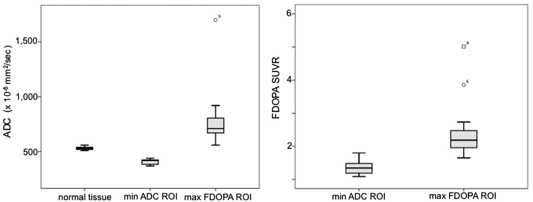
- Fig. 1.
Box-and-whisker plots outlining the distribution (mean and SD) of ADC and FDOPA SUVR measures within the minimum ADC and maximum FDOPA SUVR–defined tumor regions.
- Fig. 2.
Representative FDOPA–MR imaging fused images for patients 12 (top) and 1 (bottom). The maps from left to right are registered as contrast-enhanced T1-weighted MR imaging, ADC, and FDOPA PET, respectively. For each patient, CE MR imaging, ADC, and FDOPA maps are given without (top) and with overlaid regions of minimum ADC (red), maximum FDOPA SUVR (blue), and FDOPA-defined tumor volume (yellow). All maps are given in radiologic format.


Tables
Patient Sex/Age (yr) Pathology ADC Valuea FDOPA Valuea Normal Tissue Min ADC ROI Max FDOPA ROI Min ADC ROI Max FDOPA ROI 1 M/72 GBM 550 (30) 380 (50) 550 (130) 1.42 (0.402) 1.96 (0.18) 2 M/58 GBM 530 (28) 410 (30) 670 (70) 1.32 (0.36) 2.68 (0.22) 3 F/56 GBM 550 (25) 380 (90) 750 (170) 1.10 (0.34) 2.27 (0.19) 4 M/69 GBM 540 (28) 440 (10) 920 (170) 1.12 (0.13) 2.23 (0.33) 5 M/61 GBM 540 (30) 420 (30) 770 (130) 1.20 (0.13) 2.18 (0.21) 6 F/69 GBM 520 (25) 390 (60) 640 (80) 1.79 (0.69) 3.85 (0.36) 7 M/69 GBM 520 (30) 390 (60) 710 (140) 1.49 (0.21) 2.00 (0.22) 8 F/54 GBM 520 (30) 370 (11) 670 (140) 1.72 (0.62) 2.26 (0.36) 9 M/62 GBM 540 (25) 420 (40) 1170 (310) 1.21 (0.49) 5.01 (1.20) 10 F/47 AA 530 (30) 420 (20) 660 (190) 1.47 (0.20) 1.65 (0.18) 11 F/59 GBM 510 (30) 440 (20) 840 (130) 1.09 (0.26) 1.90 (0.22) 12 M/64 GBM 520 (30) 380 (50) 710 (110) 1.55 (0.38) 2.74 (0.34) 13 M/78 GBM 530 (34) 432 (39) 725 (165) 1.17 (0.25) 1.95 (0.17) 14 M/52 GBM 514 (40) 442 (27) 675 (162) 1.34 (0.18) 1.88 (0.23) 15 M/85 GBM 561 (30) 417 (35) 845 (167) 1.44 (0.26) 2.11 (0.28) mean (SD) 531 (29) 409 (38) 790 (151) 1.36 (0.22) 2.45 (0.88) Note:—Min indicates minimum; Max, maximum; AA, anaplastic astrocytoma.
↵a × 10−6 mm2/s; mean (SD) ADC and FDOPA values are given for each patient including for the entire cohort.


