
Article Figures & Data
Figures
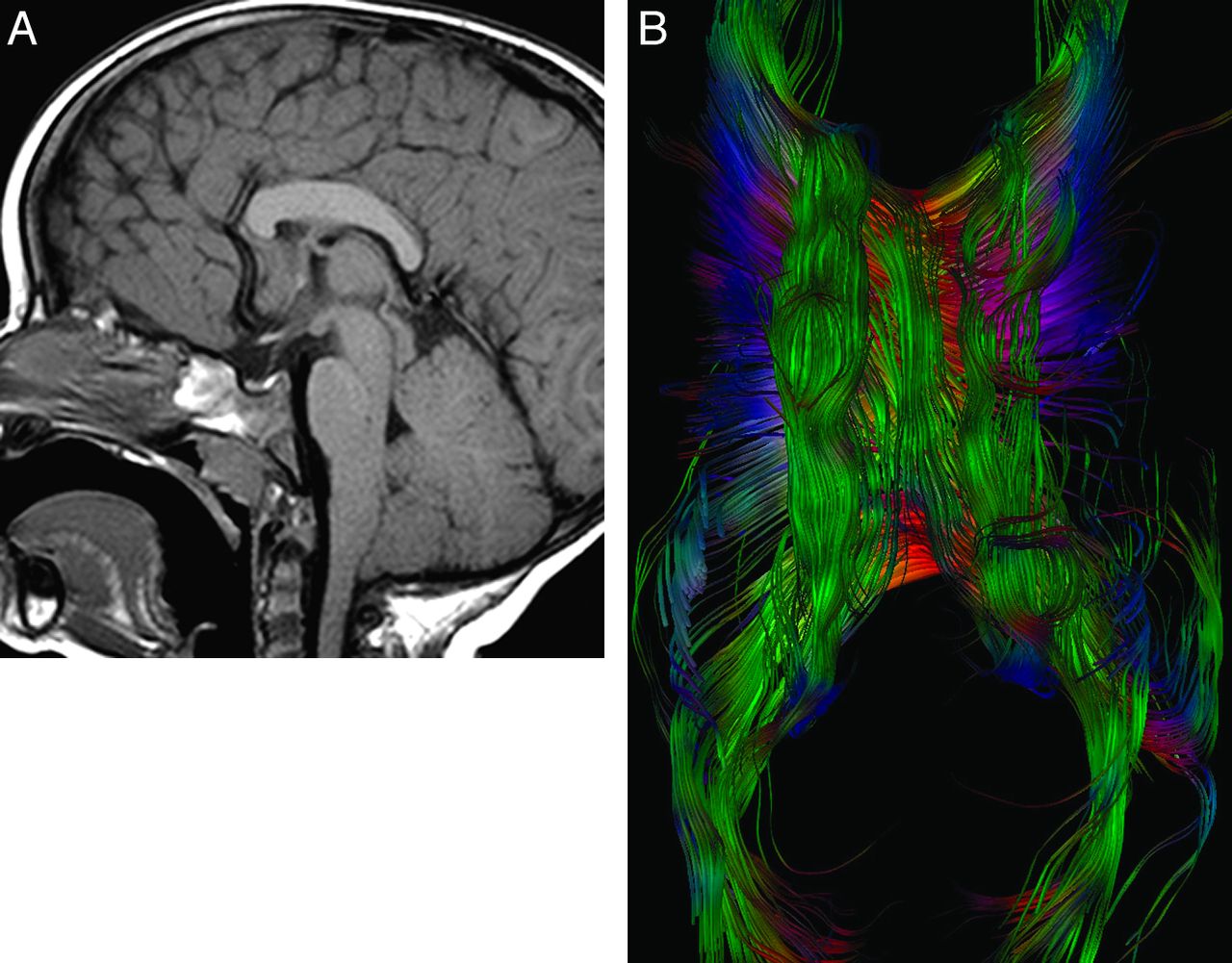
- Fig. 1.
A 2-year-old boy with pervasive developmental delay. A, Midsagittal T1 image shows marked thickening of the midcallosal body (14.2 mm) with subjective shortening of the callosal length. B, Midsagittal directionally encoded color map. The arrow indicates longitudinal supracallosal fibers, which account for the callosal thickening. Longitudinal fibers are not normally seen in the midline; like association fibers, the normal cingulum does not cross the midline. C, Coronal color map. The large midline longitudinal fibers are separate from the paired cingulum (arrows). D, Top-down projection of the supracallosal fibers on diffusion tractography. E, Axial top-down tractography from a healthy subject for comparison. The cingulum is seen as parasagittal rather than midline.
- Fig. 2.
A 20-month-old child referred for epilepsy. A, Midsagittal T1 image. There is subjective callosal shortening. The midcallosal body is thicker (8.7 mm) than the splenium and genu. The configuration of the corpus callosum at 20 months of age is unchanged from the appearance at 11 months. B, Axial top-down view from diffusion tractography shows the midline longitudinal fibers fanning out across the surface of the corpus callosum.
- Fig. 3.
An 18-month-old boy referred for pervasive developmental delay, failure to thrive, and recurrent aspiration. A, Midsagittal T1 image shows that the rostral body is thicker (9.9 mm) than the splenium and genu. B, Axial T1 inversion recovery image shows extensive frontal and peri-Sylvian polymicrogyria and periventricular gray matter heterotopia. C, Sagittal color map shows longitudinal fibers in the midline above the corpus callosum. D, Axial top-down image from diffusion tractography shows the midline longitudinal fibers.
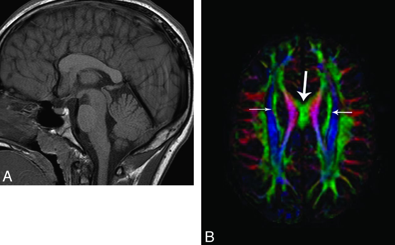
- Fig. 4.
A 12-year-old boy with chronic seizure disorder and pervasive developmental delay with autism. A, On the midsagittal T1 image, the callosal length is normal. However, the rostral body is thicker than the splenium, measuring 9.2 mm. There is optic hypoplasia. B, Axial color map shows a well-formed superior fronto-occipital (SFO) fasciculus (short arrows), and the anomalous midline fibers clearly are separable from the SFO fasciculus. In this patient, the cingulum was poorly formed.


Tables
Demographics and anomalies associated with thickened corpus callosum
Case Age/Sex Indication for MRI Maximum Callosal Thickness Fornices Cingulum Associated Abnormalities 1 25 mo/Male Seizures, DD 14.2 mm Thickened Formed bilaterally None 2 20 mo/Female Seizures, mild DD 8.7 mm Atrophic right Incompletely formed left Acquired right MTS 3 18 mo/Male Seizures, DD 9.9 mm Normal Formed bilaterally PMG, PGH 4 12 yr/Male Autism, DD, decreased vision, seizures 9.2 mm Normal Hypoplastic Optic pathway hypoplasia Note:—DD indicates developmental delay; PMG, polymicrogyria; PGH, periventricular gray matter heterotopia; MTS, mesial temporal sclerosis.


