Advertisement

Main menu

  • Home
  • Content
    • Current Issue
    • Accepted Manuscripts
    • Article Preview
    • Past Issue Archive
    • Video Articles
    • AJNR Case Collection
    • Case of the Week Archive
    • Case of the Month Archive
    • Classic Case Archive
  • Special Collections
    • Advancing NeuroMRI with High-Relaxivity Contrast Agents
    • AJNR Awards
    • Low-Field MRI
    • Alzheimer Disease
    • ASNR Foundation Special Collection
    • Photon-Counting CT
    • View All
  • Multimedia
    • AJNR Podcasts
    • AJNR SCANtastic
    • Trainee Corner
    • MRI Safety Corner
    • Imaging Protocols
  • For Authors
    • Submit a Manuscript
    • Submit a Video Article
    • Submit an eLetter to the Editor/Response
    • Manuscript Submission Guidelines
    • Statistical Tips
    • Fast Publishing of Accepted Manuscripts
    • Graphical Abstract Preparation
    • Imaging Protocol Submission
    • Author Policies
  • About Us
    • About AJNR
    • Editorial Board
    • Editorial Board Alumni
  • More
    • Become a Reviewer/Academy of Reviewers
    • Subscribers
    • Permissions
    • Alerts
    • Feedback
    • Advertisers
    • ASNR Home

User menu

  • Alerts
  • Log in

Search

  • Advanced search
American Journal of Neuroradiology

American Journal of Neuroradiology

ASHNR American Society of Functional Neuroradiology ASHNR American Society of Pediatric Neuroradiology ASSR
  • Alerts
  • Log in

Advanced Search

  • Home
  • Content
    • Current Issue
    • Accepted Manuscripts
    • Article Preview
    • Past Issue Archive
    • Video Articles
    • AJNR Case Collection
    • Case of the Week Archive
    • Case of the Month Archive
    • Classic Case Archive
  • Special Collections
    • Advancing NeuroMRI with High-Relaxivity Contrast Agents
    • AJNR Awards
    • Low-Field MRI
    • Alzheimer Disease
    • ASNR Foundation Special Collection
    • Photon-Counting CT
    • View All
  • Multimedia
    • AJNR Podcasts
    • AJNR SCANtastic
    • Trainee Corner
    • MRI Safety Corner
    • Imaging Protocols
  • For Authors
    • Submit a Manuscript
    • Submit a Video Article
    • Submit an eLetter to the Editor/Response
    • Manuscript Submission Guidelines
    • Statistical Tips
    • Fast Publishing of Accepted Manuscripts
    • Graphical Abstract Preparation
    • Imaging Protocol Submission
    • Author Policies
  • About Us
    • About AJNR
    • Editorial Board
    • Editorial Board Alumni
  • More
    • Become a Reviewer/Academy of Reviewers
    • Subscribers
    • Permissions
    • Alerts
    • Feedback
    • Advertisers
    • ASNR Home
  • Follow AJNR on Twitter
  • Visit AJNR on Facebook
  • Follow AJNR on Instagram
  • Join AJNR on LinkedIn
  • RSS Feeds

AJNR Awards, New Junior Editors, and more. Read the latest AJNR updates


Improved Turnaround Times | Median time to first decision: 12 days

Research ArticleHead and Neck Imaging

CT and MR Imaging Findings of Sinonasal Schwannoma: A Review of 12 Cases

Y.S. Kim, H.-J. Kim, C.-H. Kim and J. Kim
American Journal of Neuroradiology March 2013, 34 (3) 628-633; DOI: https://doi.org/10.3174/ajnr.A3257
Y.S. Kim
aFrom the Departments of Otorhinolaryngology (Y.S.K., C.-H.K.)
  • Find this author on Google Scholar
  • Find this author on PubMed
  • Search for this author on this site
H.-J. Kim
cDepartment of Radiology (H.-J.K.), Samsung Medical Center, Sungkyunkwan University School of Medicine, Seoul, Korea.
  • Find this author on Google Scholar
  • Find this author on PubMed
  • Search for this author on this site
C.-H. Kim
aFrom the Departments of Otorhinolaryngology (Y.S.K., C.-H.K.)
  • Find this author on Google Scholar
  • Find this author on PubMed
  • Search for this author on this site
J. Kim
bRadiology (J.K.), Severance Hospital, Yonsei University College of Medicine, Seoul, Korea
  • Find this author on Google Scholar
  • Find this author on PubMed
  • Search for this author on this site
  • Article
  • Figures & Data
  • Info & Metrics
  • Responses
  • References
  • PDF
Loading

Article Figures & Data

Figures

  • Tables
  • Fig. 1.
    • Download figure
    • Open in new tab
    • Download powerpoint
    Fig. 1.

    Case 10. Schwannoma of the nasal cavity in a 79-year-old man. A, Precontrast axial CT scan with a bone algorithm shows a polypoid mass in the left anterior nasal cavity, originating from the nasal septum. B, Contrast-enhanced axial CT scan with a soft-tissue algorithm shows mild and patchy enhancement of the mass. C, Fat-suppressed axial T2-weighted MR image shows that the mass is isointense to the brain stem. D, Contrast-enhanced fat-suppressed sagittal T1-weighted MR image shows marked contrast enhancement within the mass.
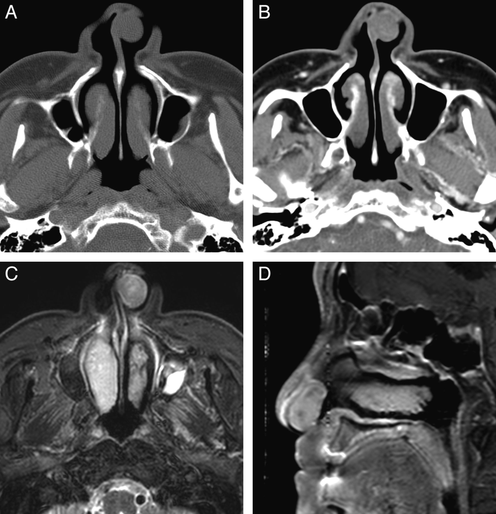
  • Fig. 2.
    • Download figure
    • Open in new tab
    • Download powerpoint
    Fig. 2.

    Case 5. Schwannoma of the nasal cavity in a 22-year-old man. A, Contrast-enhanced coronal CT image shows a tubular expansile soft-tissue mass in the left nasal cavity, demonstrating mild enhancement. B, Axial T1-weighted MR image shows the tumor extending in the anteroposterior dimension and remodeling the lateral nasal wall. C, Fat-suppressed axial T2-weighted MR image shows that the tumor is hyperintense to the brain stem. D, Contrast-enhanced fat-suppressed axial T1-weighted MR images show marked homogeneous enhancement within the tumor.

  • Fig. 3.
    • Download figure
    • Open in new tab
    • Download powerpoint
    Fig. 3.

    Case 7. Schwannoma of the nasal cavity and ethmoid sinus in a 42-year-old woman. A, Precontrast axial CT scan shows a large lobulated expansile mass isoattenuating to the brain stem and centered in the right posterior ethmoid sinus. B, Postcontrast axial CT scan shows marked enhancement of the mass, greater than that of the muscles in the masticator space. The mass extends to the ipsilateral orbit and maxillary and sphenoid sinuses with scalloping and remodeling of the bony walls of the nasal septum, maxilla, and sphenoid bone. C, Coronal T2-weighted MR image shows heterogeneous signal intensity of the mass and signal voids within the lesion, suggestive of prominent vascularity. D, Contrast-enhanced fat-suppressed coronal T1-weighted MR image shows marked and heterogeneous enhancement of the mass.

  • Fig. 4.
    • Download figure
    • Open in new tab
    • Download powerpoint
    Fig. 4.

    Case 3. Schwannoma of the maxillary sinus in a 24-year-old woman. A and B, Precontrast axial and coronal CT scan with a bone algorithm shows a lobulated expansile mass arising from the left infraorbital canal, which replaces the left maxillary sinus. Note cortical thinning and remodeling of the orbital floor and the medial and posterior maxillary sinus walls by the mass. C, Fat-suppressed axial T2-weighted MR image shows multiple fluid-fluid levels within the lesion, which are suggestive of intratumoral hemorrhage. D, Contrast-enhanced fat-suppressed coronal T1-weighted MR image shows cystic change at the lower part of the mass and marked enhancement in the upper solid part of the mass.

Tables

  • Figures
  • Clinical and imaging findings in 12 patients with sinonasal schwannoma

    Patient/Age(yr)/SexChief SymptomCT/MR ImageLocationSize (mm)Shape/Bone ErosionCTMR ImagingCystic or Hemorrhagic Degeneration
    AttenuationaEnhancementbT1-Weighted ImageaT2-Weighted ImageaEnhancementb
    1/46/FNasal obstruction for 2 moYes/yesLt. NC + ES52Tubular, expansile/yesIsodenseMild, patchyIsointenseIsointenseMarkedYes
    2/27/MAnterior cheek pain for 3 moYes/yesLt. MS29Round, expansile/yesIsodenseIsointenseIsointenseMarkedNo
    3/24/FExophthalmos for 1 moYes/yesLt. MS45Lobulated, expansile/yesIsodenseMild, patchyIsointenseIsointenseMarkedYes
    4/14/MRhinorrhea for 2 moYes/noSeptum14Round, nonexpansile/noIsodenseMild, patchyNo
    5/22/MNasal obstruction for 6 moYes/yesLt. NC55Tubular, expansile/yesIsodenseMild, patchyHypointenseHyperintenseMarkedNo
    6/51/MNasal obstruction for 9 moYes/noLt. NC50Tubular, nonexpansile/noIsodenseMild, patchyNo
    7/42/FNasal obstruction for 6 moYes/yesRt. NC + ES45Lobulated, expansile/yesIsodenseMarkedHypointenseIsointenseMarkedNo
    8/33/MNasal obstruction for 4 moYes/yesRt. NC + ES30Tubular, nonexpansile/noIsodenseMildIsointenseIsointenseMarkedNo
    9/33/FNasal obstruction for 3 moYes/yesRt. NC + ES35Tubular, expansile/yesIsodenseMildIsointenseIsointenseMarkedNo
    10/79/MRhinorrhea for 2 yrYes/yesLt. septum15Round, nonexpansile/noIsodenseMild, patchyIsointenseIsointenseMarkedNo
    11/45/MEpistaxis for 2 moYes/noRt. septum5Round, nonexpansile/noIsodenseNo
    12/30/FHeadache for 1 moYes/yesLt. NC + ES45Lobulated, expansile/yesIsodenseIsointenseIsointenseMarkedNo
    • Note:—ES indicates ethmoid sinus; NC, nasal cavity; MS, maxillary sinus; Rt., right; Lt., left.

    • ↵a Compared with the attenuation and signal intensity of the brain stem.

    • ↵b Compared with the enhancement of the masseter muscle.

PreviousNext
Back to top

In this issue

American Journal of Neuroradiology: 34 (3)
American Journal of Neuroradiology
Vol. 34, Issue 3
1 Mar 2013
  • Table of Contents
  • Index by author
Advertisement
Print
Download PDF
Email Article

Thank you for your interest in spreading the word on American Journal of Neuroradiology.

NOTE: We only request your email address so that the person you are recommending the page to knows that you wanted them to see it, and that it is not junk mail. We do not capture any email address.

Enter multiple addresses on separate lines or separate them with commas.
CT and MR Imaging Findings of Sinonasal Schwannoma: A Review of 12 Cases
(Your Name) has sent you a message from American Journal of Neuroradiology
(Your Name) thought you would like to see the American Journal of Neuroradiology web site.
CAPTCHA
This question is for testing whether or not you are a human visitor and to prevent automated spam submissions.
Cite this article
Y.S. Kim, H.-J. Kim, C.-H. Kim, J. Kim
CT and MR Imaging Findings of Sinonasal Schwannoma: A Review of 12 Cases
American Journal of Neuroradiology Mar 2013, 34 (3) 628-633; DOI: 10.3174/ajnr.A3257

Citation Manager Formats

  • BibTeX
  • Bookends
  • EasyBib
  • EndNote (tagged)
  • EndNote 8 (xml)
  • Medlars
  • Mendeley
  • Papers
  • RefWorks Tagged
  • Ref Manager
  • RIS
  • Zotero
0 Responses
Respond to this article
Share
Bookmark this article
CT and MR Imaging Findings of Sinonasal Schwannoma: A Review of 12 Cases
Y.S. Kim, H.-J. Kim, C.-H. Kim, J. Kim
American Journal of Neuroradiology Mar 2013, 34 (3) 628-633; DOI: 10.3174/ajnr.A3257
del.icio.us logo Twitter logo Facebook logo Mendeley logo
  • Tweet Widget
  • Facebook Like
  • Google Plus One
Purchase

Jump to section

  • Article
    • Abstract
    • Case Series
    • Discussion
    • Conclusions
    • Footnotes
    • References
  • Figures & Data
  • Info & Metrics
  • Responses
  • References
  • PDF

Related Articles

  • No related articles found.
  • PubMed
  • Google Scholar

Cited By...

  • Sinonasal schwannoma masquerading as an IgG4-related nasal mass
  • Crossref
  • Google Scholar

This article has not yet been cited by articles in journals that are participating in Crossref Cited-by Linking.

More in this TOC Section

  • Diagnostic Performance of US in Neck Node NIRADS 2
  • Hydrops Herniation into the Semicircular Canals
  • ASL Sensitivity for Head and Neck Paraganglioma
Show more HEAD AND NECK IMAGING

Similar Articles

Advertisement

Indexed Content

  • Current Issue
  • Accepted Manuscripts
  • Article Preview
  • Past Issues
  • Editorials
  • Editor's Choice
  • Fellows' Journal Club
  • Letters to the Editor
  • Video Articles

Cases

  • Case Collection
  • Archive - Case of the Week
  • Archive - Case of the Month
  • Archive - Classic Case

More from AJNR

  • Trainee Corner
  • Imaging Protocols
  • MRI Safety Corner
  • Book Reviews

Multimedia

  • AJNR Podcasts
  • AJNR Scantastics

Resources

  • Turnaround Time
  • Submit a Manuscript
  • Submit a Video Article
  • Submit an eLetter to the Editor/Response
  • Manuscript Submission Guidelines
  • Statistical Tips
  • Fast Publishing of Accepted Manuscripts
  • Graphical Abstract Preparation
  • Imaging Protocol Submission
  • Evidence-Based Medicine Level Guide
  • Publishing Checklists
  • Author Policies
  • Become a Reviewer/Academy of Reviewers
  • News and Updates

About Us

  • About AJNR
  • Editorial Board
  • Editorial Board Alumni
  • Alerts
  • Permissions
  • Not an AJNR Subscriber? Join Now
  • Advertise with Us
  • Librarian Resources
  • Feedback
  • Terms and Conditions
  • AJNR Editorial Board Alumni

American Society of Neuroradiology

  • Not an ASNR Member? Join Now

© 2025 by the American Society of Neuroradiology All rights, including for text and data mining, AI training, and similar technologies, are reserved.
Print ISSN: 0195-6108 Online ISSN: 1936-959X

Powered by HighWire