Advertisement

Main menu

  • Home
  • Content
    • Current Issue
    • Accepted Manuscripts
    • Article Preview
    • Past Issue Archive
    • Video Articles
    • AJNR Case Collection
    • Case of the Week Archive
    • Case of the Month Archive
    • Classic Case Archive
  • Special Collections
    • Advancing NeuroMRI with High-Relaxivity Contrast Agents
    • AJNR Awards
    • Low-Field MRI
    • Alzheimer Disease
    • ASNR Foundation Special Collection
    • Photon-Counting CT
    • View All
  • Multimedia
    • AJNR Podcasts
    • AJNR SCANtastic
    • Trainee Corner
    • MRI Safety Corner
    • Imaging Protocols
  • For Authors
    • Submit a Manuscript
    • Submit a Video Article
    • Submit an eLetter to the Editor/Response
    • Manuscript Submission Guidelines
    • Statistical Tips
    • Fast Publishing of Accepted Manuscripts
    • Graphical Abstract Preparation
    • Imaging Protocol Submission
    • Author Policies
  • About Us
    • About AJNR
    • Editorial Board
    • Editorial Board Alumni
  • More
    • Become a Reviewer/Academy of Reviewers
    • Subscribers
    • Permissions
    • Alerts
    • Feedback
    • Advertisers
    • ASNR Home

User menu

  • Alerts
  • Log in

Search

  • Advanced search
American Journal of Neuroradiology

American Journal of Neuroradiology

ASHNR American Society of Functional Neuroradiology ASHNR American Society of Pediatric Neuroradiology ASSR
  • Alerts
  • Log in

Advanced Search

  • Home
  • Content
    • Current Issue
    • Accepted Manuscripts
    • Article Preview
    • Past Issue Archive
    • Video Articles
    • AJNR Case Collection
    • Case of the Week Archive
    • Case of the Month Archive
    • Classic Case Archive
  • Special Collections
    • Advancing NeuroMRI with High-Relaxivity Contrast Agents
    • AJNR Awards
    • Low-Field MRI
    • Alzheimer Disease
    • ASNR Foundation Special Collection
    • Photon-Counting CT
    • View All
  • Multimedia
    • AJNR Podcasts
    • AJNR SCANtastic
    • Trainee Corner
    • MRI Safety Corner
    • Imaging Protocols
  • For Authors
    • Submit a Manuscript
    • Submit a Video Article
    • Submit an eLetter to the Editor/Response
    • Manuscript Submission Guidelines
    • Statistical Tips
    • Fast Publishing of Accepted Manuscripts
    • Graphical Abstract Preparation
    • Imaging Protocol Submission
    • Author Policies
  • About Us
    • About AJNR
    • Editorial Board
    • Editorial Board Alumni
  • More
    • Become a Reviewer/Academy of Reviewers
    • Subscribers
    • Permissions
    • Alerts
    • Feedback
    • Advertisers
    • ASNR Home
  • Follow AJNR on Twitter
  • Visit AJNR on Facebook
  • Follow AJNR on Instagram
  • Join AJNR on LinkedIn
  • RSS Feeds

AJNR Awards, New Junior Editors, and more. Read the latest AJNR updates


Improved Turnaround Times | Median time to first decision: 12 days

Research ArticleHead and Neck Imaging

Quantitative MRI Analysis of Craniofacial Bone Marrow in Patients with Sickle Cell Disease

E.J. Elias, J.H. Liao, H. Jara, M. Watanabe, R.N. Nadgir, Y. Sakai, K. Erbay, N. Saito, A. Ozonoff, M.H. Steinberg and O. Sakai
American Journal of Neuroradiology March 2013, 34 (3) 622-627; DOI: https://doi.org/10.3174/ajnr.A3240
E.J. Elias
aFrom the Departments of Radiology (E.J.E., J.H.L., H.J., M.W., R.N.N., Y.S., K.E., N.S., O.S.)
  • Find this author on Google Scholar
  • Find this author on PubMed
  • Search for this author on this site
J.H. Liao
aFrom the Departments of Radiology (E.J.E., J.H.L., H.J., M.W., R.N.N., Y.S., K.E., N.S., O.S.)
  • Find this author on Google Scholar
  • Find this author on PubMed
  • Search for this author on this site
H. Jara
aFrom the Departments of Radiology (E.J.E., J.H.L., H.J., M.W., R.N.N., Y.S., K.E., N.S., O.S.)
  • Find this author on Google Scholar
  • Find this author on PubMed
  • Search for this author on this site
M. Watanabe
aFrom the Departments of Radiology (E.J.E., J.H.L., H.J., M.W., R.N.N., Y.S., K.E., N.S., O.S.)
  • Find this author on Google Scholar
  • Find this author on PubMed
  • Search for this author on this site
R.N. Nadgir
aFrom the Departments of Radiology (E.J.E., J.H.L., H.J., M.W., R.N.N., Y.S., K.E., N.S., O.S.)
  • Find this author on Google Scholar
  • Find this author on PubMed
  • Search for this author on this site
Y. Sakai
aFrom the Departments of Radiology (E.J.E., J.H.L., H.J., M.W., R.N.N., Y.S., K.E., N.S., O.S.)
  • Find this author on Google Scholar
  • Find this author on PubMed
  • Search for this author on this site
K. Erbay
aFrom the Departments of Radiology (E.J.E., J.H.L., H.J., M.W., R.N.N., Y.S., K.E., N.S., O.S.)
  • Find this author on Google Scholar
  • Find this author on PubMed
  • Search for this author on this site
N. Saito
aFrom the Departments of Radiology (E.J.E., J.H.L., H.J., M.W., R.N.N., Y.S., K.E., N.S., O.S.)
  • Find this author on Google Scholar
  • Find this author on PubMed
  • Search for this author on this site
A. Ozonoff
cDepartment of Biostatistics (A.O.), Boston University School of Public Health, Boston, Massachusetts
  • Find this author on Google Scholar
  • Find this author on PubMed
  • Search for this author on this site
M.H. Steinberg
bMedicine (M.H.S.), Boston Medical Center, Boston University School of Medicine, Boston, Massachusetts
  • Find this author on Google Scholar
  • Find this author on PubMed
  • Search for this author on this site
O. Sakai
aFrom the Departments of Radiology (E.J.E., J.H.L., H.J., M.W., R.N.N., Y.S., K.E., N.S., O.S.)
  • Find this author on Google Scholar
  • Find this author on PubMed
  • Search for this author on this site
  • Article
  • Figures & Data
  • Info & Metrics
  • Responses
  • References
  • PDF
Loading

Article Figures & Data

Figures

  • Tables
  • Fig. 1.
    • Download figure
    • Open in new tab
    • Download powerpoint
    Fig. 1.

    Craniofacial bone marrow segmentation. The bone marrow of a 24.5-year-old man with sickle cell disease (outlined in blue) has been segmented on the axial proton attenuation–weighted images with 3-mm thickness. Approximately 75 images were segmented per subject. After segmentation was complete, 3D Slicer was used to layer the images over each other to create a 3D model. Shown below are sagittal (bottom left) and coronal (bottom right) images cut through the fully segmented cranial bone marrow after 3D reconstruction. After segmentation, the image data were analyzed in Mathcad for T1, T2, and secular-T2 relaxation times and volume.
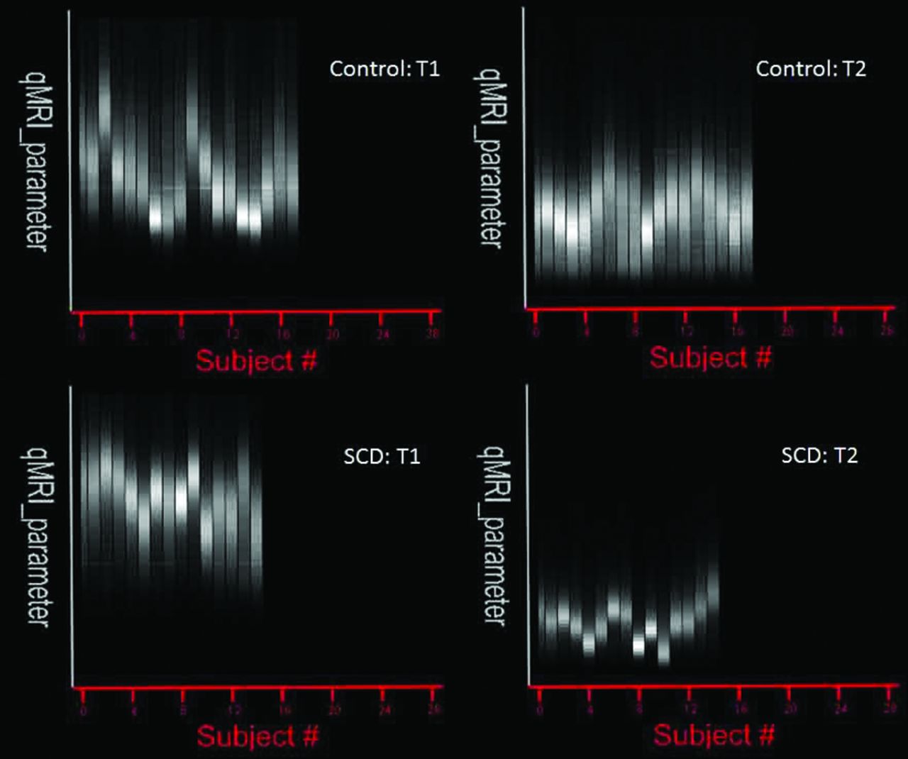
  • Fig. 2.
    • Download figure
    • Open in new tab
    • Download powerpoint
    Fig. 2.

    T1 and T2 histograms for craniofacial bone marrow. Mathcad-generated group histograms of craniofacial bone marrow T1 and T2 values for control subjects and subjects with SCD. Each vertical bar represents averaged relaxation times after segmentation. For T1, intersubject variability was high between groups, which prompted a more individualized analysis and breakdown of T1 peaks (Fig 3). For T2, the intersubject variability was minimal between the groups with more pronounced differences in relaxation times between subjects with SCD and control subjects.

  • Fig. 3.
    • Download figure
    • Open in new tab
    • Download powerpoint
    Fig. 3.

    Individual histogram analysis of T1 peaks in a 32-year-old man without SCD (A) and a 42-year-old woman with SCD (B). A bimodal T1 peak distribution is seen in both cases, shown by dotted yellow and blue lines. Note the increased T1 shift seen in the subject with SCD and a reversal of T1 peak amplitudes compared with the control subject.

  • Fig. 4.
    • Download figure
    • Open in new tab
    • Download powerpoint
    Fig. 4.

    Comparison of peak 1 (A) and peak 2 (B) components of T1 histogram. A, Significant increase of T1 peak 1 relaxation time is noted in patients with SCD, whereas there is no significant difference in peak 2 (B). Both T1 peaks in subjects with SCD showed an age-associated decline (P = .003 and P = .02 for T1 peak 1 and T1 peak 2, respectively).

  • Fig. 5.
    • Download figure
    • Open in new tab
    • Download powerpoint
    Fig. 5.

    Comparison of T2 and secular-T2 peak relaxation times. Both T2 (A) and secular-T2 peak (B) relaxation time graphs show significant shortening of relaxation times in patients with SCD compared with control subjects. Age was not associated with either T2 or secular-T2 changes in subjects with SCD.

  • Fig. 6.
    • Download figure
    • Open in new tab
    • Download powerpoint
    Fig. 6.

    T2 association with the number of transfusions received. Shortening of T2 times is seen as the number of packed red blood cell (PRBC) transfusions increase.

  • Fig. 7.
    • Download figure
    • Open in new tab
    • Download powerpoint
    Fig. 7.

    Comparison of craniofacial bone marrow volume (BMV)/intracranial volume (ICV). Significant increase in craniofacial bone marrow volume is noted in patients with SCD compared with control subjects.

Tables

  • Figures
    • View popup
    Table 1:

    Mixed TSE pulse sequence parameters

    ParameterMixed TSE
    Geometry
        Imaging planeAxial
        Acquisition matrix256 × 192
        Voxel dimensions (mm)0.94 × 0.94 × 3.00
        Intersection gapNull (2 packages)
        PE sampling (%)75
        FOV (FE) × FOV (PE) × mm2240 × 180
        Number of sections80
    Contrast
        Effective echo time (ms)
            TE1eff7.142
            TE2eff100
        TR (ms)14,882.18
        TI (ms)
            1700
            27,441
        Echo-train length18 (9 per echo)
        Phase-encoding ordersCentric 1st echo and linear 2nd echo
        Fat suppressionNo
    Acquisition
        AveragesNEX = 1
        SAR (W/kg)2.7
        Scan time (minutes)9:05
    • Note:—SAR indicates specific absorption rate; FE, frequency encoding; PE, phase encoding.

    • View popup
    Table 2:

    Summary of peak relaxation times and volumesa

    Control (n = 17)SCD (n = 14)Analysis P
    MeanSDMeanSD
    T1 Peak 1504.6122.1691.2103.8<.0001
    T1 Peak 2715.6115.5775.072.8.09
    T2119.712.777.621.4<.0001
    Secular-T2131.717.481.923.8<.0001
    BM volume429.780.7500.769.8.01
    BM/ICV0.330.070.380.06.02
    • Note:—BM indicates bone marrow; ICV, intracranial volume.

    • ↵a Not shown: there was an age association between T1 peak 1 times (P = .003) and T1 peak 2 times (P = .02).

PreviousNext
Back to top

In this issue

American Journal of Neuroradiology: 34 (3)
American Journal of Neuroradiology
Vol. 34, Issue 3
1 Mar 2013
  • Table of Contents
  • Index by author
Advertisement
Print
Download PDF
Email Article

Thank you for your interest in spreading the word on American Journal of Neuroradiology.

NOTE: We only request your email address so that the person you are recommending the page to knows that you wanted them to see it, and that it is not junk mail. We do not capture any email address.

Enter multiple addresses on separate lines or separate them with commas.
Quantitative MRI Analysis of Craniofacial Bone Marrow in Patients with Sickle Cell Disease
(Your Name) has sent you a message from American Journal of Neuroradiology
(Your Name) thought you would like to see the American Journal of Neuroradiology web site.
CAPTCHA
This question is for testing whether or not you are a human visitor and to prevent automated spam submissions.
Cite this article
E.J. Elias, J.H. Liao, H. Jara, M. Watanabe, R.N. Nadgir, Y. Sakai, K. Erbay, N. Saito, A. Ozonoff, M.H. Steinberg, O. Sakai
Quantitative MRI Analysis of Craniofacial Bone Marrow in Patients with Sickle Cell Disease
American Journal of Neuroradiology Mar 2013, 34 (3) 622-627; DOI: 10.3174/ajnr.A3240

Citation Manager Formats

  • BibTeX
  • Bookends
  • EasyBib
  • EndNote (tagged)
  • EndNote 8 (xml)
  • Medlars
  • Mendeley
  • Papers
  • RefWorks Tagged
  • Ref Manager
  • RIS
  • Zotero
0 Responses
Respond to this article
Share
Bookmark this article
Quantitative MRI Analysis of Craniofacial Bone Marrow in Patients with Sickle Cell Disease
E.J. Elias, J.H. Liao, H. Jara, M. Watanabe, R.N. Nadgir, Y. Sakai, K. Erbay, N. Saito, A. Ozonoff, M.H. Steinberg, O. Sakai
American Journal of Neuroradiology Mar 2013, 34 (3) 622-627; DOI: 10.3174/ajnr.A3240
del.icio.us logo Twitter logo Facebook logo Mendeley logo
  • Tweet Widget
  • Facebook Like
  • Google Plus One
Purchase

Jump to section

  • Article
    • Abstract
    • ABBREVIATIONS:
    • Materials and Methods
    • Results
    • Discussion
    • Conclusions
    • Footnotes
    • References
  • Figures & Data
  • Info & Metrics
  • Responses
  • References
  • PDF

Related Articles

  • No related articles found.
  • PubMed
  • Google Scholar

Cited By...

  • Cerebral Blood Flow and Marrow Diffusion Alterations in Children with Sickle Cell Anemia after Bone Marrow Transplantation and Transfusion
  • Crossref
  • Google Scholar

This article has not yet been cited by articles in journals that are participating in Crossref Cited-by Linking.

More in this TOC Section

  • Diagnostic Performance of US in Neck Node NIRADS 2
  • Hydrops Herniation into the Semicircular Canals
  • ASL Sensitivity for Head and Neck Paraganglioma
Show more HEAD AND NECK IMAGING

Similar Articles

Advertisement

Indexed Content

  • Current Issue
  • Accepted Manuscripts
  • Article Preview
  • Past Issues
  • Editorials
  • Editor's Choice
  • Fellows' Journal Club
  • Letters to the Editor
  • Video Articles

Cases

  • Case Collection
  • Archive - Case of the Week
  • Archive - Case of the Month
  • Archive - Classic Case

More from AJNR

  • Trainee Corner
  • Imaging Protocols
  • MRI Safety Corner
  • Book Reviews

Multimedia

  • AJNR Podcasts
  • AJNR Scantastics

Resources

  • Turnaround Time
  • Submit a Manuscript
  • Submit a Video Article
  • Submit an eLetter to the Editor/Response
  • Manuscript Submission Guidelines
  • Statistical Tips
  • Fast Publishing of Accepted Manuscripts
  • Graphical Abstract Preparation
  • Imaging Protocol Submission
  • Evidence-Based Medicine Level Guide
  • Publishing Checklists
  • Author Policies
  • Become a Reviewer/Academy of Reviewers
  • News and Updates

About Us

  • About AJNR
  • Editorial Board
  • Editorial Board Alumni
  • Alerts
  • Permissions
  • Not an AJNR Subscriber? Join Now
  • Advertise with Us
  • Librarian Resources
  • Feedback
  • Terms and Conditions
  • AJNR Editorial Board Alumni

American Society of Neuroradiology

  • Not an ASNR Member? Join Now

© 2025 by the American Society of Neuroradiology All rights, including for text and data mining, AI training, and similar technologies, are reserved.
Print ISSN: 0195-6108 Online ISSN: 1936-959X

Powered by HighWire