Advertisement

Main menu

  • Home
  • Content
    • Current Issue
    • Accepted Manuscripts
    • Article Preview
    • Past Issue Archive
    • Video Articles
    • AJNR Case Collection
    • Case of the Week Archive
    • Case of the Month Archive
    • Classic Case Archive
  • Special Collections
    • Advancing NeuroMRI with High-Relaxivity Contrast Agents
    • AJNR Awards
    • Low-Field MRI
    • Alzheimer Disease
    • ASNR Foundation Special Collection
    • Photon-Counting CT
    • View All
  • Multimedia
    • AJNR Podcasts
    • AJNR SCANtastic
    • Trainee Corner
    • MRI Safety Corner
    • Imaging Protocols
  • For Authors
    • Submit a Manuscript
    • Submit a Video Article
    • Submit an eLetter to the Editor/Response
    • Manuscript Submission Guidelines
    • Statistical Tips
    • Fast Publishing of Accepted Manuscripts
    • Graphical Abstract Preparation
    • Imaging Protocol Submission
    • Author Policies
  • About Us
    • About AJNR
    • Editorial Board
    • Editorial Board Alumni
  • More
    • Become a Reviewer/Academy of Reviewers
    • Subscribers
    • Permissions
    • Alerts
    • Feedback
    • Advertisers
    • ASNR Home

User menu

  • Alerts
  • Log in

Search

  • Advanced search
American Journal of Neuroradiology

American Journal of Neuroradiology

ASHNR American Society of Functional Neuroradiology ASHNR American Society of Pediatric Neuroradiology ASSR
  • Alerts
  • Log in

Advanced Search

  • Home
  • Content
    • Current Issue
    • Accepted Manuscripts
    • Article Preview
    • Past Issue Archive
    • Video Articles
    • AJNR Case Collection
    • Case of the Week Archive
    • Case of the Month Archive
    • Classic Case Archive
  • Special Collections
    • Advancing NeuroMRI with High-Relaxivity Contrast Agents
    • AJNR Awards
    • Low-Field MRI
    • Alzheimer Disease
    • ASNR Foundation Special Collection
    • Photon-Counting CT
    • View All
  • Multimedia
    • AJNR Podcasts
    • AJNR SCANtastic
    • Trainee Corner
    • MRI Safety Corner
    • Imaging Protocols
  • For Authors
    • Submit a Manuscript
    • Submit a Video Article
    • Submit an eLetter to the Editor/Response
    • Manuscript Submission Guidelines
    • Statistical Tips
    • Fast Publishing of Accepted Manuscripts
    • Graphical Abstract Preparation
    • Imaging Protocol Submission
    • Author Policies
  • About Us
    • About AJNR
    • Editorial Board
    • Editorial Board Alumni
  • More
    • Become a Reviewer/Academy of Reviewers
    • Subscribers
    • Permissions
    • Alerts
    • Feedback
    • Advertisers
    • ASNR Home
  • Follow AJNR on Twitter
  • Visit AJNR on Facebook
  • Follow AJNR on Instagram
  • Join AJNR on LinkedIn
  • RSS Feeds

AJNR Awards, New Junior Editors, and more. Read the latest AJNR updates


Improved Turnaround Times | Median time to first decision: 12 days

Research ArticleNeurointervention

Subarachnoid Hyperattenuation on Flat Panel Detector–Based Conebeam CT Immediately after Uneventful Coil Embolization of Unruptured Intracranial Aneurysms

Y. Shinohara, M. Sakamoto, H. Takeuchi, T. Uno, T. Watanabe, T. Kaminou and T. Ogawa
American Journal of Neuroradiology March 2013, 34 (3) 577-582; DOI: https://doi.org/10.3174/ajnr.A3243
Y. Shinohara
aFrom the Division of Radiology (Y.S., T.K., T.O.), Department of Pathophysiological Therapeutic Science
  • Find this author on Google Scholar
  • Find this author on PubMed
  • Search for this author on this site
M. Sakamoto
bDivision of Neurosurgery (M.S., T.U., T.W.), Department of Brain and Neurosciences, Faculty of Medicine, Tottori University, Yonago, Japan
  • Find this author on Google Scholar
  • Find this author on PubMed
  • Search for this author on this site
H. Takeuchi
cDepartment of Neurosurgery (H.T.), Nojima Hospital, Kurayoshi, Japan.
  • Find this author on Google Scholar
  • Find this author on PubMed
  • Search for this author on this site
T. Uno
bDivision of Neurosurgery (M.S., T.U., T.W.), Department of Brain and Neurosciences, Faculty of Medicine, Tottori University, Yonago, Japan
  • Find this author on Google Scholar
  • Find this author on PubMed
  • Search for this author on this site
T. Watanabe
bDivision of Neurosurgery (M.S., T.U., T.W.), Department of Brain and Neurosciences, Faculty of Medicine, Tottori University, Yonago, Japan
  • Find this author on Google Scholar
  • Find this author on PubMed
  • Search for this author on this site
T. Kaminou
aFrom the Division of Radiology (Y.S., T.K., T.O.), Department of Pathophysiological Therapeutic Science
  • Find this author on Google Scholar
  • Find this author on PubMed
  • Search for this author on this site
T. Ogawa
aFrom the Division of Radiology (Y.S., T.K., T.O.), Department of Pathophysiological Therapeutic Science
  • Find this author on Google Scholar
  • Find this author on PubMed
  • Search for this author on this site
  • Article
  • Figures & Data
  • Info & Metrics
  • Responses
  • References
  • PDF
Loading

Article Figures & Data

Figures

  • Tables
  • Fig. 1.
    • Download figure
    • Open in new tab
    • Download powerpoint
    Fig. 1.

    A 63-year-old man with an unruptured 6-mm AcomA complex aneurysm. The patient underwent endovascular embolization for the aneurysm with the guiding catheter in the left ICA. The aneurysm was coiled with balloon assistance. The total volume of contrast medium injected was approximately 300 mL. A, DSA after embolization reveals coil mesh in the left A1-A2 bifurcation (black arrow). B, CBCT immediately after the procedure shows focal subarachnoid hyperattenuation in the left superior frontal sulcus (white arrow). C, Conventional CT 45 minutes after the procedure also shows focal subarachnoid hyperattenuation in the left superior frontal sulcus (white arrow). D, DWI reveals no abnormal high-intensity area indicating acute infarction. E, FLAIR imaging shows no high signal intensity in the sulci indicating SAH. F, T2*-weighted imaging demonstrates no low signal intensity in the sulci indicating SAH. These MR images were obtained 111 minutes after the procedure.
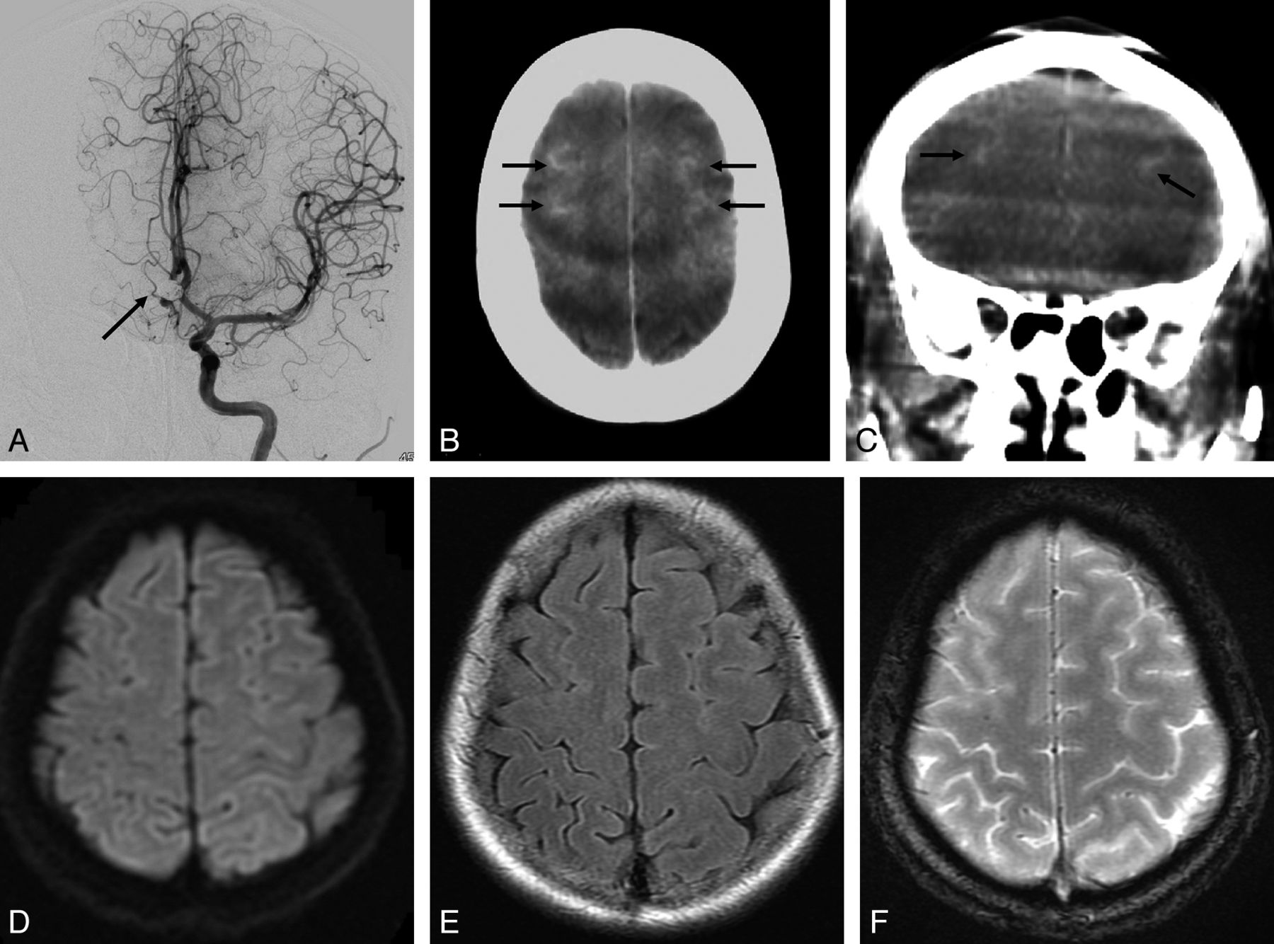
  • Fig. 2.
    • Download figure
    • Open in new tab
    • Download powerpoint
    Fig. 2.

    A 42-year-old woman with an unruptured 7-mm AcomA aneurysm. The patient underwent endovascular embolization for the aneurysm with the guiding catheter in the left ICA. The aneurysm was coiled with balloon assistance. The total volume of contrast medium injected was approximately 250 mL. A, Postembolization DSA of the left ICA injection reveals coil mesh in the AcomA region (black arrow). Notice that the bilateral distal ACA is symmetrically visualized, presumably due to the hypoplastic right A1 segment. B and C, Axial and coronal CBCT scans, respectively, immediately after the procedure show focal subarachnoid hyperattenuation in the bilateral superior frontal sulci (black arrows). D, DWI reveals no abnormal high-intensity area indicating acute infarction. E, FLAIR imaging shows no high signal intensity in the sulci indicating SAH. F, T2*-weighted imaging demonstrates no low signal intensity in the sulci indicating SAH. These MR images were obtained 2 days after the procedure.

Tables

  • Figures
  • Relationship between each parameter and subarachnoid hyperattenuation on FPD-based CBCT immediately after coil embolization

    ParameterSubarachnoid HyperattenuationP Value
    +−
    Location of aneurysm
        ICA419.0728
        MCA02
        AcomA33
        ACA20
        VB03
    Size of aneurysm (mm)
        <5012<.05
        5–10413
        10–1510
        ≥1542
    Amount of contrast medium (mL)
        <150017<.001
        150–25048
        ≥25052
    Balloon assistance
        Performed312.567
        Not performed615
    Stent assistance
        Performed45.150
        Not performed522
    • Note:—+ indicates presence; −, absence.

PreviousNext
Back to top

In this issue

American Journal of Neuroradiology: 34 (3)
American Journal of Neuroradiology
Vol. 34, Issue 3
1 Mar 2013
  • Table of Contents
  • Index by author
Advertisement
Print
Download PDF
Email Article

Thank you for your interest in spreading the word on American Journal of Neuroradiology.

NOTE: We only request your email address so that the person you are recommending the page to knows that you wanted them to see it, and that it is not junk mail. We do not capture any email address.

Enter multiple addresses on separate lines or separate them with commas.
Subarachnoid Hyperattenuation on Flat Panel Detector–Based Conebeam CT Immediately after Uneventful Coil Embolization of Unruptured Intracranial Aneurysms
(Your Name) has sent you a message from American Journal of Neuroradiology
(Your Name) thought you would like to see the American Journal of Neuroradiology web site.
CAPTCHA
This question is for testing whether or not you are a human visitor and to prevent automated spam submissions.
Cite this article
Y. Shinohara, M. Sakamoto, H. Takeuchi, T. Uno, T. Watanabe, T. Kaminou, T. Ogawa
Subarachnoid Hyperattenuation on Flat Panel Detector–Based Conebeam CT Immediately after Uneventful Coil Embolization of Unruptured Intracranial Aneurysms
American Journal of Neuroradiology Mar 2013, 34 (3) 577-582; DOI: 10.3174/ajnr.A3243

Citation Manager Formats

  • BibTeX
  • Bookends
  • EasyBib
  • EndNote (tagged)
  • EndNote 8 (xml)
  • Medlars
  • Mendeley
  • Papers
  • RefWorks Tagged
  • Ref Manager
  • RIS
  • Zotero
0 Responses
Respond to this article
Share
Bookmark this article
Subarachnoid Hyperattenuation on Flat Panel Detector–Based Conebeam CT Immediately after Uneventful Coil Embolization of Unruptured Intracranial Aneurysms
Y. Shinohara, M. Sakamoto, H. Takeuchi, T. Uno, T. Watanabe, T. Kaminou, T. Ogawa
American Journal of Neuroradiology Mar 2013, 34 (3) 577-582; DOI: 10.3174/ajnr.A3243
del.icio.us logo Twitter logo Facebook logo Mendeley logo
  • Tweet Widget
  • Facebook Like
  • Google Plus One
Purchase

Jump to section

  • Article
    • Abstract
    • ABBREVIATIONS:
    • Materials and Methods
    • Results
    • Discussion
    • Conclusions
    • Acknowledgments
    • References
  • Figures & Data
  • Info & Metrics
  • Responses
  • References
  • PDF

Related Articles

  • No related articles found.
  • PubMed
  • Google Scholar

Cited By...

  • Metal artifact reduction for flat panel detector intravenous CT angiography in patients with intracranial metallic implants after endovascular and surgical treatment
  • Mechanical thrombectomy using a combined CT/C-arm X-ray system
  • Flat Detector Angio-CT following Intra-Arterial Therapy of Acute Ischemic Stroke: Identification of Hemorrhage and Distinction from Contrast Accumulation due to Blood-Brain Barrier Disruption
  • Crossref
  • Google Scholar

This article has not yet been cited by articles in journals that are participating in Crossref Cited-by Linking.

More in this TOC Section

  • Prophylactic Endovascular Treatments for CBS
  • Smoking & Aneurysm Outcomes: A Meta-Analysis
  • “Buddy-wire anchoring” technique for TVE
Show more NEUROINTERVENTION

Similar Articles

Advertisement

Indexed Content

  • Current Issue
  • Accepted Manuscripts
  • Article Preview
  • Past Issues
  • Editorials
  • Editor's Choice
  • Fellows' Journal Club
  • Letters to the Editor
  • Video Articles

Cases

  • Case Collection
  • Archive - Case of the Week
  • Archive - Case of the Month
  • Archive - Classic Case

More from AJNR

  • Trainee Corner
  • Imaging Protocols
  • MRI Safety Corner
  • Book Reviews

Multimedia

  • AJNR Podcasts
  • AJNR Scantastics

Resources

  • Turnaround Time
  • Submit a Manuscript
  • Submit a Video Article
  • Submit an eLetter to the Editor/Response
  • Manuscript Submission Guidelines
  • Statistical Tips
  • Fast Publishing of Accepted Manuscripts
  • Graphical Abstract Preparation
  • Imaging Protocol Submission
  • Evidence-Based Medicine Level Guide
  • Publishing Checklists
  • Author Policies
  • Become a Reviewer/Academy of Reviewers
  • News and Updates

About Us

  • About AJNR
  • Editorial Board
  • Editorial Board Alumni
  • Alerts
  • Permissions
  • Not an AJNR Subscriber? Join Now
  • Advertise with Us
  • Librarian Resources
  • Feedback
  • Terms and Conditions
  • AJNR Editorial Board Alumni

American Society of Neuroradiology

  • Not an ASNR Member? Join Now

© 2025 by the American Society of Neuroradiology All rights, including for text and data mining, AI training, and similar technologies, are reserved.
Print ISSN: 0195-6108 Online ISSN: 1936-959X

Powered by HighWire