
Article Figures & Data
Figures
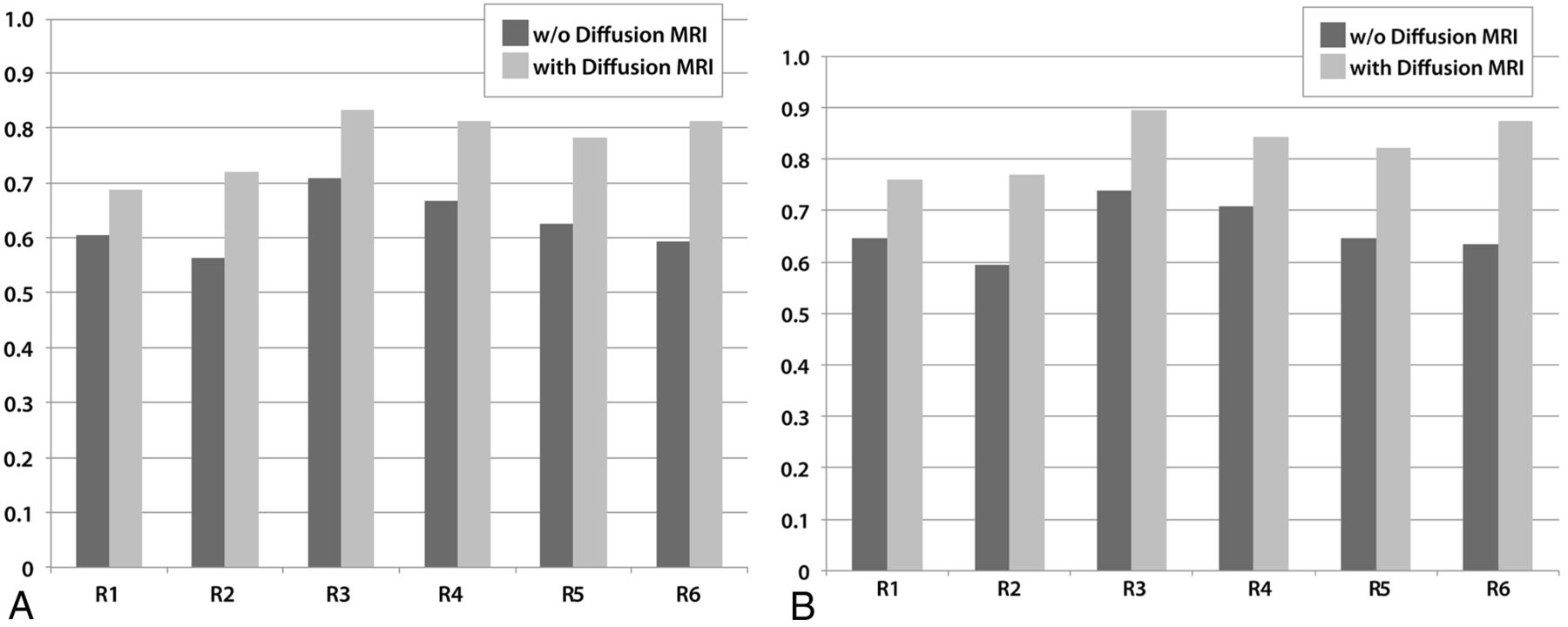
- Fig 1.
Graphs demonstrate the performances of the reviewers (R1–R4: residents, R5–R6: neuroradiologists) without (dark gray) and with (light gray) diffusion MR imaging data. A, When 4 choices are considered: PA, medulloblastoma, ependymoma, and AT/RT. B, When 3 choices are considered: embryonal tumors (medulloblastoma and AT/RT), ependymoma, and PA.
- Fig 2.
Impact of inclusion of diffusion MR imaging data to correct diagnoses. A, For 4 tumor categories. A total of 1,152 diagnoses (96 tumors × 6 reviewers × 2 sessions) were rendered. B, For embryonal tumors (medulloblastomas and AT/RTs combined), ependymomas, and PAs.
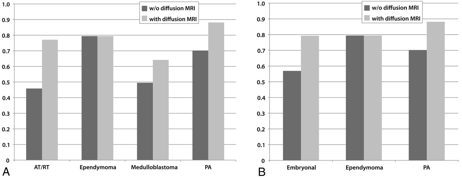
- Fig 3.
Graphs depict the percentages of correct diagnoses for tumor categories without (dark gray) and with (light gray) diffusion MR imaging data. A, When 4 choices are considered: PA, medulloblastoma, ependymoma, and AT/RT. B, When 3 choices are considered: Embryonal tumors (medulloblastoma and AT/RT), ependymoma, and PA.
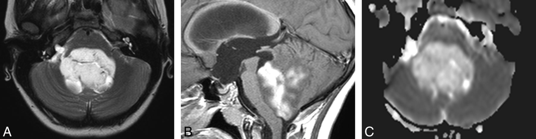
- Fig 4.
PA in a 10-year-old girl. Without diffusion MR imaging data, 5 reviewers were incorrect. Four selected ependymoma and 1 selected medulloblastoma. With diffusion MR imaging data, 5 reviewers were correct. One reviewer (a neuroradiologist) still chose ependymoma. A, Axial T2-weighted image displays a markedly hyperintense tumor. B, Sagittal gadolinium-enhanced T1-weighted image demonstrates intense and heterogeneous enhancement of the mass. C, ADC map shows facilitated diffusion within the mass compared with the uninvolved cerebellum.
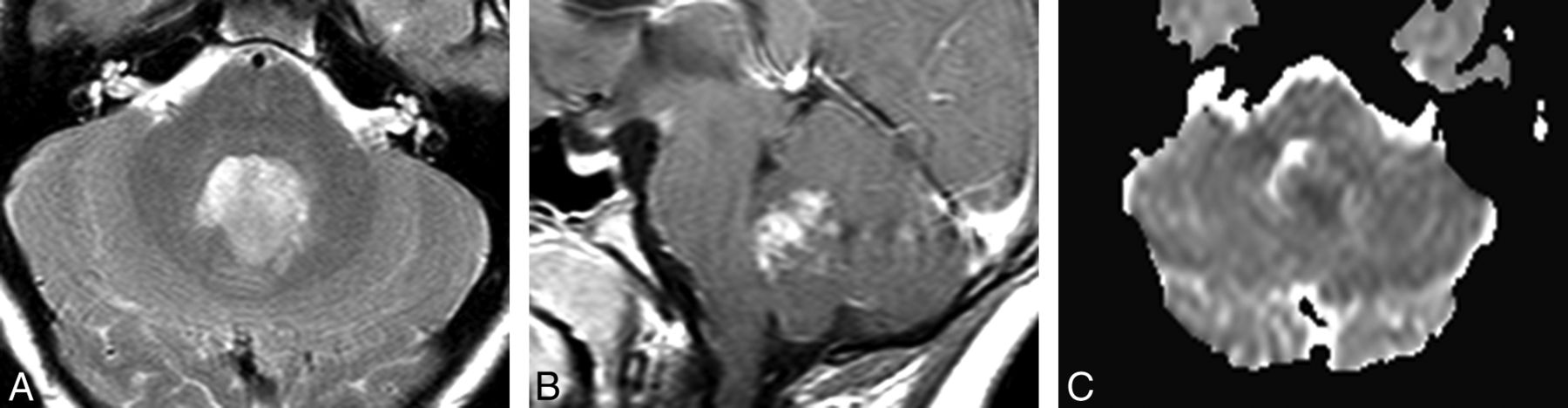
- Fig 5.
Medulloblastoma in a 12-year-old boy. Without diffusion MR imaging data, 4 reviewers (3 residents and 1 neuroradiologist who chose ependymoma) were incorrect. With diffusion MR imaging data, all reviewers were correct. A, Axial T2-weighted image shows that the tumor in the fourth ventricle is relatively hypointense. B, Sagittal gadolinium-enhanced T1-weighted image shows that the tumor enhances intensely and heterogenously. C, ADC map shows that the tumor is slightly hypointense to isoinstense compared with the normal cerebellum.
- Fig 6.
Ependymoma (WHO grade 2) in a 15-year-old boy. Without diffusion MR imaging, all reviewers were correct. With diffusion data, 3 reviewers (2 residents and 1 neuroradiologist) changed their diagnoses to medulloblastoma. A, Axial T2-weighted image demonstrates a hyperintense mass extending toward the right foramen of Luschka. B, Sagittal gadolinium-enhanced T1-weighted image shows that the tumor is heterogeneous, but intense with enhancement. C, ADC map shows that the mass is isointense to slightly hyperintense compared with normal cerebellum.


Tables


Jump to section
Related Articles
Cited By...
- No citing articles found.