Advertisement

Main menu

  • Home
  • Content
    • Current Issue
    • Accepted Manuscripts
    • Article Preview
    • Past Issue Archive
    • Video Articles
    • AJNR Case Collection
    • Case of the Week Archive
    • Case of the Month Archive
    • Classic Case Archive
  • Special Collections
    • AJNR Awards
    • Low-Field MRI
    • Alzheimer Disease
    • ASNR Foundation Special Collection
    • Photon-Counting CT
    • View All
  • Multimedia
    • AJNR Podcasts
    • AJNR SCANtastic
    • Trainee Corner
    • MRI Safety Corner
    • Imaging Protocols
  • For Authors
    • Submit a Manuscript
    • Submit a Video Article
    • Submit an eLetter to the Editor/Response
    • Manuscript Submission Guidelines
    • Statistical Tips
    • Fast Publishing of Accepted Manuscripts
    • Graphical Abstract Preparation
    • Imaging Protocol Submission
    • Author Policies
  • About Us
    • About AJNR
    • Editorial Board
    • Editorial Board Alumni
  • More
    • Become a Reviewer/Academy of Reviewers
    • Subscribers
    • Permissions
    • Alerts
    • Feedback
    • Advertisers
    • ASNR Home

User menu

  • Alerts
  • Log in

Search

  • Advanced search
American Journal of Neuroradiology

American Journal of Neuroradiology

ASHNR American Society of Functional Neuroradiology ASHNR American Society of Pediatric Neuroradiology ASSR
  • Alerts
  • Log in

Advanced Search

  • Home
  • Content
    • Current Issue
    • Accepted Manuscripts
    • Article Preview
    • Past Issue Archive
    • Video Articles
    • AJNR Case Collection
    • Case of the Week Archive
    • Case of the Month Archive
    • Classic Case Archive
  • Special Collections
    • AJNR Awards
    • Low-Field MRI
    • Alzheimer Disease
    • ASNR Foundation Special Collection
    • Photon-Counting CT
    • View All
  • Multimedia
    • AJNR Podcasts
    • AJNR SCANtastic
    • Trainee Corner
    • MRI Safety Corner
    • Imaging Protocols
  • For Authors
    • Submit a Manuscript
    • Submit a Video Article
    • Submit an eLetter to the Editor/Response
    • Manuscript Submission Guidelines
    • Statistical Tips
    • Fast Publishing of Accepted Manuscripts
    • Graphical Abstract Preparation
    • Imaging Protocol Submission
    • Author Policies
  • About Us
    • About AJNR
    • Editorial Board
    • Editorial Board Alumni
  • More
    • Become a Reviewer/Academy of Reviewers
    • Subscribers
    • Permissions
    • Alerts
    • Feedback
    • Advertisers
    • ASNR Home
  • Follow AJNR on Twitter
  • Visit AJNR on Facebook
  • Follow AJNR on Instagram
  • Join AJNR on LinkedIn
  • RSS Feeds

AJNR Awards, New Junior Editors, and more. Read the latest AJNR updates


Improved Turnaround Times | Median time to first decision: 12 days

Research ArticleHead and Neck Imaging
Open Access

Automated Quantitation of the Posterior Scleral Flattening and Optic Nerve Protrusion by MRI in Idiopathic Intracranial Hypertension

N. Alperin, A.M. Bagci, B.L. Lam and E. Sklar
American Journal of Neuroradiology December 2013, 34 (12) 2354-2359; DOI: https://doi.org/10.3174/ajnr.A3600
N. Alperin
aFrom the Department of Radiology (N.A., A.M.B., E.S.)
  • Find this author on Google Scholar
  • Find this author on PubMed
  • Search for this author on this site
A.M. Bagci
aFrom the Department of Radiology (N.A., A.M.B., E.S.)
  • Find this author on Google Scholar
  • Find this author on PubMed
  • Search for this author on this site
B.L. Lam
bBascom Palmer Eye Institute (B.L.L.), University of Miami, Miami, Florida.
  • Find this author on Google Scholar
  • Find this author on PubMed
  • Search for this author on this site
E. Sklar
aFrom the Department of Radiology (N.A., A.M.B., E.S.)
  • Find this author on Google Scholar
  • Find this author on PubMed
  • Search for this author on this site
  • Article
  • Figures & Data
  • Info & Metrics
  • Responses
  • References
  • PDF
Loading

Article Figures & Data

Figures

  • Tables
  • Fig 1.
    • Download figure
    • Open in new tab
    • Download powerpoint
    Fig 1.

    MR images of the globe reformatted in sagittal (A) and axial (B) planes. The coordinate system is defined by the center of mass of the lens and the globe (red and green points), respectively. The red contour marks the posterior sclera. Each point on the sclera is defined by the distance to the center of the orbit (dashed line), azimuth angle (θ), and the elevation angle (ϕ).
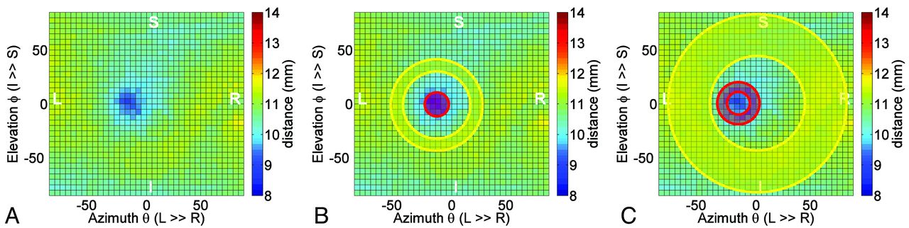
  • Fig 2.
    • Download figure
    • Open in new tab
    • Download powerpoint
    Fig 2.

    A 2D color-coded distance map visualizes distances between the center of the globe and points on the posterior sclera. The map represents the globe shown in Fig 1. The inward protrusion of the papilla is visualized as an off-center blue patch (A). The central (red) and peripheral (yellow) ROIs used for the derivation of NP and GF are shown in B and C, respectively. The angular boundaries of the central (papillar), peripappilar, and peripheral ROIs are 0–10°, 10–18°, and 40–80°, respectively.
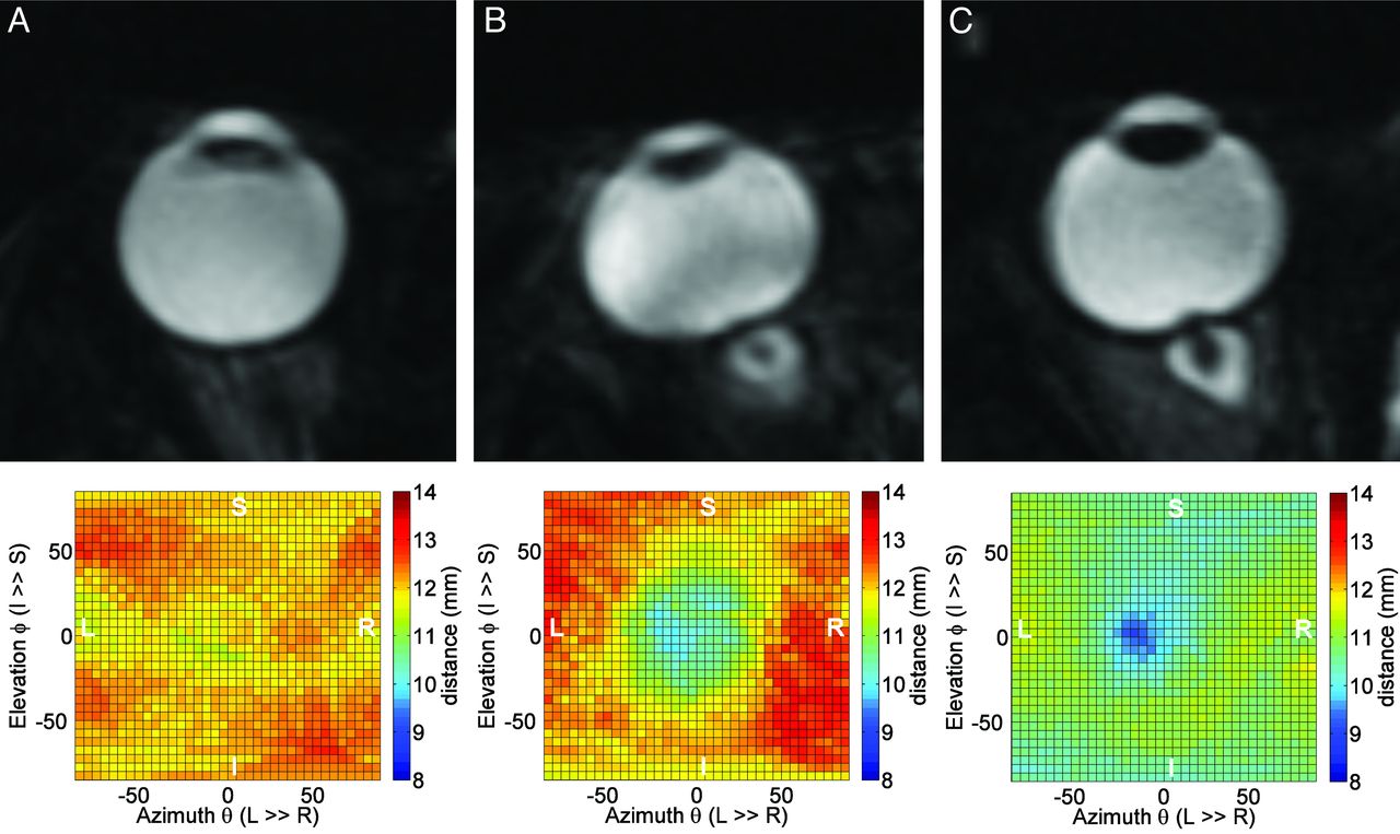
  • Fig 3.
    • Download figure
    • Open in new tab
    • Download powerpoint
    Fig 3.

    Reformatted MR images shown in axial plane for a normal globe (A), a flattened globe with minimal optic nerve protrusion (B), and a globe with minimal flattening and extensive optic nerve protrusion (C). Respective distance maps are shown in the bottom. The corresponding GF and NP values are (A) 0.97 and 0.97, (B) 0.86 and 0.91, and (C) 0.91 and 0.88.

  • Fig 4.
    • Download figure
    • Open in new tab
    • Download powerpoint
    Fig 4.

    Average left and right eyes, 2D-distance maps obtained from the control cohort (upper row) and the IIH cohort (lower row). The presence of nerve protrusion is clearly seen in the maps from the IIH cohort.

  • Fig 5.
    • Download figure
    • Open in new tab
    • Download powerpoint
    Fig 5.

    Pretreatment (upper row) and posttreatment (lower row) average distance maps of the patients with IIH who had a follow-up MR imaging scan (n = 4). A significant reversal of the extent of the optic nerve protrusion is visualized in the posttreatment maps.
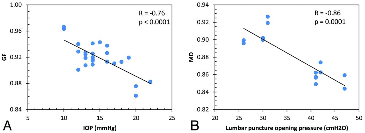
  • Fig 6.
    • Download figure
    • Open in new tab
    • Download powerpoint
    Fig 6.

    Scatterplots of the relationships between GF and intraocular pressure (A) and between MD and CSF opening pressure (B).

Tables

  • Figures
    • View popup
    Table 1:

    Mean values of deformation measures for the control and IIH cohorts

    MeasureControl (n = 7)IIH (n = 6)P Value
    NP0.96 ± 0.0130.91 ± 0.028.00002
    GF0.93 ± 0.0200.91 ± 0.022.0035
    MD0.93 ± 0.0210.88 ± 0.027.00002
    • View popup
    Table 2:

    Pretreatment and posttreatment mean values of deformation measures in the IIH subcohort

    MeasureIIH Pre (n = 4)IIH Post (n = 4)P Value
    NP0.91 ± 0.0320.94 ± 0.017.036
    GF0.90 ± 0.0210.92 ± 0.023.09
    MD0.88 ± 0.0240.91 ± 0.024.011
PreviousNext
Back to top

In this issue

American Journal of Neuroradiology: 34 (12)
American Journal of Neuroradiology
Vol. 34, Issue 12
1 Dec 2013
  • Table of Contents
  • Index by author
  • Complete Issue (PDF)
Advertisement
Print
Download PDF
Email Article

Thank you for your interest in spreading the word on American Journal of Neuroradiology.

NOTE: We only request your email address so that the person you are recommending the page to knows that you wanted them to see it, and that it is not junk mail. We do not capture any email address.

Enter multiple addresses on separate lines or separate them with commas.
Automated Quantitation of the Posterior Scleral Flattening and Optic Nerve Protrusion by MRI in Idiopathic Intracranial Hypertension
(Your Name) has sent you a message from American Journal of Neuroradiology
(Your Name) thought you would like to see the American Journal of Neuroradiology web site.
CAPTCHA
This question is for testing whether or not you are a human visitor and to prevent automated spam submissions.
Cite this article
N. Alperin, A.M. Bagci, B.L. Lam, E. Sklar
Automated Quantitation of the Posterior Scleral Flattening and Optic Nerve Protrusion by MRI in Idiopathic Intracranial Hypertension
American Journal of Neuroradiology Dec 2013, 34 (12) 2354-2359; DOI: 10.3174/ajnr.A3600

Citation Manager Formats

  • BibTeX
  • Bookends
  • EasyBib
  • EndNote (tagged)
  • EndNote 8 (xml)
  • Medlars
  • Mendeley
  • Papers
  • RefWorks Tagged
  • Ref Manager
  • RIS
  • Zotero
0 Responses
Respond to this article
Share
Bookmark this article
Automated Quantitation of the Posterior Scleral Flattening and Optic Nerve Protrusion by MRI in Idiopathic Intracranial Hypertension
N. Alperin, A.M. Bagci, B.L. Lam, E. Sklar
American Journal of Neuroradiology Dec 2013, 34 (12) 2354-2359; DOI: 10.3174/ajnr.A3600
del.icio.us logo Twitter logo Facebook logo Mendeley logo
  • Tweet Widget
  • Facebook Like
  • Google Plus One
Purchase

Jump to section

  • Article
    • Abstract
    • ABBREVIATIONS:
    • Materials and Methods
    • Results
    • Discussion
    • Conclusions
    • Footnotes
    • REFERENCES
  • Figures & Data
  • Info & Metrics
  • Responses
  • References
  • PDF

Related Articles

  • No related articles found.
  • PubMed
  • Google Scholar

Cited By...

  • Evolution of MRI Findings in Patients with Idiopathic Intracranial Hypertension after Venous Sinus Stenting
  • The effect of intraocular and intracranial pressure on retinal structure and function in rats
  • Crossref
  • Google Scholar

This article has not yet been cited by articles in journals that are participating in Crossref Cited-by Linking.

More in this TOC Section

  • Diagnostic Performance of US in Neck Node NIRADS 2
  • Hydrops Herniation into the Semicircular Canals
  • ASL Sensitivity for Head and Neck Paraganglioma
Show more HEAD AND NECK IMAGING

Similar Articles

Advertisement

Indexed Content

  • Current Issue
  • Accepted Manuscripts
  • Article Preview
  • Past Issues
  • Editorials
  • Editor's Choice
  • Fellows' Journal Club
  • Letters to the Editor
  • Video Articles

Cases

  • Case Collection
  • Archive - Case of the Week
  • Archive - Case of the Month
  • Archive - Classic Case

More from AJNR

  • Trainee Corner
  • Imaging Protocols
  • MRI Safety Corner
  • Book Reviews

Multimedia

  • AJNR Podcasts
  • AJNR Scantastics

Resources

  • Turnaround Time
  • Submit a Manuscript
  • Submit a Video Article
  • Submit an eLetter to the Editor/Response
  • Manuscript Submission Guidelines
  • Statistical Tips
  • Fast Publishing of Accepted Manuscripts
  • Graphical Abstract Preparation
  • Imaging Protocol Submission
  • Evidence-Based Medicine Level Guide
  • Publishing Checklists
  • Author Policies
  • Become a Reviewer/Academy of Reviewers
  • News and Updates

About Us

  • About AJNR
  • Editorial Board
  • Editorial Board Alumni
  • Alerts
  • Permissions
  • Not an AJNR Subscriber? Join Now
  • Advertise with Us
  • Librarian Resources
  • Feedback
  • Terms and Conditions
  • AJNR Editorial Board Alumni

American Society of Neuroradiology

  • Not an ASNR Member? Join Now

© 2025 by the American Society of Neuroradiology All rights, including for text and data mining, AI training, and similar technologies, are reserved.
Print ISSN: 0195-6108 Online ISSN: 1936-959X

Powered by HighWire