
Article Figures & Data
Figures
- Fig 1.
VEGF pathway for angiogenesis. Bcl-2, in the beginning of the pathway, is inhibited by a small molecule inhibitor, AT-101. The downstream serum marker CXCL8 is depicted by the red circle. Reprinted with permission from the American Association for Cancer Research: Karl E, Warner K, Zeitlin B, et al. Bcl-2 Acts in a Proangiogenic Signaling Pathway through Nuclear Factor-kB and CXC Chemokines. Cancer Res 2005;65:5063–69.
- Fig 2.
Peri-stomal recurrence of HNSCC (patient 5) in a 73-year-old woman. A, Base image shows arterial enhancement. Blue arrow points to the right internal carotid artery (for the arterial input signal). Circle 3 is within the tumor; circle 4 is in contralateral noninvolved musculature. B, Blood volume image shows that the region of interest (circle 3) was placed in solid non-necrotic tumor. C, Blood flow imaging shows minimally increased signal within the tumor bed (circle 3).
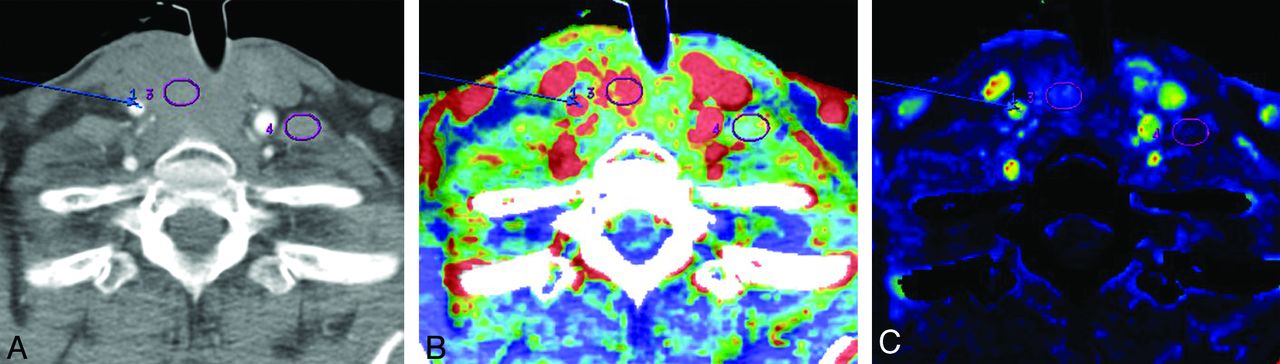
- Fig 3.
Floor-of-the-mouth HNSCC in a 67-year-old man (patient 2). A, Base image shows arterial enhancement. Blue arrow points to the right internal carotid artery (for the arterial input signal). Circle 3 is within the tumor; circle 4 is in noninvolved musculature. B, Blood volume image shows that the region of interest (circle 3) within the tumor was placed in solid non-necrotic tumor. C, Blood flow imaging shows minimally increased signal within the tumor bed (circle 3).
- Fig 4.
Scatterplot displays positive correlation between serum CXCL8 expression level (pg/mL) and rBF (mL/100 g per minute) in the 7 patients with advanced stage HNSCC.


Tables
- Table 1:
Patient data, tumor characteristics, and clinical course in 7 patients with advanced, treatment-resistant HNSCC
Patient Location Race Sex Age Metastatic Site Tobacco Clinical Course 1 Right nasal cavity White Male 54 n/a None Died 2 Left floor of mouth White Male 67 n/a 50 Pack-years Died 3 Right parotid gland African American Male 68 Lung 30 Pack-years Alive 4 Left oral tongue White Male 57 Lung, liver, lymph nodes, bone None Died 5 Peri-stomal African American Female 73 Lung 20 Pack-years Died 6 Peri-stomal White Male 54 Lung 60 Pack-years Alive 7 Left floor of mouth White Male 41 Lung, lymph nodes 15 Pack-years Alive Parameter CXCL8 Correlation Coefficient P Value BF −0.16 .67 BV 0.22 .47 CP −0.57 .19 MTT −0.03 .92 rBF 0.94 .01 rBV −0.14 .77 rCP −0.07 .88 rMTT 0.18 .68 Parameter Mean ± SD Range CXCL8 38.46 ± 30.46 6.97–99.89 BF 74.08 ± 56.41 14.78–187.93 BV 22.56 ± 35.96 5.08–103.71 CP 48.07 ± 21.56 16.73–79.84 MTT 9.46 ± 2.08 7.34–13.81 rBF 6.18 ± 6.56 1.08–20.26 rBV 2.77 ± 0.87 1.31–3.72 rCP 3.31 ± 1.88 1.46–6.70 rMTT 1.12 ± 0.41 0.78–1.94


