Advertisement

Main menu

  • Home
  • Content
    • Current Issue
    • Accepted Manuscripts
    • Article Preview
    • Past Issue Archive
    • Video Articles
    • AJNR Case Collection
    • Case of the Week Archive
    • Case of the Month Archive
    • Classic Case Archive
  • Special Collections
    • AJNR Awards
    • Low-Field MRI
    • Alzheimer Disease
    • ASNR Foundation Special Collection
    • Photon-Counting CT
    • View All
  • Multimedia
    • AJNR Podcasts
    • AJNR SCANtastic
    • Trainee Corner
    • MRI Safety Corner
    • Imaging Protocols
  • For Authors
    • Submit a Manuscript
    • Submit a Video Article
    • Submit an eLetter to the Editor/Response
    • Manuscript Submission Guidelines
    • Statistical Tips
    • Fast Publishing of Accepted Manuscripts
    • Graphical Abstract Preparation
    • Imaging Protocol Submission
    • Author Policies
  • About Us
    • About AJNR
    • Editorial Board
    • Editorial Board Alumni
  • More
    • Become a Reviewer/Academy of Reviewers
    • Subscribers
    • Permissions
    • Alerts
    • Feedback
    • Advertisers
    • ASNR Home

User menu

  • Alerts
  • Log in

Search

  • Advanced search
American Journal of Neuroradiology

American Journal of Neuroradiology

ASHNR American Society of Functional Neuroradiology ASHNR American Society of Pediatric Neuroradiology ASSR
  • Alerts
  • Log in

Advanced Search

  • Home
  • Content
    • Current Issue
    • Accepted Manuscripts
    • Article Preview
    • Past Issue Archive
    • Video Articles
    • AJNR Case Collection
    • Case of the Week Archive
    • Case of the Month Archive
    • Classic Case Archive
  • Special Collections
    • AJNR Awards
    • Low-Field MRI
    • Alzheimer Disease
    • ASNR Foundation Special Collection
    • Photon-Counting CT
    • View All
  • Multimedia
    • AJNR Podcasts
    • AJNR SCANtastic
    • Trainee Corner
    • MRI Safety Corner
    • Imaging Protocols
  • For Authors
    • Submit a Manuscript
    • Submit a Video Article
    • Submit an eLetter to the Editor/Response
    • Manuscript Submission Guidelines
    • Statistical Tips
    • Fast Publishing of Accepted Manuscripts
    • Graphical Abstract Preparation
    • Imaging Protocol Submission
    • Author Policies
  • About Us
    • About AJNR
    • Editorial Board
    • Editorial Board Alumni
  • More
    • Become a Reviewer/Academy of Reviewers
    • Subscribers
    • Permissions
    • Alerts
    • Feedback
    • Advertisers
    • ASNR Home
  • Follow AJNR on Twitter
  • Visit AJNR on Facebook
  • Follow AJNR on Instagram
  • Join AJNR on LinkedIn
  • RSS Feeds

AJNR Awards, New Junior Editors, and more. Read the latest AJNR updates

Research ArticleResearch Perspectives

Hot Topics in Functional Neuroradiology

S.H. Faro, F.B. Mohamed, J.A. Helpern, J.H. Jensen, K.R. Thulborn, I.C. Atkinson, H.I. Sair and D.J. Mikulis
American Journal of Neuroradiology December 2013, 34 (12) 2241-2249; DOI: https://doi.org/10.3174/ajnr.A3721
S.H. Faro
  • Find this author on Google Scholar
  • Find this author on PubMed
  • Search for this author on this site
F.B. Mohamed
  • Find this author on Google Scholar
  • Find this author on PubMed
  • Search for this author on this site
J.A. Helpern
  • Find this author on Google Scholar
  • Find this author on PubMed
  • Search for this author on this site
J.H. Jensen
  • Find this author on Google Scholar
  • Find this author on PubMed
  • Search for this author on this site
K.R. Thulborn
  • Find this author on Google Scholar
  • Find this author on PubMed
  • Search for this author on this site
I.C. Atkinson
  • Find this author on Google Scholar
  • Find this author on PubMed
  • Search for this author on this site
H.I. Sair
  • Find this author on Google Scholar
  • Find this author on PubMed
  • Search for this author on this site
D.J. Mikulis
  • Find this author on Google Scholar
  • Find this author on PubMed
  • Search for this author on this site
  • Article
  • Figures & Data
  • Info & Metrics
  • Responses
  • References
  • PDF
Loading

Article Figures & Data

Figures

  • Tables
  • Fig 1.
    • Download figure
    • Open in new tab
    • Download powerpoint
    Fig 1.

    Axial color FA map of a normal midcervical spine in a healthy pediatric subject.

  • Fig 2.
    • Download figure
    • Open in new tab
    • Download powerpoint
    Fig 2.

    Color FA map of a patient with SCI (right) and a corresponding T2-weighed sagittal image (left) demonstrating SCI at the C5 level (arrow).
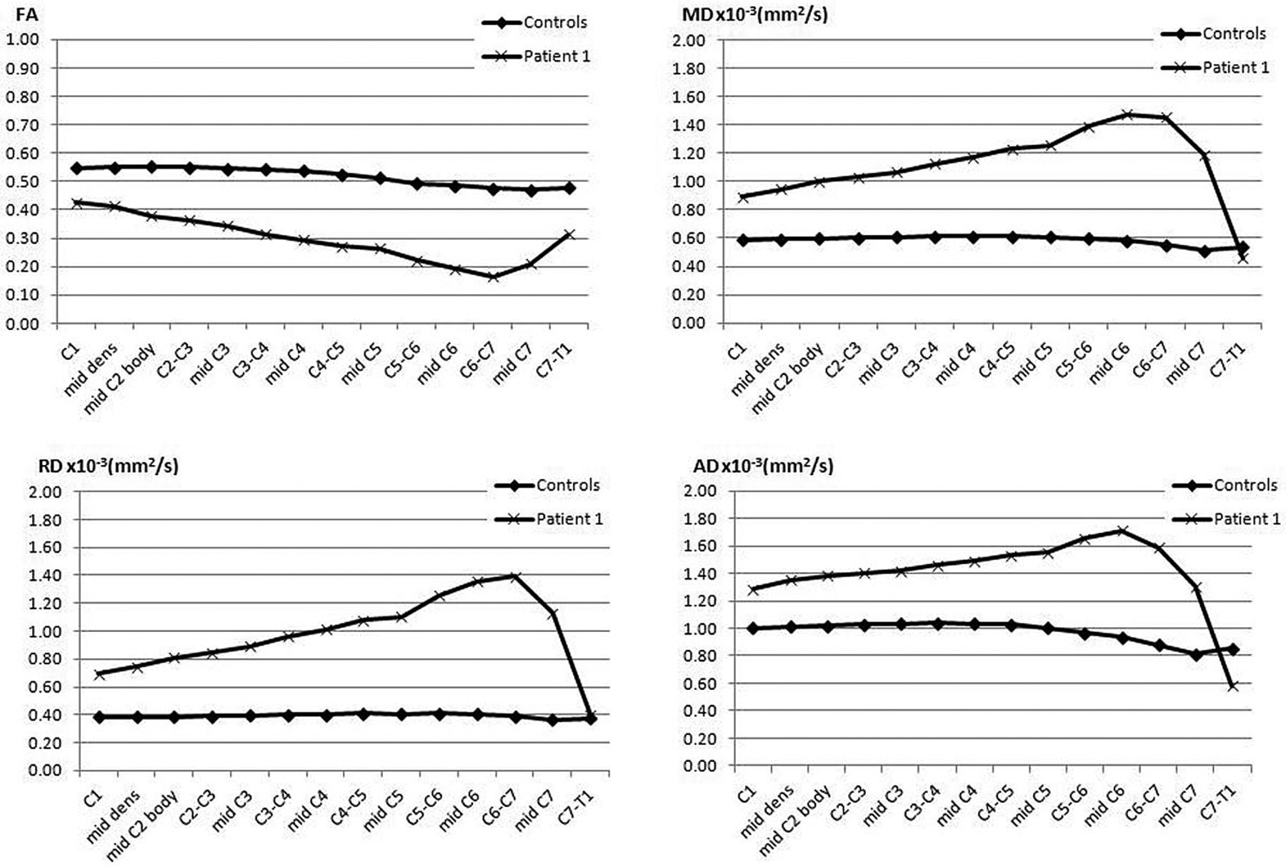
  • Fig 3.
    • Download figure
    • Open in new tab
    • Download powerpoint
    Fig 3.

    Healthy control and a patient with SCI at the mid-C6 level displaying changes in various DTI indices (FA, MD, radial diffusivity, and axial diffusivity) from C1 to C7.

  • Fig 4.
    • Download figure
    • Open in new tab
    • Download powerpoint
    Fig 4.

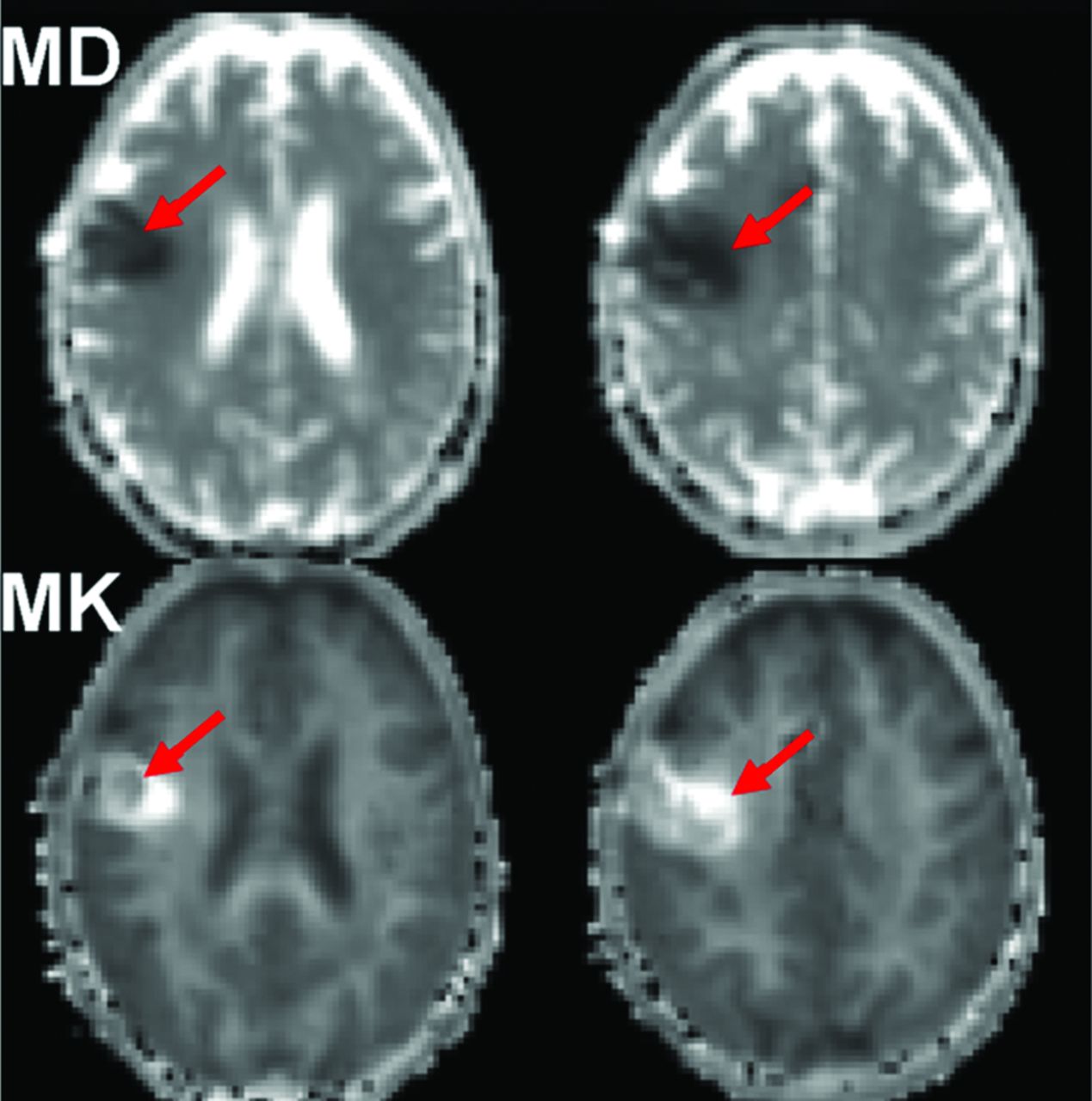
    Mean diffusivity and mean kurtosis maps of a patient with subacute ischemic stroke. Notice the distinct ischemic lesion signal heterogeneity on mean kurtosis that is not apparent on MD maps (red arrows).

  • Fig 5.
    • Download figure
    • Open in new tab
    • Download powerpoint
    Fig 5.

    Distribution of tissue cell fraction measured in brain parenchyma for the whole brain, including gray and white matter (blue, 0.81 ± 0.014) for cognitively healthy individuals (n = 15). The inset shows the individual measurements (vertical dashed lines) of each individual. The SD of the distribution of cell attenuation in healthy subjects is <2% of the total range of possible cell densities.

  • Fig 6.
    • Download figure
    • Open in new tab
    • Download powerpoint
    Fig 6.

    Tissue cell fraction as a function of age (years) in the cognitively healthy subjects in Fig 1. The fitted line has a near-zero gradient, indicating that there is no age dependence for mean kurtosis in the whole brain of cognitively healthy individuals.

  • Fig 7.
    • Download figure
    • Open in new tab
    • Download powerpoint
    Fig 7.

    Language network extracted from resting-state fMRI by using independent component analysis in a single healthy control demonstrates Broca and Wernicke areas.

  • Fig 8.
    • Download figure
    • Open in new tab
    • Download powerpoint
    Fig 8.

    The results of CVR mapping by using a controlled elevation of arterial carbon dioxide during blood oxygen level–dependent MR imaging in 2 different patients presenting with transient ischemic attacks. Both patients (A and C) have >90% carotid stenosis on MR angiography (red arrows). Corresponding CVR maps are shown with red/orange/yellow indicating increased oxyhemoglobin and therefore increased blood flow with CO2-induced vasodilation and blue indicating increased deoxyhemoglobin and therefore decreased blood flow with CO2-induced vasodilation. In the first patient, CVR is normal (B), indicating excellent collaterals, implying an embolic origin of symptoms. In the second patient (D), the CVR map shows blue in the anterior circulation, indicating exhausted vascular reserve and steal physiology, implying that the symptoms are secondary to hemodynamic compromise. As opposed to the first patient, the second patient would not benefit from medical management alone requiring a flow-restoration procedure (endarterectomy or stent placement) to alleviate symptoms and stroke risk. Note that in both cases, findings of conventional perfusion imaging with MR imaging or CT by using dynamic bolus techniques would be abnormal, showing delays in transit time in the affected hemispheres.

Tables

  • Figures
  • Features for a bioscale that enable its role as a surrogate for a clinical end point beyond the NIH definition of a biomarker

    No.Properties of a BioscaleImplications
    1Image-derived map of the disease siteMore sensitivity to earlier disease than diluted remote biomarkers
    2Spatially resolved quantitative parameterPrecise and accurate measurement
    3Small biologic variance in the healthy populationSensitivity to early disease
    4Continuously and monotonically varying with disease progressionSensitivity to disease progression or response to treatment
    5Intrinsically related to the disease mechanismHighly conserved metabolic parameter, essential for use as a surrogate of a clinical end point
    • Note:—NIH indicates National Institutes of Health.

PreviousNext
Back to top

In this issue

American Journal of Neuroradiology: 34 (12)
American Journal of Neuroradiology
Vol. 34, Issue 12
1 Dec 2013
  • Table of Contents
  • Index by author
  • Complete Issue (PDF)
Advertisement
Print
Download PDF
Email Article

Thank you for your interest in spreading the word on American Journal of Neuroradiology.

NOTE: We only request your email address so that the person you are recommending the page to knows that you wanted them to see it, and that it is not junk mail. We do not capture any email address.

Enter multiple addresses on separate lines or separate them with commas.
Hot Topics in Functional Neuroradiology
(Your Name) has sent you a message from American Journal of Neuroradiology
(Your Name) thought you would like to see the American Journal of Neuroradiology web site.
CAPTCHA
This question is for testing whether or not you are a human visitor and to prevent automated spam submissions.
Cite this article
S.H. Faro, F.B. Mohamed, J.A. Helpern, J.H. Jensen, K.R. Thulborn, I.C. Atkinson, H.I. Sair, D.J. Mikulis
Hot Topics in Functional Neuroradiology
American Journal of Neuroradiology Dec 2013, 34 (12) 2241-2249; DOI: 10.3174/ajnr.A3721

Citation Manager Formats

  • BibTeX
  • Bookends
  • EasyBib
  • EndNote (tagged)
  • EndNote 8 (xml)
  • Medlars
  • Mendeley
  • Papers
  • RefWorks Tagged
  • Ref Manager
  • RIS
  • Zotero
0 Responses
Respond to this article
Share
Bookmark this article
Hot Topics in Functional Neuroradiology
S.H. Faro, F.B. Mohamed, J.A. Helpern, J.H. Jensen, K.R. Thulborn, I.C. Atkinson, H.I. Sair, D.J. Mikulis
American Journal of Neuroradiology Dec 2013, 34 (12) 2241-2249; DOI: 10.3174/ajnr.A3721
del.icio.us logo Twitter logo Facebook logo Mendeley logo
  • Tweet Widget
  • Facebook Like
  • Google Plus One
Purchase

Jump to section

  • Article
    • Abstract
    • ABBREVIATIONS:
    • Diffusion Tensor Imaging of the Pediatric Spinal Cord
    • Diffusional Kurtosis Imaging
    • From Standardization to Quantification: Beyond Biomarkers toward Bioscales as Neuro MR Imaging Surrogates of Clinical End Points
    • Resting-State Functional MR imaging
    • Current Use of CVR Imaging in Clinical Neuroradiology
    • REFERENCES
  • Figures & Data
  • Info & Metrics
  • Responses
  • References
  • PDF

Related Articles

  • No related articles found.
  • PubMed
  • Google Scholar

Cited By...

  • Residual Tumor Volume, Cell Volume Fraction, and Tumor Cell Kill During Fractionated Chemoradiation Therapy of Human Glioblastoma using Quantitative Sodium MR Imaging
  • Crossref (1)
  • Google Scholar

This article has been cited by the following articles in journals that are participating in Crossref Cited-by Linking.

  • Quantitative sodium MR imaging: A review of its evolving role in medicine
    Keith R. Thulborn
    NeuroImage 2018 168

More in this TOC Section

  • Hot Topics in Research: Preventive Neuroradiology in Brain Aging and Cognitive Decline
  • Evidence Levels for Neuroradiology Articles: Low Agreement among Raters
  • Imaging Biomarkers in Ischemic Stroke Clinical Trials: An Opportunity for Rigor
Show more RESEARCH PERSPECTIVES

Similar Articles

Advertisement

Indexed Content

  • Current Issue
  • Accepted Manuscripts
  • Article Preview
  • Past Issues
  • Editorials
  • Editor's Choice
  • Fellows' Journal Club
  • Letters to the Editor
  • Video Articles

Cases

  • Case Collection
  • Archive - Case of the Week
  • Archive - Case of the Month
  • Archive - Classic Case

More from AJNR

  • Trainee Corner
  • Imaging Protocols
  • MRI Safety Corner
  • Book Reviews

Multimedia

  • AJNR Podcasts
  • AJNR Scantastics

Resources

  • Turnaround Time
  • Submit a Manuscript
  • Submit a Video Article
  • Submit an eLetter to the Editor/Response
  • Manuscript Submission Guidelines
  • Statistical Tips
  • Fast Publishing of Accepted Manuscripts
  • Graphical Abstract Preparation
  • Imaging Protocol Submission
  • Evidence-Based Medicine Level Guide
  • Publishing Checklists
  • Author Policies
  • Become a Reviewer/Academy of Reviewers
  • News and Updates

About Us

  • About AJNR
  • Editorial Board
  • Editorial Board Alumni
  • Alerts
  • Permissions
  • Not an AJNR Subscriber? Join Now
  • Advertise with Us
  • Librarian Resources
  • Feedback
  • Terms and Conditions
  • AJNR Editorial Board Alumni

American Society of Neuroradiology

  • Not an ASNR Member? Join Now

© 2025 by the American Society of Neuroradiology All rights, including for text and data mining, AI training, and similar technologies, are reserved.
Print ISSN: 0195-6108 Online ISSN: 1936-959X

Powered by HighWire