Advertisement

Main menu

  • Home
  • Content
    • Current Issue
    • Accepted Manuscripts
    • Article Preview
    • Past Issue Archive
    • Video Articles
    • AJNR Case Collection
    • Case of the Week Archive
    • Case of the Month Archive
    • Classic Case Archive
  • Special Collections
    • AJNR Awards
    • Low-Field MRI
    • Alzheimer Disease
    • ASNR Foundation Special Collection
    • Photon-Counting CT
    • View All
  • Multimedia
    • AJNR Podcasts
    • AJNR SCANtastic
    • Trainee Corner
    • MRI Safety Corner
    • Imaging Protocols
  • For Authors
    • Submit a Manuscript
    • Submit a Video Article
    • Submit an eLetter to the Editor/Response
    • Manuscript Submission Guidelines
    • Statistical Tips
    • Fast Publishing of Accepted Manuscripts
    • Graphical Abstract Preparation
    • Imaging Protocol Submission
    • Author Policies
  • About Us
    • About AJNR
    • Editorial Board
    • Editorial Board Alumni
  • More
    • Become a Reviewer/Academy of Reviewers
    • Subscribers
    • Permissions
    • Alerts
    • Feedback
    • Advertisers
    • ASNR Home

User menu

  • Alerts
  • Log in

Search

  • Advanced search
American Journal of Neuroradiology

American Journal of Neuroradiology

ASHNR American Society of Functional Neuroradiology ASHNR American Society of Pediatric Neuroradiology ASSR
  • Alerts
  • Log in

Advanced Search

  • Home
  • Content
    • Current Issue
    • Accepted Manuscripts
    • Article Preview
    • Past Issue Archive
    • Video Articles
    • AJNR Case Collection
    • Case of the Week Archive
    • Case of the Month Archive
    • Classic Case Archive
  • Special Collections
    • AJNR Awards
    • Low-Field MRI
    • Alzheimer Disease
    • ASNR Foundation Special Collection
    • Photon-Counting CT
    • View All
  • Multimedia
    • AJNR Podcasts
    • AJNR SCANtastic
    • Trainee Corner
    • MRI Safety Corner
    • Imaging Protocols
  • For Authors
    • Submit a Manuscript
    • Submit a Video Article
    • Submit an eLetter to the Editor/Response
    • Manuscript Submission Guidelines
    • Statistical Tips
    • Fast Publishing of Accepted Manuscripts
    • Graphical Abstract Preparation
    • Imaging Protocol Submission
    • Author Policies
  • About Us
    • About AJNR
    • Editorial Board
    • Editorial Board Alumni
  • More
    • Become a Reviewer/Academy of Reviewers
    • Subscribers
    • Permissions
    • Alerts
    • Feedback
    • Advertisers
    • ASNR Home
  • Follow AJNR on Twitter
  • Visit AJNR on Facebook
  • Follow AJNR on Instagram
  • Join AJNR on LinkedIn
  • RSS Feeds

AJNR Awards, New Junior Editors, and more. Read the latest AJNR updates

Review ArticleReview Articles
Open Access

CT Angiography for Surgical Planning in Face Transplantation Candidates

S. Soga, B. Pomahac, N. Wake, K. Schultz, R.F. Prior, K. Kumamaru, M.L. Steigner, D. Mitsouras, J. Signorelli, E.M. Bueno, D.S. Enterline and F.J. Rybicki
American Journal of Neuroradiology October 2013, 34 (10) 1873-1881; DOI: https://doi.org/10.3174/ajnr.A3268
S. Soga
aFrom the Department of Radiology (S.S., N.W., R.F.P., K.K., M.L.S., D.M., J.S., F.J.R.), Applied Imaging Science Laboratory
eDr Soga is currently affiliated with Department of Radiology, National Defense Medical College, Tokorozawa, Saitama, Japan.
  • Find this author on Google Scholar
  • Find this author on PubMed
  • Search for this author on this site
B. Pomahac
bDivision of Plastic and Reconstructive Surgery (B.P., E.M.B.), Brigham and Women's Hospital, Boston, Massachusetts
  • Find this author on Google Scholar
  • Find this author on PubMed
  • Search for this author on this site
N. Wake
aFrom the Department of Radiology (S.S., N.W., R.F.P., K.K., M.L.S., D.M., J.S., F.J.R.), Applied Imaging Science Laboratory
  • Find this author on Google Scholar
  • Find this author on PubMed
  • Search for this author on this site
K. Schultz
cToshiba Medical Research Institute (K.S.), Vernon Hills, Illinois
  • Find this author on Google Scholar
  • Find this author on PubMed
  • Search for this author on this site
R.F. Prior
aFrom the Department of Radiology (S.S., N.W., R.F.P., K.K., M.L.S., D.M., J.S., F.J.R.), Applied Imaging Science Laboratory
  • Find this author on Google Scholar
  • Find this author on PubMed
  • Search for this author on this site
K. Kumamaru
aFrom the Department of Radiology (S.S., N.W., R.F.P., K.K., M.L.S., D.M., J.S., F.J.R.), Applied Imaging Science Laboratory
  • Find this author on Google Scholar
  • Find this author on PubMed
  • Search for this author on this site
M.L. Steigner
aFrom the Department of Radiology (S.S., N.W., R.F.P., K.K., M.L.S., D.M., J.S., F.J.R.), Applied Imaging Science Laboratory
  • Find this author on Google Scholar
  • Find this author on PubMed
  • Search for this author on this site
D. Mitsouras
aFrom the Department of Radiology (S.S., N.W., R.F.P., K.K., M.L.S., D.M., J.S., F.J.R.), Applied Imaging Science Laboratory
  • Find this author on Google Scholar
  • Find this author on PubMed
  • Search for this author on this site
J. Signorelli
aFrom the Department of Radiology (S.S., N.W., R.F.P., K.K., M.L.S., D.M., J.S., F.J.R.), Applied Imaging Science Laboratory
  • Find this author on Google Scholar
  • Find this author on PubMed
  • Search for this author on this site
E.M. Bueno
bDivision of Plastic and Reconstructive Surgery (B.P., E.M.B.), Brigham and Women's Hospital, Boston, Massachusetts
  • Find this author on Google Scholar
  • Find this author on PubMed
  • Search for this author on this site
D.S. Enterline
dDepartment of Radiology (D.S.E.), Duke University Medical Center, Durham, North Carolina.
  • Find this author on Google Scholar
  • Find this author on PubMed
  • Search for this author on this site
F.J. Rybicki
aFrom the Department of Radiology (S.S., N.W., R.F.P., K.K., M.L.S., D.M., J.S., F.J.R.), Applied Imaging Science Laboratory
  • Find this author on Google Scholar
  • Find this author on PubMed
  • Search for this author on this site
  • Article
  • Figures & Data
  • Info & Metrics
  • Responses
  • References
  • PDF
Loading

Article Figures & Data

Figures

  • Tables
  • Fig 1.
    • Download figure
    • Open in new tab
    • Download powerpoint
    Fig 1.

    A 59-year-old man who underwent partial face transplantation. A, Severe disfigurement of the midface caused by a high-voltage burn injury is demonstrated, despite multiple conventional reconstructive attempts. B, Two-year postoperative follow-up illustrates restoration of form and function.

  • Fig 2.
    • Download figure
    • Open in new tab
    • Download powerpoint
    Fig 2.

    Candidate for full face transplantation. A, After catastrophic loss of facial tissues, muscle flaps and skin grafts placed during >20 surgeries rendered the patient's face featureless. B, Surgical-planning volume-rendered CT angiography depicts residual arteries after previous reconstructions using bilateral free latissimus muscle and serratus muscle flap arteries (dashed arrows), which are anastomosed end-to-end to the bilateral facial artery stumps (arrows). While not ideal, those facial and/or flap arteries are technically available and considered for microsurgical anastomoses. Prior surgical clips are rendered in green using the multiobject segmentation described in the text. C, Venous images from the same CT acquisition show occluded or absent bilateral anterior, posterior facial, and left external jugular veins. Patency of the right external jugular vein (arrow) and bilateral internal jugular veins (dashed arrows) is confirmed.
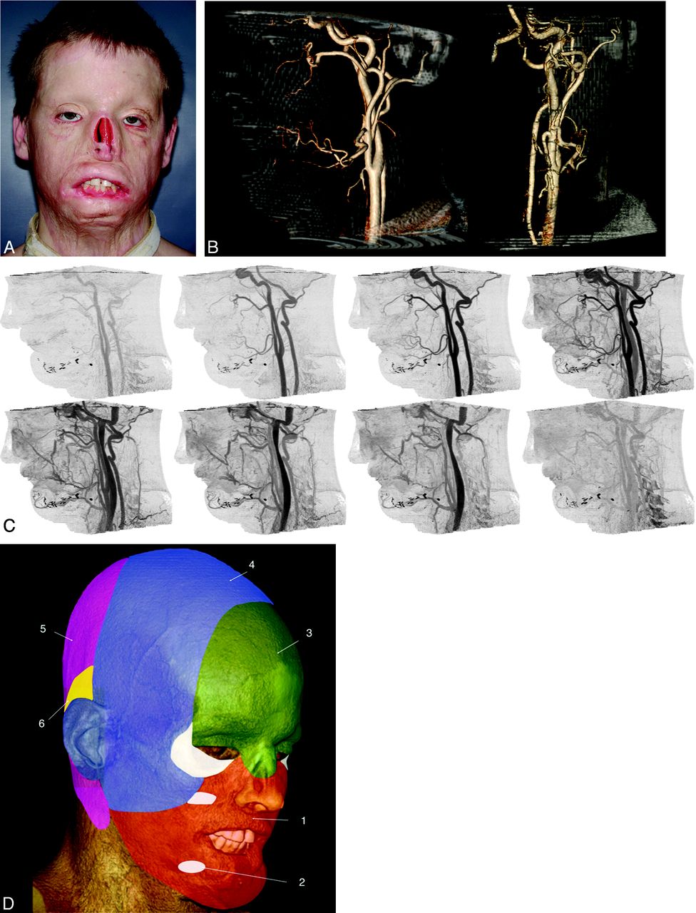
  • Fig 3.
    • Download figure
    • Open in new tab
    • Download powerpoint
    Fig 3.

    Candidate for full face transplantation. A, After a high-voltage injury, several flap and graft procedures resulted in persistent disfigurement. The patient wore a nasal prosthesis, removed for this photograph. B, Representative sagittal (left) and anteroposterior (right) projections from CT volume rendering. Volumes are viewed from an arbitrary angle to characterize branch and smaller vessels—for example, those from the external carotid artery that may be available for anastomoses. The nasal prosthesis was included in the CT acquisition. C, Sagittal cine CT images provide time resolution and enable separation of arteries and veins so that each dataset can be individually postprocessed. D, Angiosomes of the face overlaid on a volume-rendered CT image, including all soft-tissue components and numbered according to the source artery: 1) facial, 2) internal maxillary, 3) ophthalmic and internal carotid, 4) superficial temporal, 5) vertebral, and 6) posterior auricular. Lower and midface (orange) allografts can be perfused solely by facial arteries. Although the lower two-thirds of the face includes internal maxillary artery angiosomes, this territory can also be perfused by the facial artery via neighboring angiosome collateral vessels. For procedures in which the allograft includes the upper face and scalp (green and blue), the facial and superficial temporal arteries should be included, with the external carotid artery as the source vessel.

  • Fig 4.
    • Download figure
    • Open in new tab
    • Download powerpoint
    Fig 4.

    Volume-rendered venous-only reformatted images from a candidate who had a blast injury. A, Anterior and posterior facial veins and the external jugular vein are absent on the right, presumably from the injury. Imaging confirms the patency of the anterior jugular vein (arrow), a potential alternative for flap drainage. B, On the left, the external jugular vein (dashed arrow) and anterior facial vein (arrowhead) are available for flap drainage.

  • Fig 5.
    • Download figure
    • Open in new tab
    • Download powerpoint
    Fig 5.

    Major variations in branching patterns of the external carotid artery (3 types) and variations of the confluence of the facial, lingual, and superior thyroid veins with the internal jugular vein (5 types), described by Shima et al.24 Descriptions of each variation are found in the Table.

  • Fig 6.
    • Download figure
    • Open in new tab
    • Download powerpoint
    Fig 6.

    Timing diagram for a 320–detector row CT acquisition for face transplantation candidates. Each bar refers to 1 phase of the multiphasic axial acquisition; each volume includes the entire anatomy required for surgical planning.
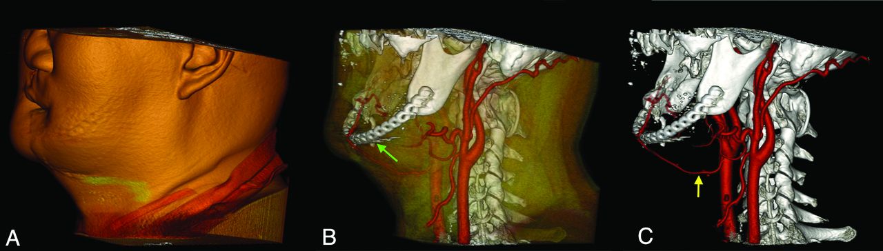
  • Fig 7.
    • Download figure
    • Open in new tab
    • Download powerpoint
    Fig 7.

    Candidate for full face transplantation. A, Volume-rendered image including full soft tissues. B, Skin and superficial soft tissues have been shadowed. This view depicts the relationship of the bones, postsurgical hardware (green arrow), and segmented arteries. C, Volume rendering that exclusively shows the bones, postsurgical hardware, and arteries. The superior thyroid artery (yellow arrow) is used for the anastomosis of free flap, and its surgically altered course is demonstrated.

  • Fig 8.
    • Download figure
    • Open in new tab
    • Download powerpoint
    Fig 8.

    Face transplantation candidate shown with fusion of the preoperative photograph and CT venography images. Meticulous CT segmentation and feature mapping are used to depict preoperative structures critical for rapid, precise, surgical dissection.

  • Fig 9.
    • Download figure
    • Open in new tab
    • Download powerpoint
    Fig 9.

    Candidate for full face transplantation who was attacked by a chimpanzee. A, Photograph of the victim after multiple conventional reconstructive surgeries for catastrophic facial injury, demonstrating the limitation of conventional surgical options. B, Volume-rendered reformations of CT images by using multiobject segmentation to rapidly communicate information to the surgical team. In red, the arteries, including the course of right facial artery, are clearly demonstrated. The facial artery was used for the anterolateral thigh flap immediately after the injury. The anastomosis can be identified via surgical clips rendered in green.

Tables

  • Figures
  • Major variations in branching patterns of the external carotid artery (3 types) and variations in the confluence of the facial, lingual, and superior thyroid veins with internal jugular vein (5 types)

    Variant TypeDescription
    Arterial
        Noncommon trunkFacial, lingual, and superior thyroid arteries arise separately from the ECA
        Truncus linguofacialisFacial and lingual arteries arise from the ECA in a common trunk
        Truncus thyrolingualisSuperior thyroid and lingual arteries arise from the ECA in a common trunk
    Venous
        ThyrolinguofacialisFacial, lingual, and superior thyroid veins form a thyrolinguofacialis vein
        LinguofacialisFacial and lingual veins form a venous stem
        ThyrofacialisFacial and superior thyroid veins join together, and separate lingua l vein joins into the IJV
        Nonfacial veinSuperior thyroid and lingual veins join together into the IJV
        SeparationLingual and superior thyroid veins fuse independently with the IJV
    • Note:—ECA indicates external carotid artery; IJV, internal jugular vein.

PreviousNext
Back to top

In this issue

American Journal of Neuroradiology: 34 (10)
American Journal of Neuroradiology
Vol. 34, Issue 10
1 Oct 2013
  • Table of Contents
  • Index by author
  • Complete Issue (PDF)
Advertisement
Print
Download PDF
Email Article

Thank you for your interest in spreading the word on American Journal of Neuroradiology.

NOTE: We only request your email address so that the person you are recommending the page to knows that you wanted them to see it, and that it is not junk mail. We do not capture any email address.

Enter multiple addresses on separate lines or separate them with commas.
CT Angiography for Surgical Planning in Face Transplantation Candidates
(Your Name) has sent you a message from American Journal of Neuroradiology
(Your Name) thought you would like to see the American Journal of Neuroradiology web site.
CAPTCHA
This question is for testing whether or not you are a human visitor and to prevent automated spam submissions.
Cite this article
S. Soga, B. Pomahac, N. Wake, K. Schultz, R.F. Prior, K. Kumamaru, M.L. Steigner, D. Mitsouras, J. Signorelli, E.M. Bueno, D.S. Enterline, F.J. Rybicki
CT Angiography for Surgical Planning in Face Transplantation Candidates
American Journal of Neuroradiology Oct 2013, 34 (10) 1873-1881; DOI: 10.3174/ajnr.A3268

Citation Manager Formats

  • BibTeX
  • Bookends
  • EasyBib
  • EndNote (tagged)
  • EndNote 8 (xml)
  • Medlars
  • Mendeley
  • Papers
  • RefWorks Tagged
  • Ref Manager
  • RIS
  • Zotero
0 Responses
Respond to this article
Share
Bookmark this article
CT Angiography for Surgical Planning in Face Transplantation Candidates
S. Soga, B. Pomahac, N. Wake, K. Schultz, R.F. Prior, K. Kumamaru, M.L. Steigner, D. Mitsouras, J. Signorelli, E.M. Bueno, D.S. Enterline, F.J. Rybicki
American Journal of Neuroradiology Oct 2013, 34 (10) 1873-1881; DOI: 10.3174/ajnr.A3268
del.icio.us logo Twitter logo Facebook logo Mendeley logo
  • Tweet Widget
  • Facebook Like
  • Google Plus One
Purchase

Jump to section

  • Article
    • Abstract
    • VASCULAR CONSIDERATIONS IN FACIAL TRANSPLANTATION
    • IMAGING FOR PREOPERATIVE SURGICAL PLANNING
    • CONCLUSIONS
    • Footnotes
    • REFERENCES
  • Figures & Data
  • Info & Metrics
  • Responses
  • References
  • PDF

Related Articles

  • No related articles found.
  • PubMed
  • Google Scholar

Cited By...

  • Early Postoperative Imaging and Image-Guided Procedures on Patients with Face Transplants
  • Facial and Hand Allotransplantation
  • Crossref (27)
  • Google Scholar

This article has been cited by the following articles in journals that are participating in Crossref Cited-by Linking.

  • Measuring and Establishing the Accuracy and Reproducibility of 3D Printed Medical Models
    Elizabeth George, Peter Liacouras, Frank J. Rybicki, Dimitrios Mitsouras
    RadioGraphics 2017 37 5
  • Face transplantation-current status and future developments
    Sotirios Tasigiorgos, Branislav Kollar, Nicco Krezdorn, Ericka M. Bueno, Stefan G. Tullius, Bohdan Pomahac
    Transplant International 2018 31 7
  • Noninvasive Monitoring of Immune Rejection in Face Transplant Recipients
    Maximilian Kueckelhaus, Amir Imanzadeh, Sebastian Fischer, Kanako Kumamaru, Muayyad Alhefzi, Ericka Bueno, Nicole Wake, Marie D. Gerhard-Herman, Frank J. Rybicki, Bohdan Pomahac
    Plastic and Reconstructive Surgery 2015 136 5
  • Transformation of Face Transplants: Volumetric and Morphologic Graft Changes Resemble Aging After Facial Allotransplantation
    M. Kueckelhaus, M. Turk, K.K. Kumamaru, L. Wo, E.M. Bueno, C.G. Lian, M. Alhefzi, M.A. Aycart, S. Fischer, U. De Girolami, G.F. Murphy, F.J. Rybicki, B. Pomahac
    American Journal of Transplantation 2016 16 3
  • Fat Embolism After Autologous Facial Fat Grafting
    Nicolas S Dhooghe, Sebastiaan Maes, Bernard Depypere, Karel E Y Claes, Renaat Coopman, Bela Kubat, Michel H Piette, Stan Monstrey
    Aesthetic Surgery Journal 2022 42 3
  • Facial Restoration by Transplantation
    Tormod S. Westvik, Alexander Dermietzel, Bohdan Pomahac
    Annals of Plastic Surgery 2015 74 Supplement 1
  • Vascular Communications Between Donor and Recipient Tissues After Successful Full Face Transplantation
    K.K. Kumamaru, G.C. Sisk, D. Mitsouras, K. Schultz, M.L. Steigner, E. George, D.S. Enterline, E.M. Bueno, B. Pomahac, F.J. Rybicki
    American Journal of Transplantation 2014 14 3
  • Evaluation of velopharyngeal closure by 4D imaging using 320-detector-row computed tomography
    Yoshiaki Sakamoto, Shigeyoshi Soga, Masahiro Jinzaki, Yoshitake Yamada, Hisao Ogata, Kazuo Kishi
    Journal of Plastic, Reconstructive & Aesthetic Surgery 2015 68 4
  • Analysis of collateral lymphatic circulation in patients with lower limb lymphedema using magnetic resonance lymphangiography
    Shigeyoshi Soga, Fumio Onishi, Masahiro Jinzaki, Ayako Mikoshi, Toshiharu Minabe, Hiroshi Shinmoto
    Journal of Vascular Surgery: Venous and Lymphatic Disorders 2021 9 2
  • Facial Transplantation
    Jack E. Russo, Eric M. Genden
    Facial Plastic Surgery Clinics of North America 2016 24 3

More in this TOC Section

  • An Atlas of Neonatal Neurovascular Imaging Anatomy as Depicted with Microvascular Imaging: The Intracranial Arteries
  • An Atlas of Neonatal Neurovascular Imaging Anatomy as Depicted with Microvascular Imaging: The Intracranial Veins
  • Clinical Translation of Hyperpolarized 13C Metabolic Probes for Glioma Imaging
Show more Review articles

Similar Articles

Advertisement

Indexed Content

  • Current Issue
  • Accepted Manuscripts
  • Article Preview
  • Past Issues
  • Editorials
  • Editor's Choice
  • Fellows' Journal Club
  • Letters to the Editor
  • Video Articles

Cases

  • Case Collection
  • Archive - Case of the Week
  • Archive - Case of the Month
  • Archive - Classic Case

More from AJNR

  • Trainee Corner
  • Imaging Protocols
  • MRI Safety Corner
  • Book Reviews

Multimedia

  • AJNR Podcasts
  • AJNR Scantastics

Resources

  • Turnaround Time
  • Submit a Manuscript
  • Submit a Video Article
  • Submit an eLetter to the Editor/Response
  • Manuscript Submission Guidelines
  • Statistical Tips
  • Fast Publishing of Accepted Manuscripts
  • Graphical Abstract Preparation
  • Imaging Protocol Submission
  • Evidence-Based Medicine Level Guide
  • Publishing Checklists
  • Author Policies
  • Become a Reviewer/Academy of Reviewers
  • News and Updates

About Us

  • About AJNR
  • Editorial Board
  • Editorial Board Alumni
  • Alerts
  • Permissions
  • Not an AJNR Subscriber? Join Now
  • Advertise with Us
  • Librarian Resources
  • Feedback
  • Terms and Conditions
  • AJNR Editorial Board Alumni

American Society of Neuroradiology

  • Not an ASNR Member? Join Now

© 2025 by the American Society of Neuroradiology All rights, including for text and data mining, AI training, and similar technologies, are reserved.
Print ISSN: 0195-6108 Online ISSN: 1936-959X

Powered by HighWire