
Article Figures & Data
Figures
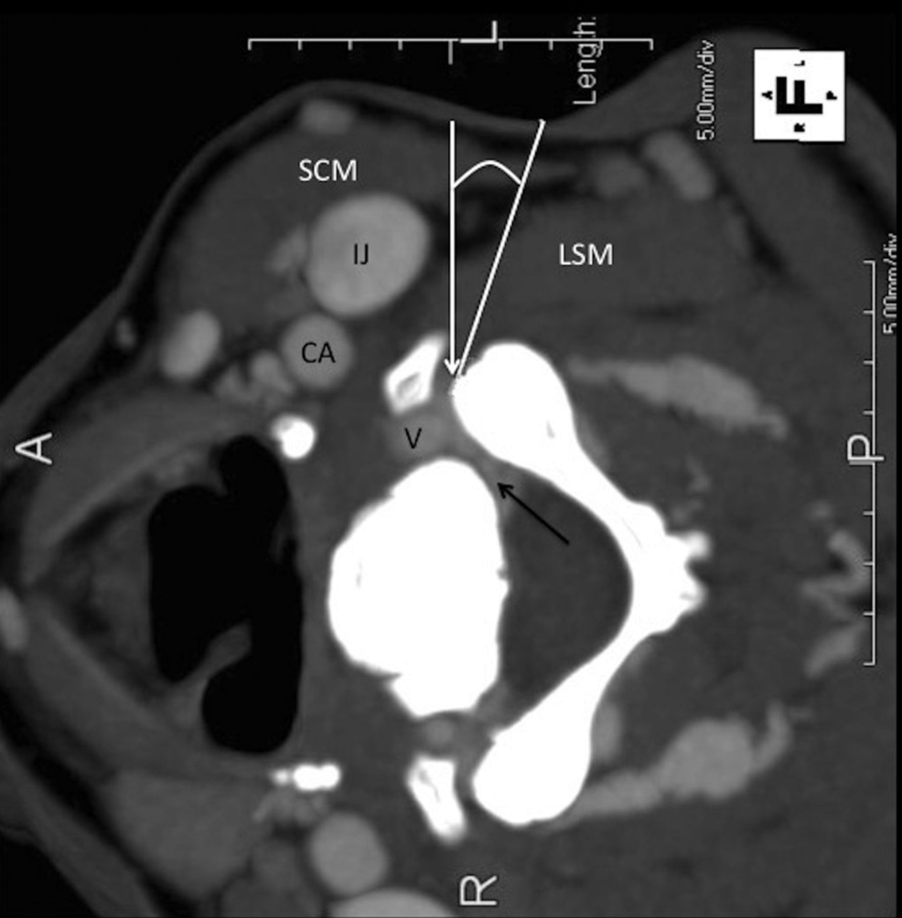
- Fig 1.
Lateral decubitus position: anatomy and targeting. CT scan with contrast. The white line indicates the deviation from the perpendicular approach; the black arrow, the perineural venous plexus; the white arrow, the target at the anterior margin of the facet. SCM indicates the sternocleidomastoid muscle; IJ, internal jugular vein; CA, carotid artery; V, vertebral artery; LSM, levator scapulae muscle.
- Fig 2.
Injection procedure. A, Needle nearly perpendicular to the floor. The dotted line indicates the theoretic dorsal approach to the same target. The dashed line is parallel to the CT table line. The vertical solid line is a plumb line. B, Injection procedure. Enlarged view shows the 3.8-cm, 25-ga needle (N), with the tip at the lateral aspect of the neural foramen (white arrow). Contrast has flowed into the neural foramen (black arrow) and outlined the vertebral artery (V).
- Fig 3.
ImPACT dose software. The shaded area represents a volume of tissue imaged for a scan series. The area is between the skull base and thyroid gland. This was used to calculate constants for the effective dose based on different amounts of thyroid gland in the imaging volume. The grid indicates the section location of the scan and correlates with the On-line Table ImPACT results.
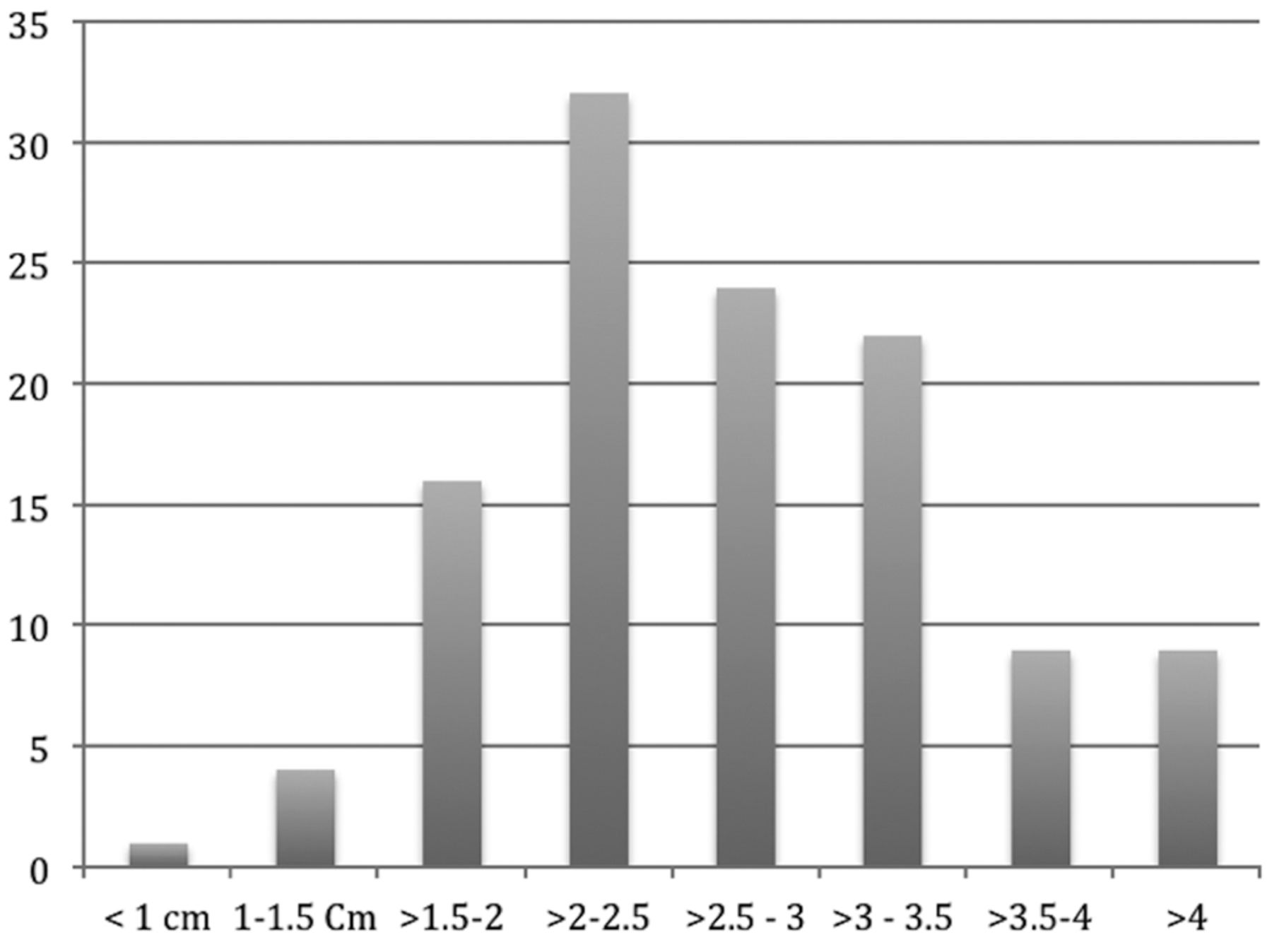
- Fig 4.
Histogram demonstrating needle-insertion lengths.
- Fig 5.
Needle-insertion attempts correlated with target depth and angle deviation from the perpendicular. More insertion attempts increase the procedural time and radiation dose (P < .001).


