
Article Figures & Data
Figures
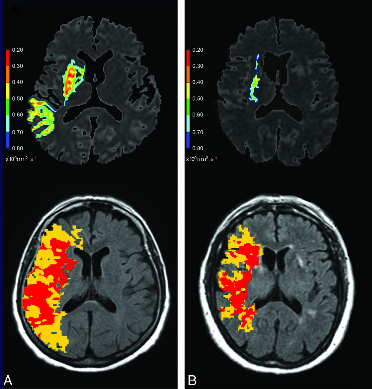
- Fig 1.
Tmax maps used for nCCD quantification and corresponding ADC maps from 2 patients with different recanalization pattern. Upper images: ADC maps with lesion delineated by visual selection of the most appropriate ADC threshold value (the hotter the color, the lower the ADC value). Lower images: Tmax maps (superimposed on FLAIR images) with colored areas corresponding to moderately (yellow area) and severely (red area) hypoperfused parenchyma. A, 75-year-old woman, proximal MCA-M1 occlusion, NIHSS = 11, TTT = 135 minutes, no MCA recanalization at 24 hours. Volume of DWI abnormalities detected on all brain sections = 54.78 mL. nCCD = 105.14. B, 74-year-old woman, distal MCA-M1 occlusion, NIHSS = 15, TTT = 94 minutes, complete MCA recanalization at 24 hours. Volume of DWI abnormalities detected on all brain sections = 2.74 mL. nCCD = 68.16. Calculation method for nCCD: The yellow area corresponds to moderately hypoperfused parenchyma (Tmax values between lower and upper Tmax thresholds). The red area corresponds to severely hypoperfused parenchyma (Tmax exceeding the upper threshold). Lower and upper thresholds were defined as the Tmax mean value of the whole contralateral hemisphere (calculated from a brain section located at the centrum semi ovale level with exclusion of skull and ventricles). The lower threshold is contralateral Tmax mean value +2 seconds and the upper threshold is contralateral Tmax mean value +6 seconds. The volume of ipsilateral and supratentorial, moderately (yellow area) or severely (red area), hypoperfused parenchyma is calculated from all brain sections. nCCD = (red area volume/yellow area volume) × (red area volume + yellow area volume).
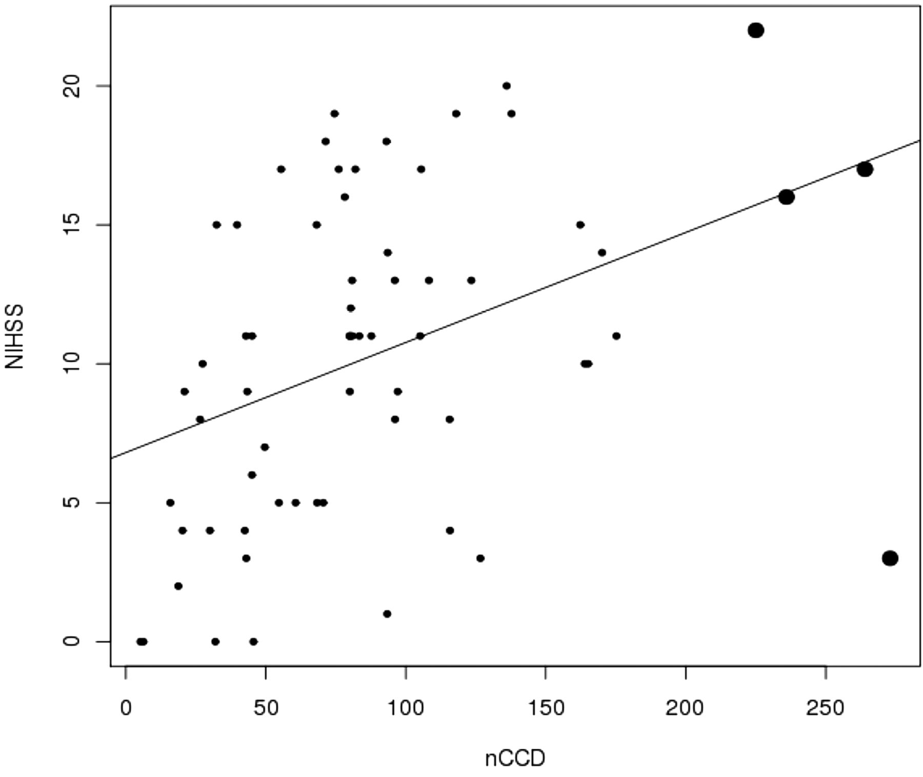
- Fig 2.
Values dispersion illustrating the significant correlation between nCCD and baseline NIHSS. The bold dots represent the outliers (Pearson correlation test, positive estimated correlation = 0.40, P = .00089).
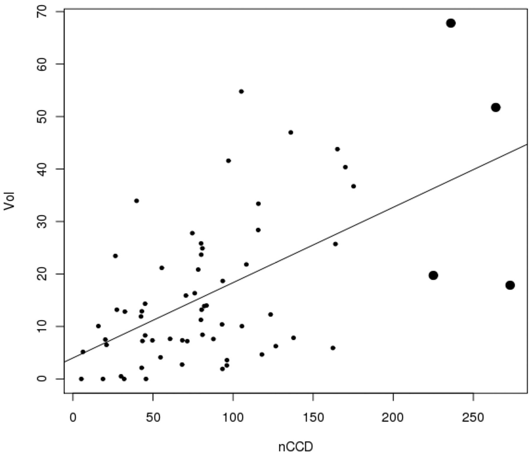
- Fig 3.
Values dispersion illustrating the significant correlation between nCCD and lesional volume on DWI. The bold dots represent the outliers (Pearson correlation test, positive estimated correlation = 0.57, P < .0001.
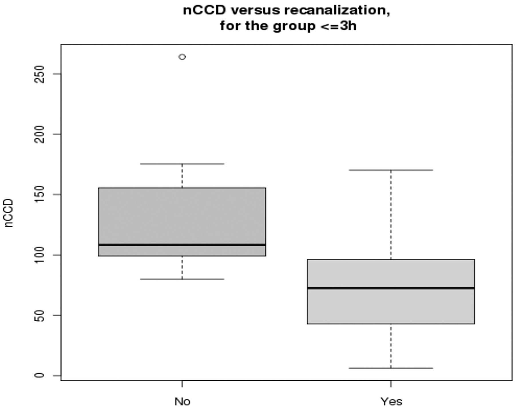
- Fig 4.
nCCD values in patients treated within 3 hours from stroke onset, function of the recanalization status (partial or null versus full). The nCCD is significantly higher in patients who did not achieve full recanalization (P = .007).
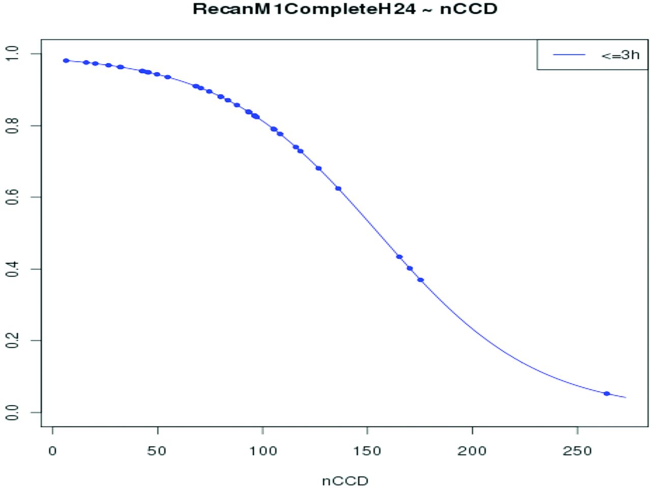
- Fig 5.
Probability of full MCA-M1 recanalization at 24 hours, function of nCCD value in patients treated within 3 hours after stroke onset. The probability of recanalization significantly decreases with the increase of nCCD (P = .021).


Tables
Expert vs Nonexpert 1 Expert vs Nonexpert 2 Nonexpert 1 vs Nonexpert 2 ICC (95% CI) Automatic AIF selection r2 0.84 0.90 0.94 0.92 (0.88–0.96) Manual AIF selection r2 0.77 0.79 0.84 0.88 (0.82–0.92) Note:—ICC indicates intraclass correlation.
- Table 2:
Reproducibility of collateral circulation deficit measurements: Intrarater (automatic vs manual AIF selection)
Expert Nonexpert 1 Nonexpert 2 r2 0.72 0.86 0.85 ICC (95% CI) 0.81 (0.69–0.89) 0.82 (0.71–0.89) 0.84 (0.74–0.90) Note:—ICC indicates intraclass correlation.
Age Sex Ratio (M/F) Diabetes (%) Antiplatelet Therapy (%) Stroke Etiology (CE/Atherom/Other) (%) Recanalizers + Nonrecanalizers Distal MCA-M1 occlusion (n = 23) 67.5 (15.1) 7/16 0 26 60.8/8.7/30.4 Proximal MCA-M1 occlusion (n = 26) 63 (16.7) 8/18 11.5 34.6 69.2/3.8/26.9 ICA-MCA M1 occlusion (n = 15) 66 (10.2) 14/1 0 33 00/93.3/6.6 All (n = 64) 65 (14.8) 30/34 4.7 31.2 50/26.5/23.5 Recanalizers Distal MCA-M1 occlusion (n = 19) 66 (16.2) 7/12 0 21 57.9/10.5/31.5 Proximal MCA-M1 occlusion (n = 16) 59.8 (20) 6/10 12.5 43.7 68.7/00/31.2 ICA-MCA M1 occlusion (n = 11) 66.7 (10.2) 11/0 0 36.3 00/100/00 All (n = 46) 62.5 (18) 13/33 4 22 46/28/24 Nonrecanalizers Distal MCA-M1 occlusion (n = 4) 74.7 (4.6) 1/3 0 50 75/00/25 Proximal MCA-M1 occlusion (n = 10) 68 (8.4) 2/8 10 20 70/10/20 ICA-MCA M1 occlusion (n = 4) 64 (11.5) 3/1 0 25 00/75/25 All (n = 18) 69 (8.88) 6/12 6 28 56/22/22 Note:—Recanalizers = arterial occlusive lesion (AOL) 3 recanalization, ie, full recanalization; nonrecanalizers = AOL 0–2 recanalization, ie, partial or null recanalization. Univariate statistical analysis comparing recanalizers with nonrecanalizers. Results are expressed as mean (standard deviation), ratio, or percentages. CE/Atherom/Other indicates cardioembolic, atheromatous, and other etiology.
- Table 4:
Baseline characteristics, nCCD values, and outcome in recanalizers and nonrecanalizers
TTT (min) NIHSS Score DWI Vol (ml) Collateral Circulation Deficit (nCCD) % mRS 0–2/6 at 3 Months Recanalizers + Nonrecanalizers Distal MCA-M1 occlusion (n = 23) 181.5 (91.2) 8.6 (5.9) 13.9 (16.4) 73.3 (53.12) 78.2/4.3 Proximal MCA-M1 occlusion (n = 26) 175.4 (58.4) 12 (5.7) 16.7 (14) 92.1 (51.3) 54/3.8 ICA-MCA M1 occlusion (n = 15) 158.86 (65.6) 9.86 (5.15) 20.77 (14) 104.1 (77.3) 60/6.6 All (n = 64) 174 (72.7) 10 (5.8) 17 (14.9) 88.17 (59.21) 64/4.7 Recanalizers Distal MCA-M1 occlusion (n = 19, 82.6%) 164.4 (88.7) 8 (5.8) 12.1 (12.2) 68.2 (42.3) 89.5/0 Proximal MCA-M1 occlusion (n = 16, 61.5%) 158.4 (46.1) 11.4 (5.4) 13.8 (11.5) 92.7 (47.3) 68.7/0 ICA-MCA M1 occlusion (n = 11, 73.3%) 155 (71.2) 9.27 (5.31) 17.6 (11.1) 86.6 (69.2) 63.6/9.1 All (n = 46, 71.8%) 157.5 (68.2) 9.3 (5.7) 13 (12) 79.6 (46.4) 76/2 Nonrecanalizers Distal MCA-M1 occlusion (n = 4, 17.4%) 262.5 (56.4) 11.5 (6.1) 22.3 (30.8) 97.85 (96.7) 25/25 Proximal MCA-M1 occlusion (n = 10, 38.5%) 202.7 (67.8) 13.1 (6.3) 21.4 (17) 91 (59.7) 30/10 ICA-MCA M1 occlusion (n = 4, 26.7%) 172 (53.4) 11.5 (5) 29.5 (19) 152 (88.2) 50/0 All (n = 18, 28.1%) 209.1 (67.2)a 12.4 (5.7)a 23.4 (19.9)a 106.1 (74.1) 33a/11 Note:—Recanalizers = arterial occlusive lesion (AOL) 3 recanalization, ie, full recanalization; nonrecanalizers = AOL 0–2 recanalization, ie, partial or null recanalization. Univariate statistical analysis comparing recanalizers versus nonrecanalizers. Student t test or Fisher exact test for comparison of percentages. Results are expressed as mean (standard deviation), ratio, or percentages. mRS indicates modified Rankin Scale; DWI Vol, volume of lesions detected on DWI.
↵a P < 0.05.
In this issue


Jump to section
Related Articles
Cited By...
- Collateral status and recanalization after endovascular treatment for acute ischemic stroke
- Collateral status and recanalization after endovascular treatment for acute ischemic stroke
- Clinical prognosis of FLAIR hyperintense arteries in ischaemic stroke patients: a systematic review and meta-analysis
- Incidence and Predictors of Early Recanalization After Intravenous Thrombolysis: A Systematic Review and Meta-Analysis
- Acute ischaemia after subarachnoid haemorrhage, relationship with early brain injury and impact on outcome: a prospective quantitative MRI study
- Time and Diffusion Lesion Size in Major Anterior Circulation Ischemic Strokes
- Hypoperfusion Intensity Ratio Predicts Infarct Progression and Functional Outcome in the DEFUSE 2 Cohort