
Article Figures & Data
Figures
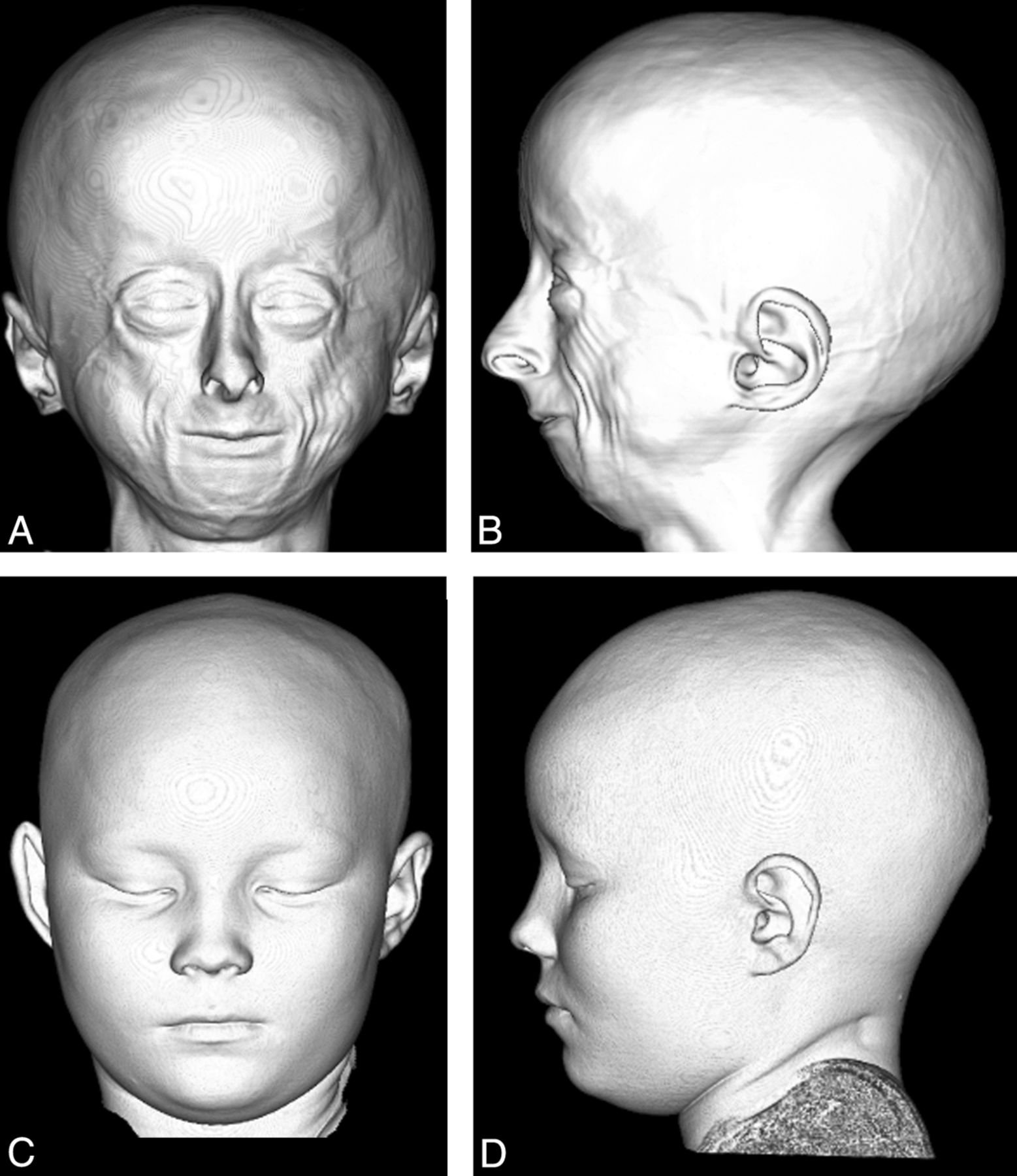
- Fig 1.
Anterior (A) and lateral (B) projection of a 3D CT shaded-surface display of the head demonstrates craniofacial disproportion, prominent eyes, hypotelorism, narrow nasal bridge with broad tipped nose, small face and mandible, lack of facial fat, and prominent veins in a 9-year-old child with HGPS compared with an age-matched control (C and D).
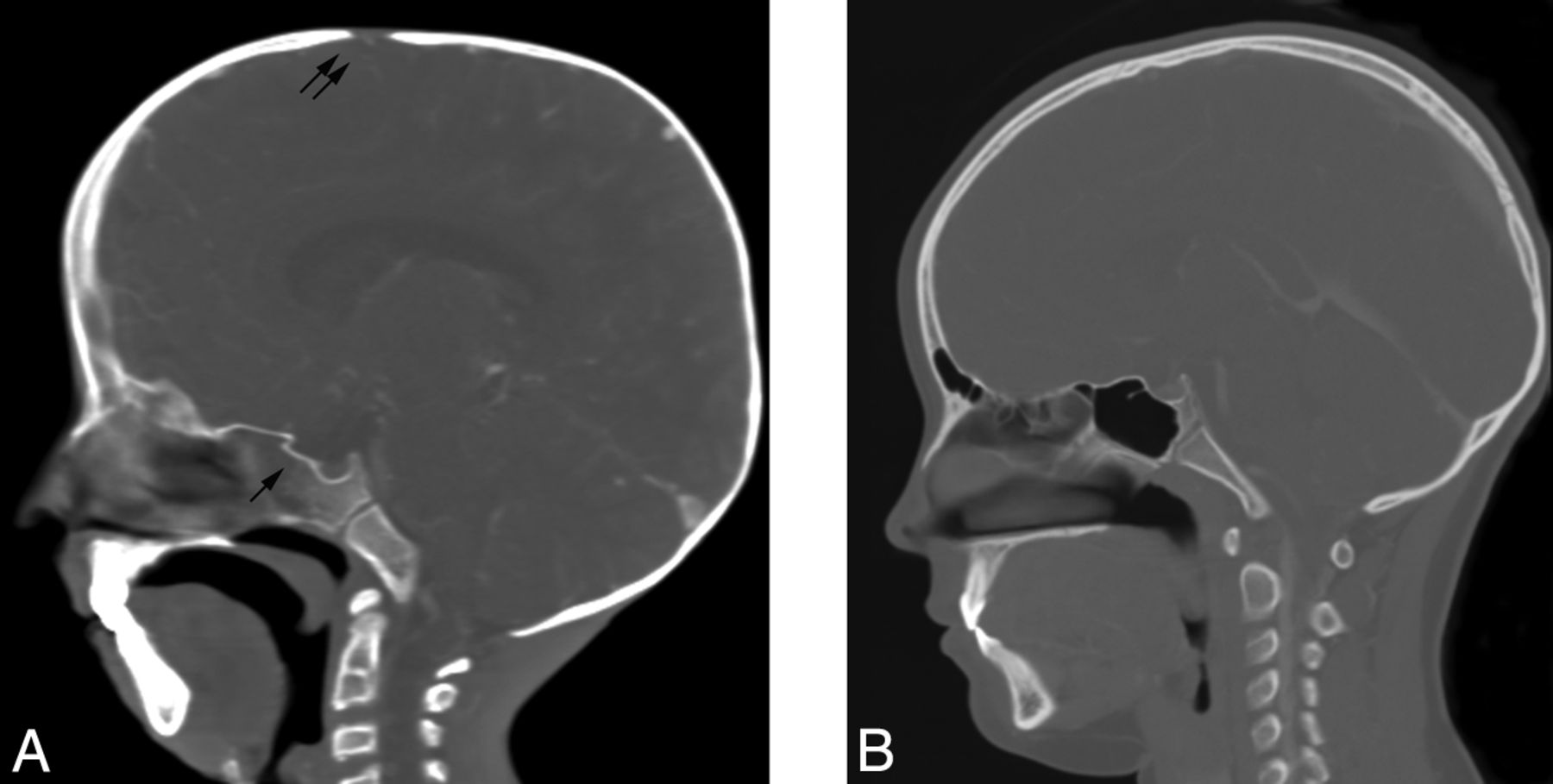
- Fig 2.
Sagittal CT reformatted image of a 9-year-old child with HGPS (A) demonstrates a thin calvarium, paucity of scalp fat, a J-shaped sella (single arrow), and a patent anterior fontanel (double arrow) compared with the control (B).
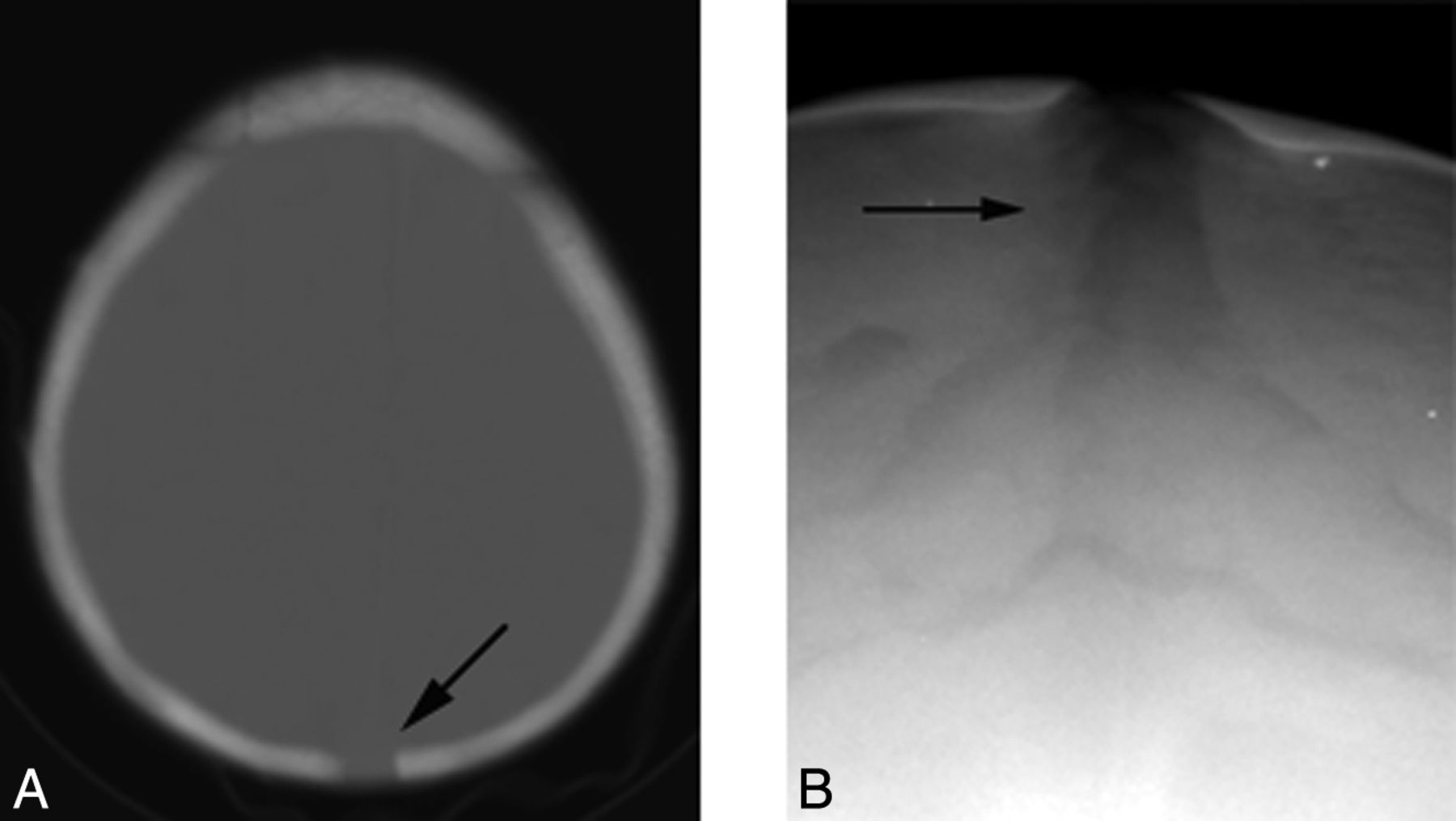
- Fig 3.
A 10-year-old child with HGPS. Axial CT image reveals a mottled appearance of the calvarium (A) and prominent vascular markings (arrow, B).
- Fig 4.
A 2-year-old child with a persistent patent posterior fontanel (arrow, A) on axial CT and a widened sagittal suture (arrow, B) on the anteroposterior radiograph of the skull.
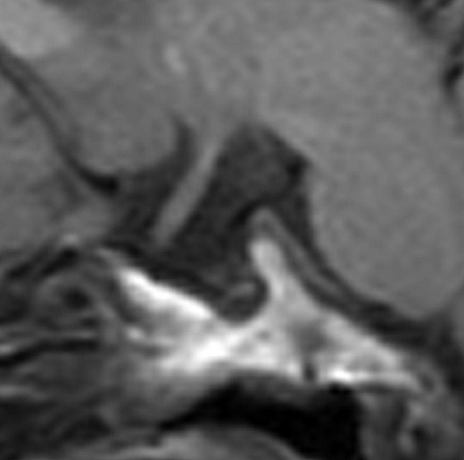
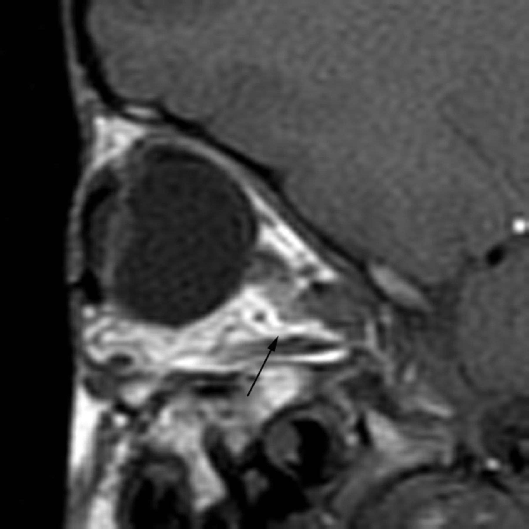
- Fig 5.
Sagittal T1-weighted MR image shows a J-shaped sella.
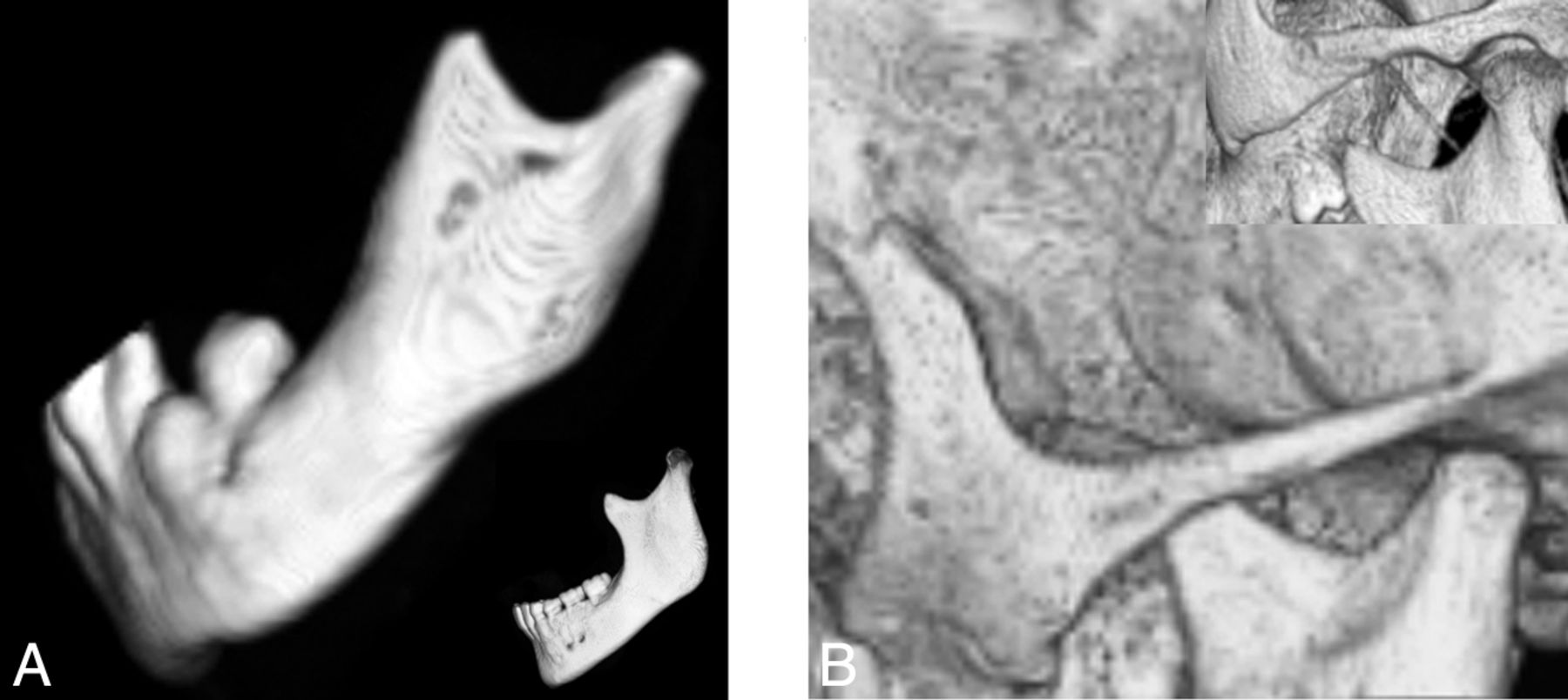
- Fig 6.
Lateral projection of a 3D CT shaded-surface display reveals a short mandibular ramus, a steep mandibular angle with disorganized dentition (A), and a thin gracile zygomatic arch in a child with HGPS (B) compared with a healthy age-matched control (insets).
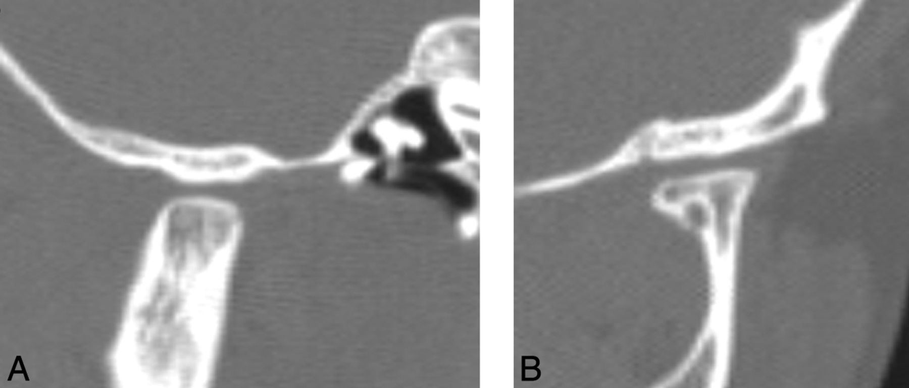
- Fig 7.
A, Lateral CT reformatted image demonstrates flattening of the condylar head, a shallow glenoid fossa, and a hypoplastic articular eminence. B, Coronal CT reformatted image of a child with HGPS shows a flattened condylar head and a shallow glenoid fossa.
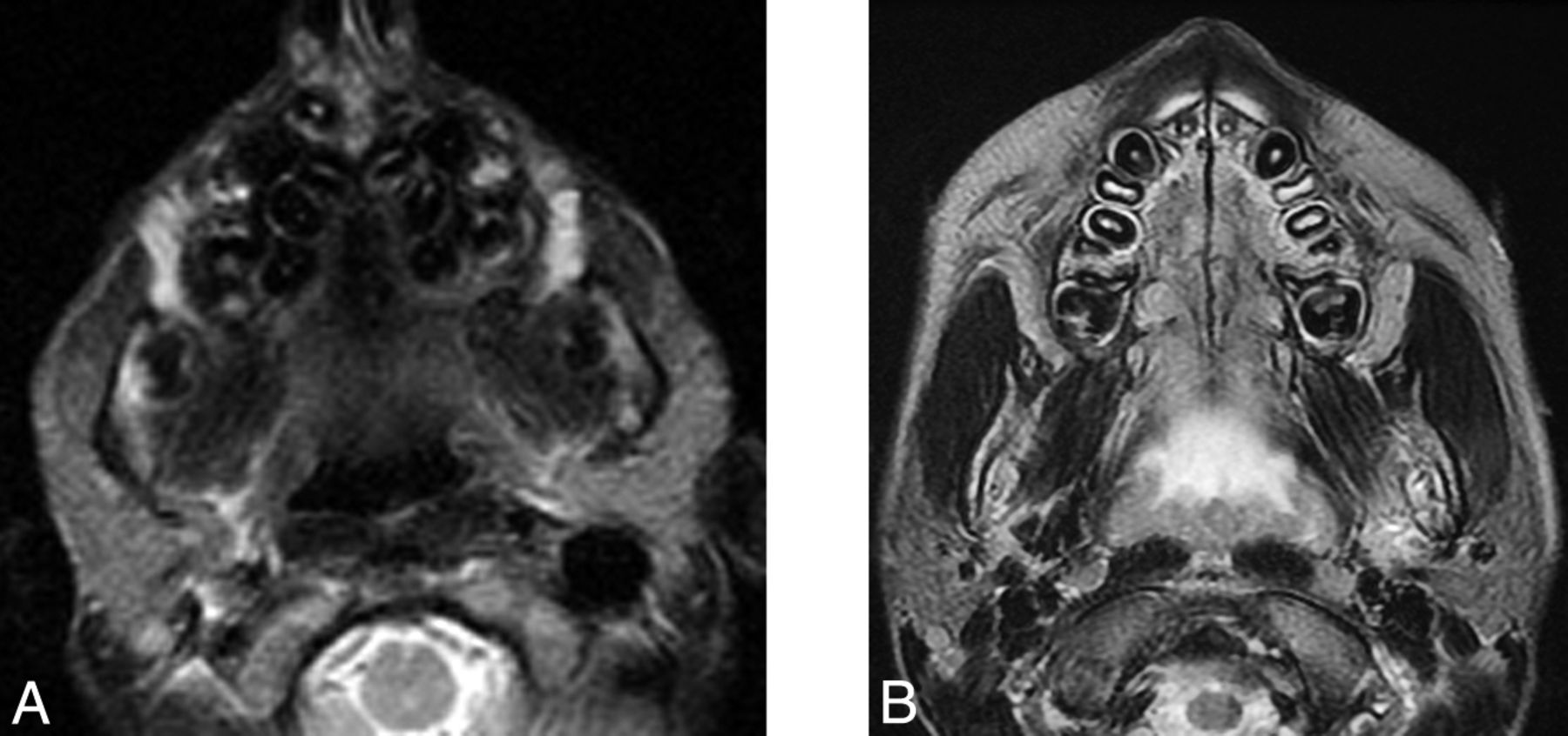
- Fig 8.
Axial T2-weighted MR imaging demonstrates a V-shaped palate, disorganized dentition, and prominent parotid glands in child with HGPS (A) compared with the control (B).
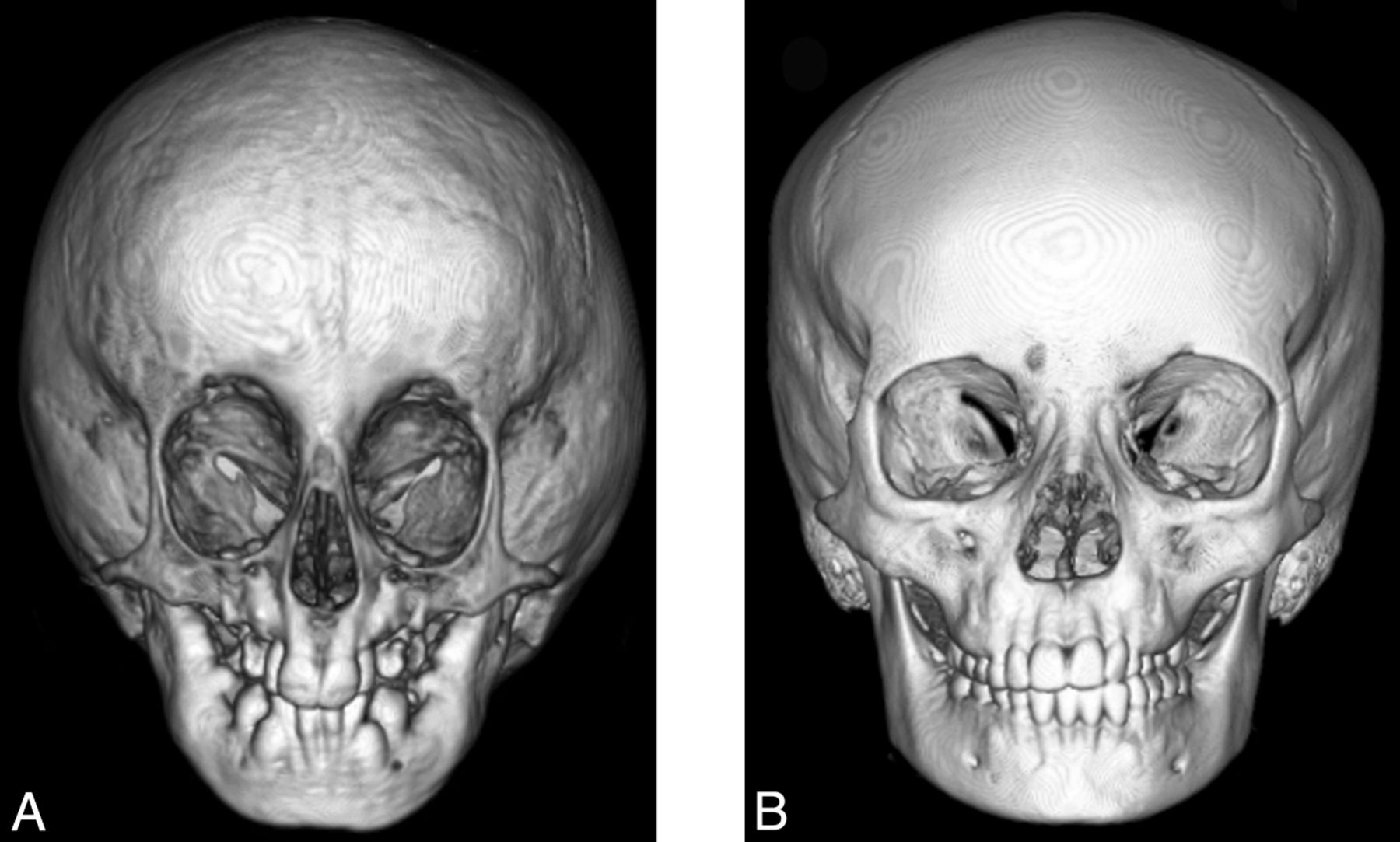
- Fig 9.
Anterior projection of a 3D CT shaded surface display of the head in a child with HGPS (A) shows hypotelorism, small mid- and lower face, and a disorganized dentition compared with the control (B).
- Fig 10.
Sagittal T1-weighted MR image demonstrates kinking of the optic nerve (arrow).


Tables
Characteristics Total (No.) 25 Female 11 Male 14 HGPS mutation types Classic by phenotype and genotype 18 Classic by phenotype only 4 Nonclassic by phenotype and genotype 3a Total No. of scans 98 Mean age at time of scan 6.5 years (range, birth–14.1 yr) Anthropomorphic datab Height-age <3rd percentile Height-age/chronologic age (mean) 0.5 Head circumference (mean) 56th percentile Types of neuroimaging (No.) 98 CT head 43 MR imaging head 51 Skull radiographs 4 Present (No.) (% of visualized) Absent (No.) N/V(No.) Scalp, calvarium, and skull base features Craniofacial disproportion 23 (92) 2 0 Thinned calvarium 20 (95) 1 4 Mottled bone appearancea 10 (59) 7 8 Prominent vascular markingsa 9 (90) 1 15 Skull fractures 2 (8) 22 1 Delayed closure of the anterior fontanelb 9 (56) 7 5 Widened sutures 7 (41) 10 1 J-shaped sellaa 17 (89) 2 6 Lack of scalp fat 21 (91) 2 2 Oral maxillary and parotid gland features Shortened ramus/mandibular hypoplasia 15 (83) 3 7 Flattened mandibular condylea 6 (43) 8 11 Hypoplastic articular eminence and shallow glenoid fossaa 6 (43) 8 11 Thin zygomatic archa 6 (50) 6 13 V-shaped palate 9 (45) 11 5 Disorganized dentition 10 (50) 10 5 Prominent parotid glanda 13 (100) 0 12 Orbital/facial features Narrow nasal bridge with pointed tip 14 (61) 9 2 Hypotelorism 19 (86) 3 3 Optic nerve kinkinga 17 (89) 2 6


