
Article Figures & Data
Figures
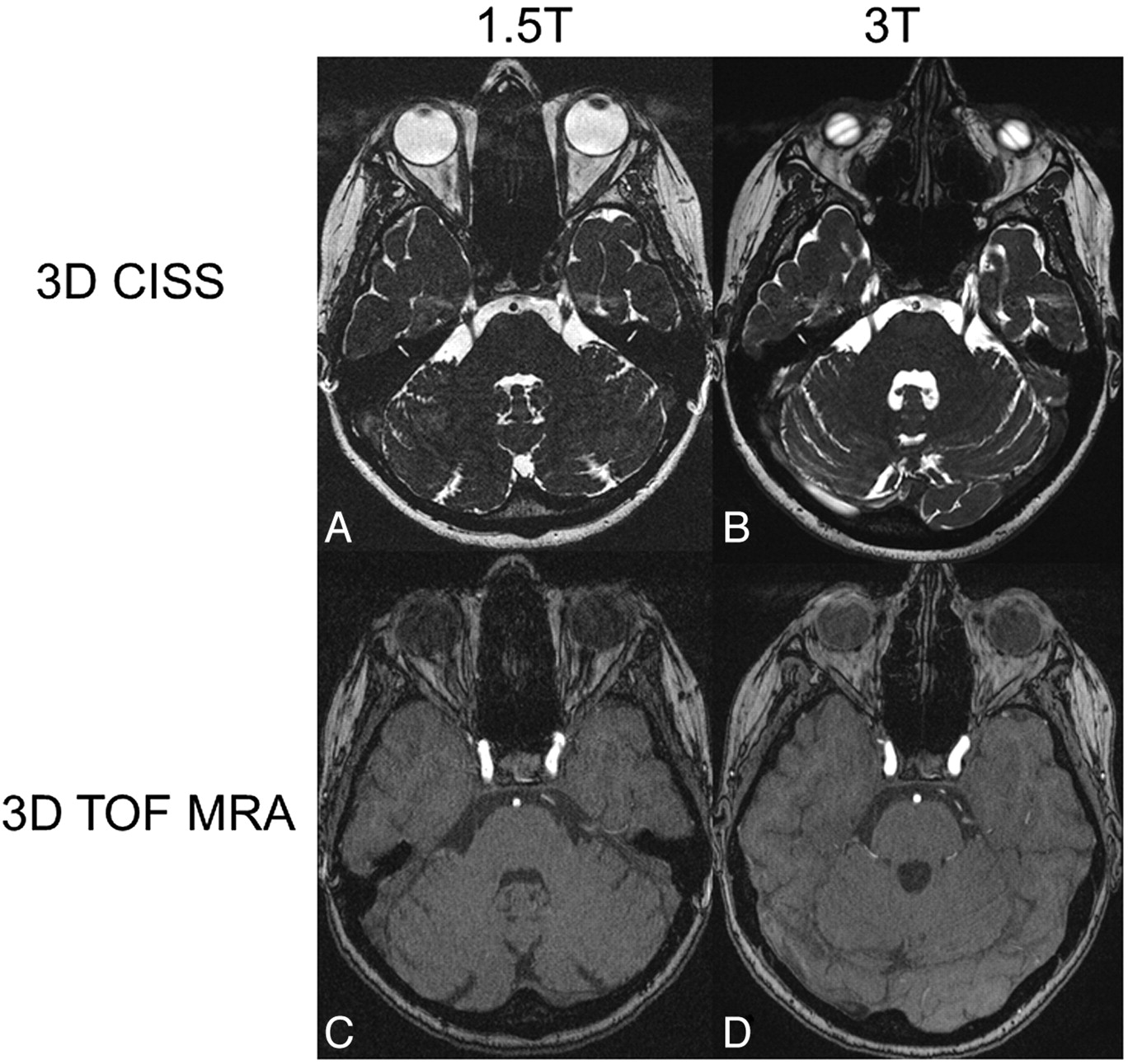
- Fig 1.
Axial source images of 3D-CISS sequences at the level of the trigeminal nerve root (A and B) and 3D-TOF MRAs at the level of the cavernous sinus (C and D) at 1.5T (A and C) and 3T (B and D) in the same patient. Note the ramification of the nerve root into its branches at some distance from the REZ. The branches are much better outlined at 3T than at 1.5T in the 3D-CISS sequence (A and B). The vessels and the boundary between the cavernous sinus and ICA can be distinguished more accurately at 3T than at 1.5T in the 3D-TOF MRA (C and D). Note the banding artifacts over the globes in the 3D-CISS at 3T (B). Ghosting artifacts are seen anterior to the CSF in the 3D-CISS at both field strengths (A and B).
- Fig 2.
Axial source images of 3D-CISS sequences (A and B) and MIP of 3D-TOF MRAs (C and D) obtained at 1.5T (A and C) and 3T (B and D). The contours of the anatomic structures can be more accurately delineated at 3T compared with 1.5T. Note that more vessels can be delineated in the MIP obtained at 3T (D) compared with 1.5T (B).
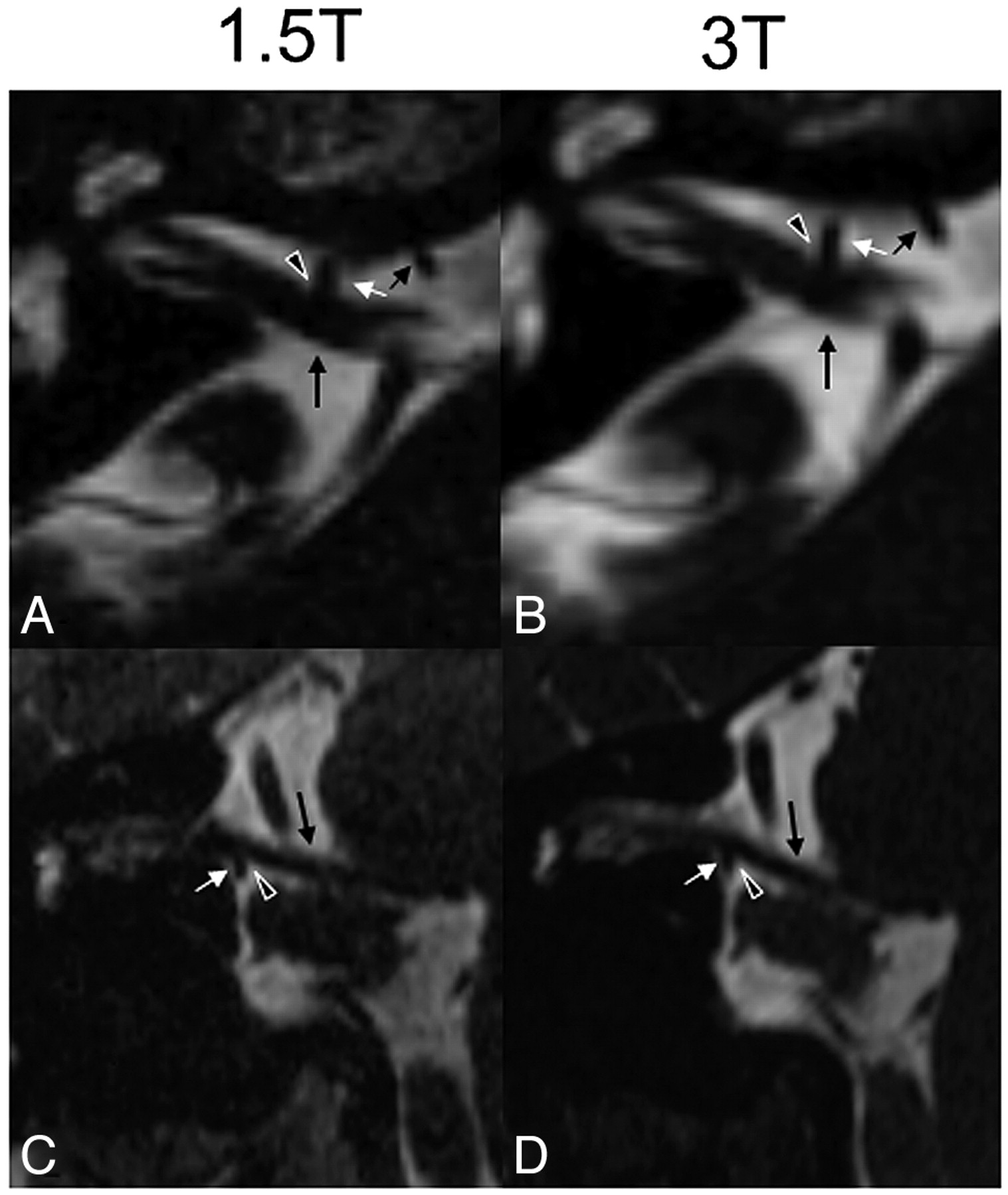
- Fig 3.
Axial source images of the 3D-CISS sequence at 1.5T (A) and 3T (B) of a patient with trigeminal neuralgia on the left side showing a close relationship (arrowhead) between the upper part of the trigeminal nerve (trigeminal neuralgia, long arrow) and a larger vein (arrow with dot) and a smaller artery (short arrow) on the left side at some distance from the REZ (asterisk, shown on the contralateral side). The proximate adjacency between the trigeminal neuralgia and the small artery is visible at both field strengths. However, the direct contact between the trigeminal neuralgia and vein is questionable at 1.5T (arrowhead in A), whereas the direct nerve-vein contact is clearly visible at 3T (arrowhead in B), confirming the diagnosis of NVC.
- Fig 4.
Axial source (A and B) and coronal (C and D) reformatted CISS images at 1.5T (A and C) and 3T (B and D) of a patient with hemifacial spasm on the right side due a direct contact (black arrowheadwith white margins) between the VII-VIII nerve complex (long black arrow) and the meatal segment of the anterior inferior cerebellar artery (short white arrow), forming a loop near the nerve complex (short black and white arrows in A and B, in which the short black arrow indicates the proximal part and the short white arrow, the distal part of the anterior inferior cerebellar artery). In this patient, the NVC is approximately equally well seen at both field strengths.
- Fig 5.
Bar graph showing the mean (± standard error) of the SNR of the basilar artery, CNR between the basilar artery and brain stem, and CNR between the CSF and ICA for both the 3D-CISS and 3D-TOF MRA. Note the higher SNR and CNR for all items at 3T, though they are more pronounced for the 3D-CISS than for the 3D-TOF MRA.


Tables
Parameters 1.5T 3T Acquisition time (min:sec) 7:03 8:26 Voxel size (mm3) 0.4 0.4 No. of slabs 1 1 Sections per slab 144 144 Distance factor (%) 20 20 FOV read (mm) 230 200 FOV phase (%) 62.5 100 Section thickness (mm) 0.4 0.4 Base resolution 512 512 Phase resolution (%) 100 100 Section resolution (%) 64 50 TR (ms) 9.65 7.48 TE (ms) 4.83 3.23 No. of averages 1 1 Flip angle (°) 90 45 Bandwidth (Hz/px) 163 250 No. of averages 1 1 No. of measurements 1 1 SNR 1 1 Phase oversampling (mm3) 0 0 Section oversampling (%) 0 22.2 Phase partial Fourier 7/8 7/8 Section partial Fourier 7/8 7/8 Parameters 1.5T 3T Acquisition time (min:sec) 15:07 15:51 Voxel size (mm3) 0.4 0.4 No. of slabs 1 1 Sections per slab 144 144 Distance factor (%) 50 50 FOV read (mm) 230 200 FOV phase (%) 62.5 100 Section thickness (mm) 0.4 0.4 Base resolution 512 512 Phase resolution (%) 100 100 Section resolution (%) 67 50 TR (ms) 40 21 TE (ms) 7.15 3.77 Flip angle (°) 25 18 Bandwidth (Hz/px) 65 212 MTC Yes No No. of averages 1 1 No. of concatenations 1 1 No. of measurements 1 1 SNR 1 1 Phase oversampling (mm3) 0 0 Section oversampling (%) 0 22.2 Phase partial Fourier 7/8 Off Section partial Fourier 7/8 Off -
Note:—MTC indicates magnetization transfer contrast.
-


