Advertisement

Main menu

  • Home
  • Content
    • Current Issue
    • Accepted Manuscripts
    • Article Preview
    • Past Issue Archive
    • Video Articles
    • AJNR Case Collection
    • Case of the Week Archive
    • Case of the Month Archive
    • Classic Case Archive
  • Special Collections
    • AJNR Awards
    • Low-Field MRI
    • Alzheimer Disease
    • ASNR Foundation Special Collection
    • Photon-Counting CT
    • View All
  • Multimedia
    • AJNR Podcasts
    • AJNR SCANtastic
    • Trainee Corner
    • MRI Safety Corner
    • Imaging Protocols
  • For Authors
    • Submit a Manuscript
    • Submit a Video Article
    • Submit an eLetter to the Editor/Response
    • Manuscript Submission Guidelines
    • Statistical Tips
    • Fast Publishing of Accepted Manuscripts
    • Graphical Abstract Preparation
    • Imaging Protocol Submission
    • Author Policies
  • About Us
    • About AJNR
    • Editorial Board
    • Editorial Board Alumni
  • More
    • Become a Reviewer/Academy of Reviewers
    • Subscribers
    • Permissions
    • Alerts
    • Feedback
    • Advertisers
    • ASNR Home

User menu

  • Alerts
  • Log in

Search

  • Advanced search
American Journal of Neuroradiology

American Journal of Neuroradiology

ASHNR American Society of Functional Neuroradiology ASHNR American Society of Pediatric Neuroradiology ASSR
  • Alerts
  • Log in

Advanced Search

  • Home
  • Content
    • Current Issue
    • Accepted Manuscripts
    • Article Preview
    • Past Issue Archive
    • Video Articles
    • AJNR Case Collection
    • Case of the Week Archive
    • Case of the Month Archive
    • Classic Case Archive
  • Special Collections
    • AJNR Awards
    • Low-Field MRI
    • Alzheimer Disease
    • ASNR Foundation Special Collection
    • Photon-Counting CT
    • View All
  • Multimedia
    • AJNR Podcasts
    • AJNR SCANtastic
    • Trainee Corner
    • MRI Safety Corner
    • Imaging Protocols
  • For Authors
    • Submit a Manuscript
    • Submit a Video Article
    • Submit an eLetter to the Editor/Response
    • Manuscript Submission Guidelines
    • Statistical Tips
    • Fast Publishing of Accepted Manuscripts
    • Graphical Abstract Preparation
    • Imaging Protocol Submission
    • Author Policies
  • About Us
    • About AJNR
    • Editorial Board
    • Editorial Board Alumni
  • More
    • Become a Reviewer/Academy of Reviewers
    • Subscribers
    • Permissions
    • Alerts
    • Feedback
    • Advertisers
    • ASNR Home
  • Follow AJNR on Twitter
  • Visit AJNR on Facebook
  • Follow AJNR on Instagram
  • Join AJNR on LinkedIn
  • RSS Feeds

AJNR Awards, New Junior Editors, and more. Read the latest AJNR updates

Research ArticleNeurointerventionE
Open Access

Endovascular Treatment of Ruptured Brain AVMs in the Acute Phase of Hemorrhage

W.J. van Rooij, S. Jacobs, M. Sluzewski, G.N. Beute and B. van der Pol
American Journal of Neuroradiology June 2012, 33 (6) 1162-1166; DOI: https://doi.org/10.3174/ajnr.A2995
W.J. van Rooij
  • Find this author on Google Scholar
  • Find this author on PubMed
  • Search for this author on this site
S. Jacobs
  • Find this author on Google Scholar
  • Find this author on PubMed
  • Search for this author on this site
M. Sluzewski
  • Find this author on Google Scholar
  • Find this author on PubMed
  • Search for this author on this site
G.N. Beute
  • Find this author on Google Scholar
  • Find this author on PubMed
  • Search for this author on this site
B. van der Pol
  • Find this author on Google Scholar
  • Find this author on PubMed
  • Search for this author on this site
  • Article
  • Figures & Data
  • Info & Metrics
  • Responses
  • References
  • PDF
Loading

Abstract

BACKGROUND AND PURPOSE: Patients with ruptured brain AVMs are at considerable risk of repeat hemorrhage, particularly when associated intranidal or flow-related aneurysms are present. There is controversy about the timing of diagnosis and treatment of patients with hemorrhagic stroke. We present our results of endovascular treatment of ruptured AVMs in the acute phase.

MATERIALS AND METHODS: Between January 2008 and March 2011, 23 patients (16 men, 7 women; mean age 42 years) with AVM-related hemorrhagic stroke were treated with endovascular techniques within 10 days of the ictus. There were 10 micro-AVMs (< 1 cm) and 1 single-hole pial fistula. In 9 patients, an intranidal or flow-related aneurysm was the likely cause of hemorrhage.

RESULTS: Complete obliteration of the AVM with Onyx was achieved in 13 of 23 patients (57%). Eight of the 13 AVMs were micro-AVMs and 3 had an intranidal aneurysm. Partial obliteration of the AVM was achieved in 10 of 23 patients (43%). In 6 of these 10 patients, an intranidal (n = 1) or flow-related aneurysm (n = 5) was obliterated with Onyx or coils. There were no complications of treatment. During a mean follow-up of 21 months in 22 surviving patients, no repeat hemorrhage occurred.

CONCLUSIONS: Endovascular treatment with Onyx in the acute phase cured most ruptured AVMs. All 9 AVM-associated aneurysms that were considered the source of hemorrhage could be excluded from the circulation. In patients with AVM-related hemorrhagic stroke, prompt angiographic diagnosis and treatment may improve prognosis by reducing repeat hemorrhage rate.

Treatment of brain AVMs remains, to some extent, controversial because of the numerous variables affecting the results and uncertainties about the natural course.1⇓⇓⇓⇓⇓⇓⇓–9 This is true not only for unruptured AVMs but also after a hemorrhagic episode. Patients with an AVM initially presenting with hemorrhagic stroke have a substantially higher risk of subsequent bleeding than those presenting with seizures or other symptoms. The risk is much higher in men than in women.10 Some angioarchitectural features of brain AVMs are associated with hemorrhage, such as the presence of intranidal aneurysms or flow-related aneurysms and venous outflow restriction by stenoses. Specific elimination of these weak spots or reduction of flow through the AVM by endovascular or surgical therapy is thought to decrease the rate of repeated hemorrhage and to improve prognosis, even when the AVM is only partially obliterated.11⇓⇓⇓⇓–16

With modern endovascular techniques, targeted treatment of AVM-associated aneurysms, partial nidus obliteration with flow reduction, or complete obliteration is possible in most patients with AVMs.16⇓⇓–19 In this article, we evaluate the results of a treatment strategy for brain AVMs that present with hemorrhage that includes early angiographic diagnosis and endovascular treatment in the acute phase.

Materials and Methods

General Management of Intracranial Hemorrhage

All patients admitted with intracranial hemorrhage not obviously appreciated as hypertensive hematomas were referred for cerebral angiography as soon as possible. Only in patients admitted in poor clinical condition who were possible candidates for emergency surgical intervention, was CTA performed first to inform about possible vascular pathology and to decide whether emergent endovascular treatment preceding surgery would be desirable.20 Cooperative patients in stable clinical condition underwent complete cerebral angiography, and when a vascular disorder amenable for endovascular therapy was diagnosed (AVM, aneurysm, or dural fistula), embolization under general anesthesia was planned on short notice. In uncooperative or intubated patients, general anesthesia was induced before angiography and embolization could follow in the same session. Angiography and embolization were performed on a biplane angiographic system (Integra BN3000 Neuro or Allura Neuro; Philips Healthcare, Best, the Netherlands) equipped with 3D rotational angiography and high-speed filming.

Patients with Acutely Ruptured AVMs

In acutely ruptured AVMs, where an intranidal- or a flow-aneurysm could be identified as the likely source of bleeding, we aimed the endovascular treatment primarily at elimination of this source from the circulation with coils or Onyx (ev3, Irvine, California), when possible, followed by partial or complete obliteration of the nidus.

In acutely ruptured AVMs without an obvious source of bleeding, we intended to completely obliterate the nidus in small or intermediate-size brain AVMs that are accessible by the microcatheter and with feeders suitable for the injection with Onyx, with allowance for at least 2–3 cm of reflux. In ruptured, large AVMs, where complete endovascular obliteration is unlikely, we primarily aimed at flow reduction and partial nidus obliteration presurgery or radiosurgery. High-flow pial or intranidal fistulas were occluded with Onyx under temporary balloon occlusion of the feeding pedicle to decrease the flow and prevent migration of Onyx through the fistula into the venous side.21

After embolization, patients were transferred to an intensive care unit and cared for by a multidisciplinary team of intensivists, neurologists, neurosurgeons, anesthetists, and neuroradiologists. After clinical recovery, patients with incompletely obliterated AVMs were scheduled for additional radiosurgery or neurosurgery. Patients with completely obliterated AVMs by embolization alone were scheduled for a follow-up angiogram at 3 months.

Between January 2008 and March 2011, 23 patients presented with AVM-related intracranial hemorrhage and were treated with endovascular techniques within 10 days of the ictus. There were 16 men and 7 women, with a mean age of 42 years (median 39, range 6–74 years). All 23 patients had parenchymal hematomas with or without intraventricular or subarachnoid extension. There were 10 micro-AVMs (nidus size <1 cm; Fig 1) and 1 single-hole pial fistula (Fig 2). The remaining 12 AVMs had a mean size of 2.8 cm (range 2–4 cm). AVM location was frontal in 11 patients, occipital in 5, parietal in 2, temporal in 2, and cerebellar in 3. In 5 patients, a flow aneurysm was the likely cause of hemorrhage, and 4 patients had an intranidal aneurysm that marked the bleeding spot.

Fig 1.
  • Download figure
  • Open in new tab
  • Download powerpoint
Fig 1.

71-year-old man with sudden headache and hemianopsia. A, CT scan shows posterior temporal hematoma with an outlined venous aneurysm indicating the source of the hemorrhage (arrow). B, Lateral view of vertebral angiography shows micro-AVM with intranidal aneurysm as the source of hemorrhage (arrow). C, Nonsubtracted lateral view shows Onyx cast after embolization. The intranidal aneurysm is partly filled with Onyx (arrow). D, Complete obliteration of the AVM with Onyx.

Fig 2.
  • Download figure
  • Open in new tab
  • Download powerpoint
Fig 2.

6-year-old girl with sudden headache and left-sided weakness. A, T2-weighted MR imaging demonstrates intraparenchymal hematoma and flow voids indicating vascular malformation. B, Frontal view of right internal carotid angiography shows single-hole, high-flow pial fistula with dilated draining veins. C, Fluoroscopic frontal image during embolization. The fistula is occluded with Onyx (short arrow) during balloon occlusion of the feeding artery (long arrow) to block the flow and prevent migration of Onyx to the venous side of the fistula. D, Complete obliteration of the pial fistula.

Results

Results of Embolization

Complete embolization of the AVM with Onyx was achieved in 13 of 23 patients (57%; Fig 3). Eight of the 13 AVMs were micro-AVMs, and 3 had an intranidal aneurysm as the likely source of the bleeding. Partial obliteration of the AVM was achieved in 10 of 23 patients (43%). In 6 of these 10 patients, an intranidal (n = 1) or flow-aneurysm (n = 5) as the source of hemorrhage was obliterated with coils (n = 3) or Onyx (n = 3) (Fig 4). There were no complications of endovascular treatment.

Fig 3.
  • Download figure
  • Open in new tab
  • Download powerpoint
Fig 3.

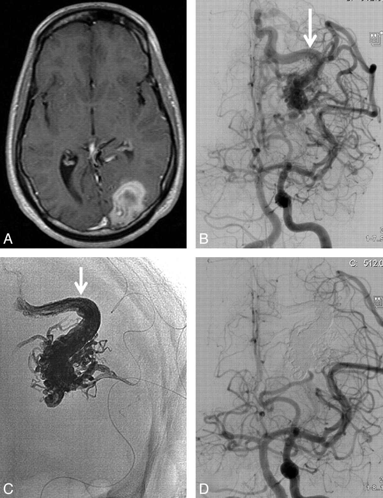
23-year-old man with sudden headache and hemianopsia. A, T1-weighted MR imaging demonstrates left occipital hematoma. B, Frontal view of concomitant left internal carotid and vertebral angiogram shows small occipital AVM with a single draining vein (arrow). C, Nonsubtracted image of Onyx cast after embolization: complete filling of the nidus and proximal part of the draining vein (arrow). D, Angiography demonstrating complete obliteration of the AVM.

Fig 4.
  • Download figure
  • Open in new tab
  • Download powerpoint
Fig 4.

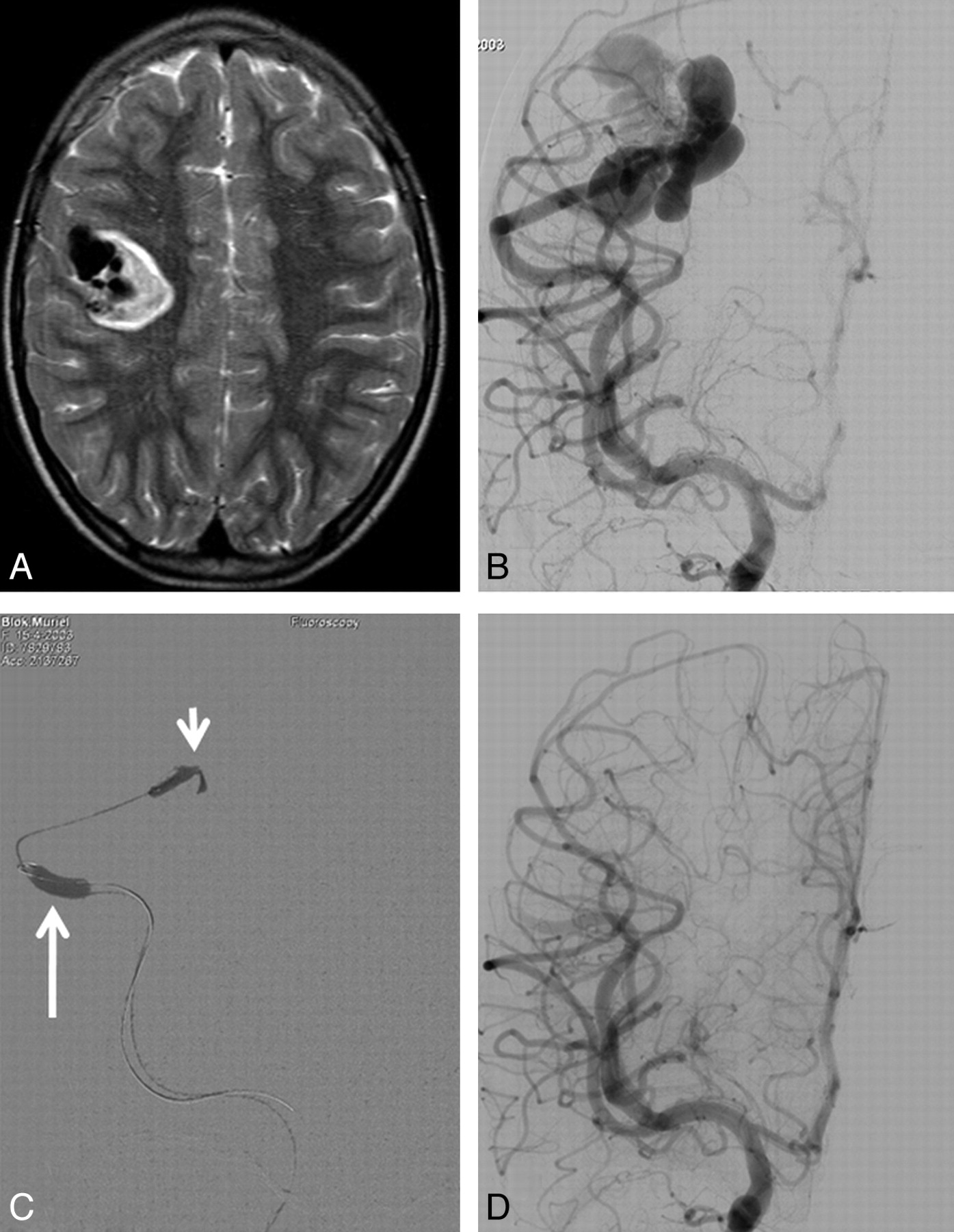
49-year-old man with sudden headache and dysphasia and frontal hematoma on CT scan. A, Frontal view of left internal carotid angiogram shows small left frontal AVM with intranidal aneurysm as the source of hemorrhage (arrow). B, Fluoroscopic image during embolization with the tip of the microcatheter inside the aneurysm (arrow). C, Nonsubtracted image with Onyx cast filling the intranidal aneurysm completely. D, Small remnant of the AVM after embolization without filling of the aneurysm. The remnant was subsequently treated with gamma knife radiosurgery.

Additional Treatments and Follow-Up

One patient died of an uncontrollable rise of intracranial pressure after surgical evacuation of a frontal hematoma after complete embolization of a micro-AVM. Additional treatments of 10 patients with partially obliterated AVMs consisted of surgery in 2 and gamma knife radiosurgery in 8. In all 13 patients with completely obliterated AVMs, follow-up angiography confirmed lasting occlusion. The 22 surviving patients had a mean follow-up of 21 months (median 21, range 3–48 months). There were no new episodes of hemorrhage during this follow-up. At last follow-up, 3 patients were dependent in a nursing home and 19 patients were functioning independently.

Discussion

In this study of 23 patients with AVM-related hemorrhagic stroke, endovascular treatment in the acute phase resulted in complete obliteration of most AVMs. All 9 AVM-associated aneurysms that were considered weak spots, and the source of hemorrhage, could be excluded from the circulation. No repeated hemorrhage occurred during a mean 21 months of follow-up. Embolization of AVMs has taken a large step forward in the last decade. The introduction of the nonadhesive polymer Onyx has largely replaced the use of acrylic glue for the obliteration of the nidus in many centers. With growing experience, advanced biplane imaging with rapid subtraction fluoroscopy, and refinements of technique in the use of Onyx,17,21 most small and intermediate-size brain AVMs can be completely obliterated at a low complication rate, on the condition that the nidus is accessible by the microcatheter and with feeders suitable for the injection with Onyx with allowance of at least 2–3 cm of reflux.16⇓⇓–19 Better recognition of certain angioarchitectural features of the AVM as likely weak spots prone to recurrent hemorrhage, such as intranidal- and flow-related aneurysms and venous stenoses, have prompted many operators to perform a partial targeted embolization treatment when complete AVM obliteration is unlikely or impossible.7,11⇓⇓⇓–15 Intranidal- and flow-related aneurysms can be excluded from the circulation with Onyx, acrylic glue, or coils, and venous outflow restrictions can be exonerated by occlusion of high-flow intranidal fistula with Onyx under flow arrest with a microballoon.21

These new developments in the treatment of brain AVMs, in combination with the concurrent and similar advancement of endovascular treatment with Onyx of dural arteriovenous fistulas with drainage to the cortical veins,22,23 have led our hospital to an adaptation of the diagnosis and treatment strategy in patients with hemorrhagic stroke. We now perform early angiography in patients who present with an intraparenchymal hematoma, except older hypertensive patients with basal ganglia or thalamus hematomas and patients with known malignant disease or impaired coagulation. Only in patients in poor clinical condition where surgical evacuation of the hematoma is considered, is CTA performed first to inform the neurosurgeon about the presence, location, and size of a possible vascular disorder. In all other patients, angiography is performed without preceding CTA, because with CTA micro-AVMs and dural fistulas, the most common causes of hemorrhagic stroke in this selected patient group, can easily be missed.20,24 Additional 3D rotational angiography provides precise information about presence, angioarchitecture, and flow of all AVMs, dural fistulas, and aneurysms, and endovascular treatment can follow immediately. For several years, we have only used Onyx for the embolization of brain AVMs, because Onyx is injected very slowly and can be better controlled then acrylic glue.

Patients with a ruptured AVM have a substantial risk of repeat hemorrhage. In a study by Mast et al,10 comprising 142 patients with a ruptured AVM, the annual rate of recurrent hemorrhage was 33% for men and 10% for women. A similar annual rate of hemorrhage is associated with dural fistula with drainage to the cortical veins. The early application of the new endovascular treatment possibilities for these aggressive lesions may improve prognosis for patients with hemorrhagic stroke by reducing or even eliminating recurrent hemorrhage. In our limited patient series, no recurrent hemorrhages occurred after early endovascular treatment. In view of this, we advocate early angiography in patients with hemorrhagic stroke in the appropriate clinical setting to diagnose or exclude, with certainty, AVMs and dural fistula as the cause of hemorrhage. At present, American and European guidelines are unclear about both the timing and the type of imaging technique for diagnosing underlying vascular pathology in adults with hemorrhagic stroke.25,26

Conclusions

Endovascular treatment with Onyx in the acute phase of hemorrhagic stroke cured most ruptured AVMs, and all AVM-associated aneurysms that were considered the source of hemorrhage could be excluded from the circulation. In patients with AVM-related hemorrhagic stroke, prompt diagnosis and treatment may improve prognosis by reducing repeat hemorrhage rate.

Indicates open access to non-subscribers at www.ajnr.org

References

  1. 1.↵
    1. Graf CJ,
    2. Perret GE,
    3. Torner JC
    . Bleeding from cerebral arteriovenous malformations as part of their natural history. J Neurosurg 1983; 58: 331–37
    PubMed
  2. 2.↵
    1. Crawford PM,
    2. West CR,
    3. Chadwick DW,
    4. et al
    . Arteriovenous malformations of the brain: natural history in unoperated patients. J Neurol Neurosurg Psychiatry 1986; 49: 1–10
    Abstract/FREE Full Text
  3. 3.↵
    1. Brown RD Jr.,
    2. Wiebers DO,
    3. Forbes G,
    4. et al
    . The natural history of unruptured intracranial arteriovenous malformations. J Neurosurg 1988; 68: 352–57
    CrossRefPubMed
  4. 4.↵
    1. Ondra SL,
    2. Troupp H,
    3. George ED,
    4. et al
    . The natural history of symptomatic arteriovenous malformations of the brain: a 24-year follow-up assessment. J Neurosurg 1990; 73: 387–91
    CrossRefPubMed
  5. 5.↵
    1. Stapf C,
    2. Mohr JP,
    3. Choi JH,
    4. et al
    . Invasive treatment of unruptured brain arteriovenous malformations is experimental therapy. Curr Opin Neurol 2006; 19: 63–68
    CrossRefPubMed
  6. 6.↵
    1. Stapf C
    . The rationale behind “A Randomized Trial of Unruptured Brain AVMs” (ARUBA). Acta Neurochir Suppl 2010; 107: 83–85
    CrossRefPubMed
  7. 7.↵
    1. da Costa L,
    2. Wallace MC,
    3. Ter Brugge KG,
    4. et al
    . The natural history and predictive features of hemorrhage from brain arteriovenous malformations. Stroke 2009; 40: 100–05
    Abstract/FREE Full Text
  8. 8.↵
    1. Hernesniemi JA,
    2. Dashti R,
    3. Juvela S,
    4. et al
    . Natural history of brain arteriovenous malformations: a long-term follow-up study of risk of hemorrhage in 238 patients. Neurosurgery 2008; 63: 823–29
    CrossRefPubMed
  9. 9.↵
    1. Meisel HJ,
    2. Mansmann U,
    3. Alvarez H,
    4. et al
    . Effect of partial targeted N-butyl-cyano-acrylate embolization in brain AVM. Acta Neurochir (Wien) 2002; 144: 879–87
    CrossRefPubMed
  10. 10.↵
    1. Mast H,
    2. Young WL,
    3. Koennecke HC,
    4. et al
    . Risk of spontaneous haemorrhage after diagnosis of cerebral arteriovenous malformation. Lancet 1997; 350: 1065–68
    CrossRefPubMed
  11. 11.↵
    1. Alexander MJ,
    2. Tolbert ME
    . Targeting cerebral arteriovenous malformations for minimally invasive therapy. Neurosurgery 2006; 59: 178–83
  12. 12.↵
    1. Hademenos GJ,
    2. Massoud TF
    . Risk of intracranial arteriovenous malformation rupture due to venous drainage impairment. A theoretical analysis. Stroke 1996; 27: 1072–83
    Abstract/FREE Full Text
  13. 13.↵
    1. Mansmann U,
    2. Meisel J,
    3. Brock M,
    4. et al
    . Factors associated with intracranial hemorrhage in cases of cerebral arteriovenous malformation. Neurosurgery 2000; 46: 272–79
    CrossRefPubMed
  14. 14.↵
    1. Stefani MA,
    2. Porter PJ,
    3. terBrugge KG,
    4. et al
    . Angioarchitectural factors present in brain arteriovenous malformations associated with hemorrhagic presentation. Stroke 2002; 33: 920–24
    Abstract/FREE Full Text
  15. 15.↵
    1. Krings T,
    2. Hans FJ,
    3. Geibprasert S,
    4. et al
    . Partial “targeted” embolisation of brain arteriovenous malformations. Eur Radiol 2010; 20: 2723–31
    CrossRefPubMed
  16. 16.↵
    1. Katsaridis V,
    2. Papagiannaki C,
    3. Aimar E
    . Curative embolization of cerebral arteriovenous malformations (AVMs) with Onyx in 101 patients. Neuroradiology 2008; 50: 589–97
    CrossRefPubMed
  17. 17.↵
    1. Saatci I,
    2. Geyik S,
    3. Yavuz K,
    4. et al
    . Endovascular treatment of brain arteriovenous malformations with prolonged intranidal Onyx injection technique: long-term results in 350 consecutive patients with completed endovascular treatment course. J Neurosurg 2011; 115: 78–88
    CrossRefPubMed
  18. 18.↵
    1. Andreou A,
    2. Ioannidis I,
    3. Lalloo S,
    4. et al
    . Endovascular treatment of intracranial microarteriovenous malformations. J Neurosurg 2008; 109: 1091–97
    CrossRefPubMed
  19. 19.↵
    1. van Rooij WJ,
    2. Sluzewski M,
    3. Beute GN
    . Brain AVM embolization with Onyx. AJNR Am J Neuroradiol 2007; 28: 172–77
    Abstract/FREE Full Text
  20. 20.↵
    1. Delgado Almandoz JE,
    2. Romero JM
    . Advanced CT imaging in the evaluation of hemorrhagic stroke. Neuroimaging Clin N Am 2011; 21: 197–213
    CrossRefPubMed
  21. 21.↵
    1. van Rooij WJ,
    2. Sluzewski M
    . Endovascular occlusion of high-flow intracranial arteriovenous shunts: technical note. Neuroradiology 2007; 49: 1029–31
    CrossRefPubMed
  22. 22.↵
    1. van Rooij WJ,
    2. Sluzewski M
    . Curative embolization with Onyx of dural arteriovenous fistulas with cortical venous drainage. AJNR Am J Neuroradiol 2010; 31: 1516–20
    Abstract/FREE Full Text
  23. 23.↵
    1. Cognard C,
    2. Januel AC,
    3. Silva NA Jr.,
    4. et al
    . Endovascular treatment of intracranial dural arteriovenous fistulas with cortical venous drainage: new management using Onyx. AJNR Am J Neuroradiol 2008; 29: 235–41
    Abstract/FREE Full Text
  24. 24.↵
    1. Smith SD,
    2. Eskey CJ
    . Hemorrhagic stroke. Radiol Clin North Am 2011; 49: 27–45
    CrossRefPubMed
  25. 25.↵
    1. Steiner T,
    2. Kaste M,
    3. Forsting M,
    4. et al
    . Recommendations for the management of intracranial haemorrhage—part I: spontaneous intracerebral haemorrhage. The European Stroke Initiative Writing Committee and the Writing Committee for the EUSI Executive Committee. Cerebrovasc Dis 2006; 22: 294–316
    CrossRefPubMed
  26. 26.↵
    1. Masdeu JC,
    2. Irimia P,
    3. Asenbaum S,
    4. et al
    . EFNS guideline on neuroimaging in acute stroke. Report of an EFNS task force. Eur J Neurol 2006; 13: 1271–83
    CrossRefPubMed
  • Received August 6, 2011.
  • Accepted after revision September 26, 2011.
  • © 2012 by American Journal of Neuroradiology
View Abstract
PreviousNext
Back to top

In this issue

American Journal of Neuroradiology: 33 (6)
American Journal of Neuroradiology
Vol. 33, Issue 6
1 Jun 2012
  • Table of Contents
  • Index by author
Advertisement
Print
Download PDF
Email Article

Thank you for your interest in spreading the word on American Journal of Neuroradiology.

NOTE: We only request your email address so that the person you are recommending the page to knows that you wanted them to see it, and that it is not junk mail. We do not capture any email address.

Enter multiple addresses on separate lines or separate them with commas.
Endovascular Treatment of Ruptured Brain AVMs in the Acute Phase of Hemorrhage
(Your Name) has sent you a message from American Journal of Neuroradiology
(Your Name) thought you would like to see the American Journal of Neuroradiology web site.
CAPTCHA
This question is for testing whether or not you are a human visitor and to prevent automated spam submissions.
Cite this article
W.J. van Rooij, S. Jacobs, M. Sluzewski, G.N. Beute, B. van der Pol
Endovascular Treatment of Ruptured Brain AVMs in the Acute Phase of Hemorrhage
American Journal of Neuroradiology Jun 2012, 33 (6) 1162-1166; DOI: 10.3174/ajnr.A2995

Citation Manager Formats

  • BibTeX
  • Bookends
  • EasyBib
  • EndNote (tagged)
  • EndNote 8 (xml)
  • Medlars
  • Mendeley
  • Papers
  • RefWorks Tagged
  • Ref Manager
  • RIS
  • Zotero
0 Responses
Respond to this article
Share
Bookmark this article
Endovascular Treatment of Ruptured Brain AVMs in the Acute Phase of Hemorrhage
W.J. van Rooij, S. Jacobs, M. Sluzewski, G.N. Beute, B. van der Pol
American Journal of Neuroradiology Jun 2012, 33 (6) 1162-1166; DOI: 10.3174/ajnr.A2995
del.icio.us logo Twitter logo Facebook logo Mendeley logo
  • Tweet Widget
  • Facebook Like
  • Google Plus One
Purchase

Jump to section

  • Article
    • Abstract
    • Materials and Methods
    • Results
    • Discussion
    • Conclusions
    • References
  • Figures & Data
  • Info & Metrics
  • Responses
  • References
  • PDF

Related Articles

  • No related articles found.
  • PubMed
  • Google Scholar

Cited By...

  • Endovascular treatment in the multimodality management of brain arteriovenous malformations: report of the Society of NeuroInterventional Surgery Standards and Guidelines Committee
  • Indications for the Performance of Intracranial Endovascular Neurointerventional Procedures: A Scientific Statement From the American Heart Association
  • Management of Brain Arteriovenous Malformations: A Scientific Statement for Healthcare Professionals From the American Heart Association/American Stroke Association
  • Crossref (42)
  • Google Scholar

This article has been cited by the following articles in journals that are participating in Crossref Cited-by Linking.

  • Management of Brain Arteriovenous Malformations: A Scientific Statement for Healthcare Professionals From the American Heart Association/American Stroke Association
    Colin P. Derdeyn, Gregory J. Zipfel, Felipe C. Albuquerque, Daniel L. Cooke, Edward Feldmann, Jason P. Sheehan, James C. Torner
    Stroke 2017 48 8
  • Indications for the Performance of Intracranial Endovascular Neurointerventional Procedures: A Scientific Statement From the American Heart Association
    Clifford J. Eskey, Philip M. Meyers, Thanh N. Nguyen, Sameer A. Ansari, Mahesh Jayaraman, Cameron G. McDougall, J. Kevin DeMarco, William A. Gray, David C. Hess, Randall T. Higashida, Dilip K. Pandey, Constantino Peña, Hermann C. Schumacher
    Circulation 2018 137 21
  • Endovascular treatment in the multimodality management of brain arteriovenous malformations: report of the Society of NeuroInterventional Surgery Standards and Guidelines Committee
    Reade De Leacy, Sameer A Ansari, Clemens M Schirmer, Daniel L Cooke, Charles J Prestigiacomo, Ketan R Bulsara, Steven W Hetts
    Journal of NeuroInterventional Surgery 2022 14 11
  • Clinical features and endovascular treatment of intracranial arteriovenous malformations in pediatric patients
    Tao Zheng, Qiu-Jing Wang, Ya-Qi Liu, Xu-Bo Cui, Yu-Yuan Gao, Ling-Feng Lai, Shi-Xing Su, Xin Zhang, Xi-Feng Li, Xu-Ying He, Chuan-Zhi Duan
    Child's Nervous System 2014 30 4
  • Arterial aneurysms associated with arteriovenous malformations of the brain: classification, incidence, risk of hemorrhage, and treatment—a systematic review
    Federico Cagnazzo, Waleed Brinjikji, Giuseppe Lanzino
    Acta Neurochirurgica 2016 158 11
  • Ruptured brain arteriovenous malformations associated with aneurysms: safety and efficacy of selective embolization in the acute phase of hemorrhage
    Francesco Signorelli, Benjamin Gory, Isabelle Pelissou-Guyotat, Jacques Guyotat, Roberto Riva, Frédéric Dailler, Francis Turjman
    Neuroradiology 2014 56 9
  • Double Arterial Catheterization Technique for Embolization of Brain Arteriovenous Malformations With Onyx
    Leonardo Renieri, Arturo Consoli, Giulia Scarpini, Giulia Grazzini, Sergio Nappini, Salvatore Mangiafico
    Neurosurgery 2013 72 1
  • Onyx® in endovascular treatment of cerebral arteriovenous malformations – a review
    Małgorzata Szczerbo-Trojanowska
    Polish Journal of Radiology 2013 78 3
  • Interval angioarchitectural evolution of brain arteriovenous malformations following rupture
    Hengwei Jin, Stephanie Lenck, Timo Krings, Ronit Agid, Yibin Fang, Youxiang Li, Alex Kostynskyy, Michael Tymianski, Vitor Mendes Pereira, Ivan Radovanovic
    Journal of Neurosurgery 2019 131 1
  • Comparison of Grading Scales Regarding Perioperative Complications and Clinical Outcomes of Brain Arteriovenous Malformations After Endovascular Therapy—Multicenter Study
    Hengwei Jin, Yuhua Jiang, Huijian Ge, Jing Luo, Conghui Li, Hongxing Wu, Youxiang Li
    World Neurosurgery 2017 106

More in this TOC Section

  • Effect of SARS-CoV2 on Endovascular Thrombectomy
  • Neuroform Atlas Stent for Intracranial Aneurysms
  • Transophthalmic Artery Embolization of Meningiomas
Show more NEUROINTERVENTION

Similar Articles

Advertisement

Indexed Content

  • Current Issue
  • Accepted Manuscripts
  • Article Preview
  • Past Issues
  • Editorials
  • Editor's Choice
  • Fellows' Journal Club
  • Letters to the Editor
  • Video Articles

Cases

  • Case Collection
  • Archive - Case of the Week
  • Archive - Case of the Month
  • Archive - Classic Case

More from AJNR

  • Trainee Corner
  • Imaging Protocols
  • MRI Safety Corner
  • Book Reviews

Multimedia

  • AJNR Podcasts
  • AJNR Scantastics

Resources

  • Turnaround Time
  • Submit a Manuscript
  • Submit a Video Article
  • Submit an eLetter to the Editor/Response
  • Manuscript Submission Guidelines
  • Statistical Tips
  • Fast Publishing of Accepted Manuscripts
  • Graphical Abstract Preparation
  • Imaging Protocol Submission
  • Evidence-Based Medicine Level Guide
  • Publishing Checklists
  • Author Policies
  • Become a Reviewer/Academy of Reviewers
  • News and Updates

About Us

  • About AJNR
  • Editorial Board
  • Editorial Board Alumni
  • Alerts
  • Permissions
  • Not an AJNR Subscriber? Join Now
  • Advertise with Us
  • Librarian Resources
  • Feedback
  • Terms and Conditions
  • AJNR Editorial Board Alumni

American Society of Neuroradiology

  • Not an ASNR Member? Join Now

© 2025 by the American Society of Neuroradiology All rights, including for text and data mining, AI training, and similar technologies, are reserved.
Print ISSN: 0195-6108 Online ISSN: 1936-959X

Powered by HighWire