
Article Figures & Data
Figures
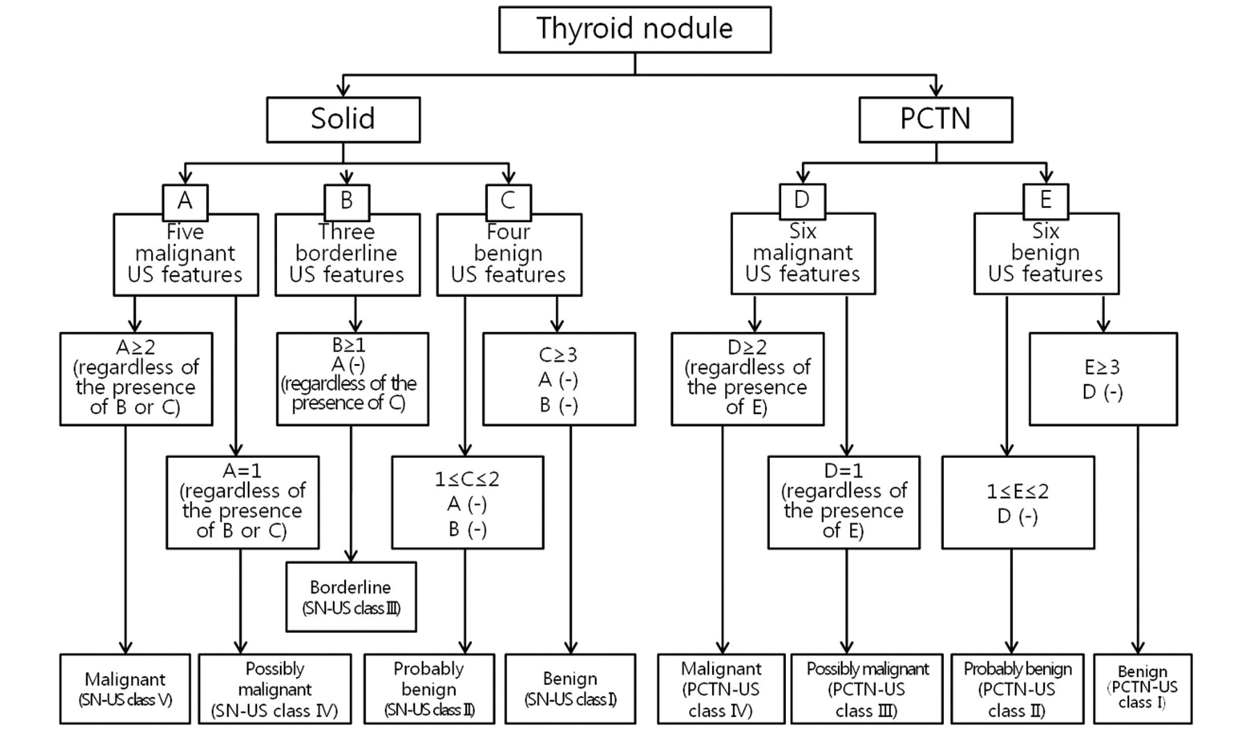
- Fig 1.
Classification system for US-based diagnosis of thyroid nodules.
- Fig 2.
Representative sonographic images of the 5 diagnostic categories for solid thyroid nodules. A, Benign: longitudinal US image, in a 62-year-old man, of a right thyroid nodule (2.2 × 2.7 × 3.1 cm) with an ovoid shape, isoechogenicity, and smooth margin (nodular hyperplasia by pathology). B, Probably benign: transverse US image, in a 31-year-old woman, of a large left thyroid nodule (3.2 × 4.9 × 7.3 cm) with an ovoid shape, inhomogeneous isoechogenicity, and macrolobulated margin (trabecular variant of follicular adenoma by pathology). C, Borderline: longitudinal US image, in a 47-year-old woman, of a right thyroid nodule (1.0 × 1.2 × 1.6 cm) with hypoechogenicity, smooth margin, and an ovoid shape (oncocytic variant of follicular adenoma by pathology). D, Possibly malignant: longitudinal US image, in a 60-year-old woman, of a right thyroid nodule (2.0 × 2.5 × 2.7 cm) with isoechogenicity, macrolobulated margin, and taller-than-wide shape (follicular variant of papillary thyroid carcinoma by pathology). E, Malignant: longitudinal US image in a 36-year-old woman of a left thyroid nodule (1.1 × 1.2 × 1.3 cm) with a round shape, marked hypoechogenicity, microlobulated margin, microcalcifications, and associated lymph nodes with intranodal cystic component or microcalcifications in the left inferior perithyroidal region (arrows) (classic type of papillary thyroid carcinoma by pathology).
- Fig 3.
Representative sonographic images of the 4 diagnostic categories for PCTNs. A, Benign: transverse US image of a right isthmic thyroid nodule (1.1 × 1.7 × 1.8 cm) showing an ovoid shape, isoechogenic and smooth-margined solid component, and a centrally and eccentrically located cystic component in a 30-year-old woman. The nodule was surgically resected because of coexisting thyroid malignancy (nodular hyperplasia by pathology). B, Probably benign: transverse US image of a right thyroid nodule (1.9 × 2.4 × 2.5 cm) showing an eccentric configuration with a blunt angle between the solid and cystic components (arrows) and an isoechoic solid component with a smooth margin in a 36-year-old woman. The nodule was surgically resected because of coexisting thyroid malignancy (nodular hyperplasia by pathology). C, Possibly malignant: transverse US image of a right thyroid nodule (1.9 × 2.1 × 2.2 cm) showing an eccentric configuration with an acute angle between the solid and cystic components (arrows) and an isoechoic solid component with a microlobulated margin in a 41-year-old man (nodular hyperplasia by pathology). D, Malignant: longitudinal US image of a left thyroid nodule (0.9 × 1.0 × 1.1 cm) showing an eccentric configuration with an acute angle between the solid and cystic components (arrows) and some microcalcifications in the solid component in a 36-year-old woman (classic type of papillary thyroid carcinoma by pathology).


Tables
- Table 1:
US diagnoses, cytologic diagnoses in initial US-FNA, and histopathologic results in 505 resected thyroid nodulesa
US Diagnoses Cytologic Diagnoses (Bethesda Classes I–VI) Histopathologic Results Solid nodules Benign (81) I (7), II (55), III (5), IV (3), V (9), VI (2) PTC (9), metastasis from renal cell carcinoma (1), FA (5), NH (66) Probably benign (82) I (3), II (38), III (8), IV (10), V (8), VI (15) PTC (22), FTC (4), poorly differentiated carcinoma (1), FA (14), NH (41) Borderline (44) I (3), II (15), III (9), V (6), VI (11) PTC (27), FTC (1), ATC (1), FA (1), NH (14) Possibly malignant (76) I (1), II (12), III (7), V (17), VI (39) PTC (60), FTC (1), MTC (2), FA (2), NH (11) Malignant (168) I (2), II (5), III (5), IV (1), V (22), VI (133) PTC (164), FTC (2), MTC (1), NH (1) Total (451) PCTN Benign (33) I (5), II (24), V (4) PTC (4), FA (1), NH (28) Probably benign (5) I (1), II (1), IV (1), V (1), VI (1) PTC (1), FTC (1), NH (3) Possibly malignant (4) II (1), III (1), V (2) PTC (1), NH (3) Malignant (12) II (1), V (2), VI (9) PTC (11), NH (1) Total (54) -
Note:—PTC indicates papillary thyroid carcinoma; FA, follicular adenoma; NH, nodular hyperplasia; FTC, follicular thyroid carcinoma; MTC, medullary thyroid carcinoma; ATC, anaplastic thyroid carcinoma.
-
↵a Data presented in parentheses are number of nodules.
-
Solid Nodules (n = 451) PCTNs (n = 54) Exclusion of Borderline US Diagnosis (%) Inclusion of Borderline US Diagnosis into Benign Category (%) Inclusion of Borderline US Diagnosis into Malignant Category (%) Sensitivity 230/267 (86.1) 230/296 (77.7) 259/296 (87.5) 12/18 (66.7) Specificity 126/140 (90) 141/155 (91.0) 126/155 (81.2) 32/36 (88.9) PPV 230/244 (94.3) 230/244 (94.3) 259/288 (89.9) 12/16 (75.0) NPV 126/163 (77.3) 141/207 (68.1) 126/163 (77.3) 32/38 (84.2) Accuracy 356/407 (87.5) 371/451 (82.3) 385/451 (85.4) 44/54 (81.5) -
Note:—PPV indicates positive predictive value; NPV, negative predictive value.
-
Sensitivity (%) Specificity (%) PPV (%) NPV (%) Accuracy (%) Solid nodules Malignant 167/296 154/155 167/168 154/283 321/451 (56.4) (99.4) (99.4) (54.4) (71.2) Possibly malignant 63/296 142/155 63/76 142/375 205/451 (21.3) (91.6) (82.9) (37.9) (45.5) Borderlinea 15/155 267/296 15/44 267/407 282/451 (9.7) (90.2) (34.1) (65.6) (62.5) Probably benign 55/155 269/296 55/82 269/369 324/451 (35.5) (90.9) (67.1) (72.9) (71.8) Benign 71/155 286/296 71/81 286/370 357/451 (45.8) (96.6) (87.7) (77.3) (79.2) PCTNs Malignant 11/18 35/36 11/12 35/42 46/54 (61.1) (97.2) (91.7) (83.3) (85.2) Possibly malignant 1/18 33/36 1/4 33/50 34/54 (5.6) (91.7) (25.0) (66.0) (63.0) Probably benign 3/36 16/18 3/5 16/49 19/54 (8.3) (88.9) (60.0) (32.7) (35.2) Benign 29/36 14/18 29/33 14/21 43/54 (80.6) (77.8) (87.9) (66.7) (79.6) -
Note:—PPV indicates positive predictive value; NPV, negative predictive value.
-
↵a Diagnostic indices with inclusion of borderline US diagnosis into the benign category.
-
- Table 4:
US findings and histopathologic results in 44 resected nodules with borderline US categorya
US Features Histopathologic Results Hypoechogenicity (14) PTC (8), FTC (1), ATC (1), FA (1), NH (3) Eggshell calcification (23) PTC (14), NH (9) Intranodular macrocalcifications (5) PTC (3), NH (2) Centrally predominant vascularity (3) PTC (2), NH (1) -
Note:—One borderline nodule showed hypoechogenicity and centrally predominant vascularity on US. PTC indicates papillary thyroid carcinoma; FTC, follicular thyroid carcinoma; ATC, anaplastic thyroid carcinoma; FA, follicular adenoma; NH, nodular hyperplasia.
-
↵a Data presented in parentheses are the number of nodules.
-


