
Article Figures & Data
Figures
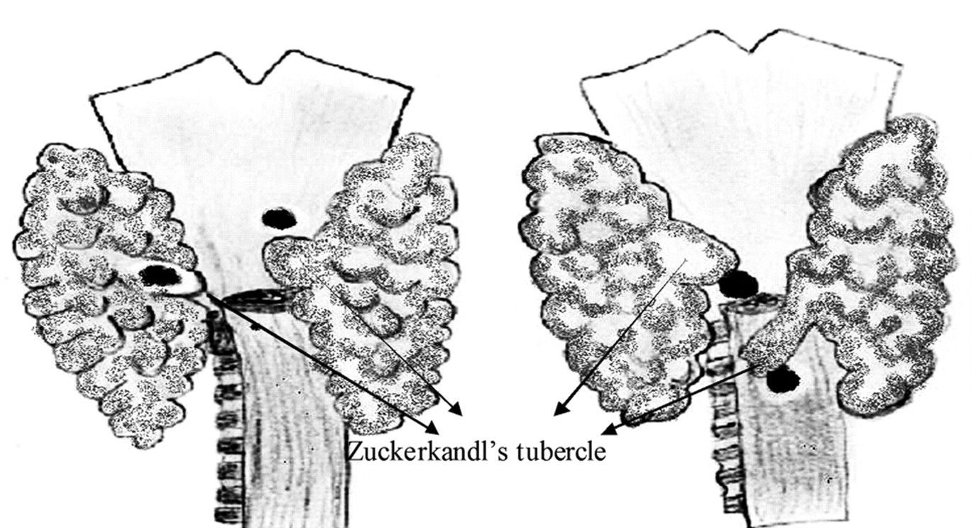
- Fig 1.
Drawings of 2 different thyroid glands as seen from a posterior point of view, with their relationships to the thyroid cartilage and a cut portion of the esophagus. The arrows indicate ZT and black dots indicate parathyroid glands.
- Fig 2.
Axial schematic representation of the thyroid (T = trachea, E = esophagus, black dot represents the right recurrent laryngeal nerve). A, Thyroid without apparent ZT. B, Thyroid with apparent right ZT. C, Thyroid with nodular right ZT (CCA = right common carotid artery within a lateral indentation of the thyroid contour).
- Fig 3.
Axial CT scan images demonstrate 3 different appearances of the ZT (arrows).
- Fig 4.
Patient A, with known ovarian carcinoma recurrence in the abdomen, presented with a few weeks' history of voice hoarseness and was noted to have a nodular area posterior to the right thyroid lobe. There was clinical concern for a metastasis or exophytic thyroid nodule.
- Fig 5.
CT-guided biopsy of Patient A in 2010 with questionable thyroid lesion (A) was performed because a recent sonography-guided fine-needle aspiration was read as indeterminate by pathology and there was persistent clinical concern despite stability from a CT in 2006. The patient was placed on the CT table with neck extended, and a metallic grid marker was placed on the section, showing the epicenter of the questionable lesion. B, A metallic grid marker is in place for biopsy planning. There is shoulder streak artifact through the questionable lesion, which lowers the attenuation. C, CT-guided percutaneous core biopsy needle was placed in the epicenter. Pathology results were consistent with normal thyroid tissue, with no malignant cells.
- Fig 6.
Patient A's pulmonary embolism chest CT from 2006 shows similar appearance of the questionable thyroid lesion.
- Fig 7.
Patient B has similar appearance of the right thyroid lobe on a CT from December 2009, with a posterior extension on the right.
- Fig 8.
Patient B's thyroid appeared similar as far back as 2005, allowing for slight difference in the obliquity of the axial section.
- Fig 9.
A, Thyroid gland with a nodular ZT at the posterior aspect of the right lobe (arrow). B, Follow-up CT study demonstrating development of a discrete hypoattenuated nodule in the right lobe and a hypoattenuated nodule in the previously identified ZT (arrow).
- Fig 10.
A and B, CT images on a patient status post total thyroidectomy for thyroid cancer show a small enhancing lesion near the right tracheoesophageal groove (arrows), which likely represents a tubercle of Zuckerhandl. C and E, Axial and coronal CT demonstrates enlargement of a paratracheal lesion after an interval of 6 months (arrows in C and E); a nearby lymph node also showed enlargement (arrow in D) and proved to be recurrent disease.


Tables
Number of CTs with demographic profile and distribution of ZT in our review
n CT studies included 96 Patients with ZT 67 Mean age (years) 56.1 Males 45 Females 31 Right 60 (89%) Left 49 (73%) Percentage of patients with nodular appearance of ZT 42.1%


