
Article Figures & Data
Figures
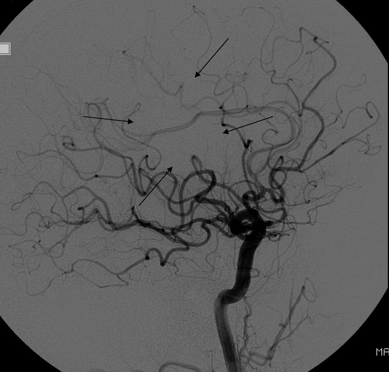
- Fig 1.
A, Lateral DSA following acute stroke intervention shows delay in flow in the posterior division of the MCA (arrows) compared with the anterior cerebral artery branches. B, Late arterial phase shows opacification of nearly all of the MCA. Because this represents more than two-thirds of the MCA (and thus not TICI 2a) but not “complete filling” (and thus not TICI 2b), it cannot be categorized by using the original TICI classifications.
- Fig 2.
Lateral DSA following acute stroke intervention shows a normal rate of antegrade flow in most of the MCA territory, with only a small amount of nonperfused parenchyma (arrows). The branches with normal antegrade flow would go into the TICI 3 category, while those with absent antegrade flow would go in the TICI 0 category, but there is no single TICI category for this angiogram. Some reviewers might want to put it into TICI 2, but it would not fit for 2 reasons: First, there is no portion of the territory with slow perfusion; second, the perfused area is greater than two-thirds of the MCA territory (making it incompatible with TICI 2a) but less than 100% of the MCA territory (making it incompatible with TICI 2b).
- Fig 3.
A, Anteroposterior DSA following acute stroke intervention shows persistent occlusion of the MCA. B, Late arterial phase DSA in the same patient as in A shows robust, retrograde, anterior cerebral artery−MCA leptomeningeal flow with opacification of most of the MCA territory.


In this issue


Jump to section
Related Articles
Cited By...
- Interobserver Agreement in Scoring Angiographic Results of Basilar Artery Occlusion Stroke Therapy
- eTICI reperfusion: defining success in endovascular stroke therapy
- To be or not 2b? To see or not 2c? Alas, the clock is ticking on TICI
- The Revascularization Scales Dilemma: Is It Right to Apply the Treatment in Cerebral Ischemia Scale in Posterior Circulation Stroke?
- Correlation of AOL recanalization, TIMI reperfusion and TICI reperfusion with infarct growth and clinical outcome
- Inter- and Intraobserver Agreement in Scoring Angiographic Results of Intra-Arterial Stroke Therapy
- 2C or not 2C: defining an improved revascularization grading scale and the need for standardization of angiography outcomes in stroke trials
- Recommendations on Angiographic Revascularization Grading Standards for Acute Ischemic Stroke: A Consensus Statement
- What Is Meant by "TICI"?
- Safety and Efficacy of Endovascular Sonolysis Using the EkoSonic Endovascular System in Patients with Acute Stroke