
Article Figures & Data
Figures
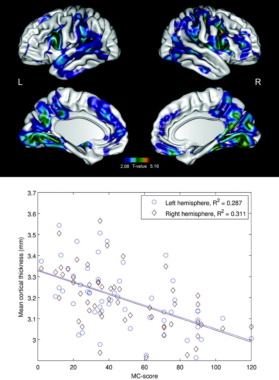
- Fig 1.
Group analysis of CTH. Brain regions demonstrate significantly (P < .0001, FDR corrected) reduced CTH in patients with EPM1 compared with healthy controls.
- Fig 2.
Brain regions with significant (P < .01, FDR-corrected) negative correlations between age and CTH in patients with EPM1 (A) and controls (B). Scatterplots indicate correlations between age and mean CTH in individual subjects. The individual mean thicknesses have been calculated on the basis of the regions surviving the statistical threshold.
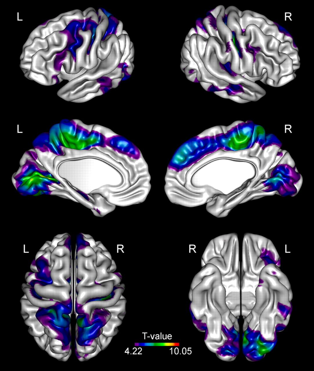
- Fig 3.
Brain regions with significant (P < .05, FDR-corrected) negative correlations between the “Myoclonus with Action” score and CTH in patients with EPM1. Scatterplots indicate correlations between the “Myoclonus with Action” score and mean CTH in individual subjects. The individual mean thicknesses have been calculated on the basis of the regions surviving the statistical threshold.


Tables
- Table 1:
Basic clinical characteristics and demographics of 53 patients with EPM1 and 70 controlsa)
Patients with EPM1 Controls Age (yr) 35 ± 11.5 33 ± 10.7 Sex (M/F) 29/24 34/36 Handedness (R/L/B/unknown) 35/6/1/11 62/4/3/1 Age at EPM1 onset (yr) 10.7 ± 2.9 (6–25) Duration of the disease (yr) 24.2 ± 10.4 (4–44) UMRS: stimulus sensitivity 1.92 ± 1.9 (0–8) UMRS: myoclonus with action 48.5 ± 28.4 (2–120) UMRS: functional tests 10.35 ± 7 (0–28) -
Note:—R indicates right; L, left; B, bilateral.
-
↵a a Data are presented as means unless otherwise indicated.
-
Brain Region Hemisphere Maximum T Value MNI Coordinates, (x, y, z)a Central sulcus Right 8.42 42, −16, 36 Left 6.69 −39, −19, 46 Precentral sulcus Left 6.05 −40, 7, 27 Paracentral lobule Right 8.11 5, −27, 50 Postcentral gyrus Right 7.32 6, −49, 65 Cingulate sulcus Left 8.06 −10, −19, 46 Postcentral sulcus Left 6.79 −24, −40, 60 Superior frontal gyrus Right 6.48 7, 45, 29 Calcarine sulcus Left 8.99 −20, −70, 6 Lingual gyrus Right 6.52 5, −68, 3 Left 8.20 −3, −76, 5 Superior temporal sulcus Right 5.32 54, −16, −14 Transverse temporal gyrus Right 5.33 43, −28, 10 Middle temporal gyrus Left 5.98 −48, −66, 4 Inferior occipital gyrus Left 7.39 −27, −89, −17 Note:—MNI indicates Montreal Neurological Institute.
-
↵a MNI coordinates are based on a standard brain template defined by using multiple MR images of healthy controls.


