
Article Figures & Data
Figures
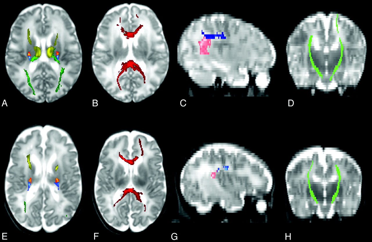
- Fig 1.
Tracts are shown on T2-weighted images in neonate with no WMA, scanned at 36 weeks corrected gestational age (A–D) and in neonate with moderate WMA, scanned at 38 weeks (E–H). Axial images (A, E) show the TRs (ATR in yellow, motor STR in yellow-red, sensory STR in blue and PTR in dark green). Axial images (B, F) show the corpus callosum (dark red). Sagittal images (C, G) show the frontoparietal SLF (dark blue) and parietotemporal SLF (pink). Coronal (D, H) images show the CSTs (light green). Images were obtained from a 1.5T magnet (Philips Achieva, Best, The Netherlands).
- Fig 2.
The diffusion indices in the left sensory STR are shown by scatter plots with mean (line) in neonates with no WMA, mild WMA and moderate WMA. Statistically significant differences (taking into account the gestational age as a covariate and after controlling for false discovery rate) were found for FA (P < .001), MD (P = .001), and λ⊥ (P < .001).


Tables
Characteristics Score 1 Score 2 Score 3 White matter signal abnormality Normal Focal regions Multiple regions (<2 regions per hemisphere) (>2 regions) 34/70 (48.6%) 15/70 (21.4%) 21/70 (30.0%) White matter volume loss Normal Mild reduction with increased ventricular size (EI = 0.3–0.36) Marked reduction with increased ventricular size (EI > 0.36) 61/70 (87.1%) 9/70 (12.9%) 0/70 (0%) Cystic abnormalities Normal <2-mm single focal cyst Multiple cysts or a single larger (>2 mm) focal cyst 66/70 (94.3%) 0/70 (0%) 4/70 (5.7%) Ventricular dilation Normal Mild-moderate enlargement of the frontal, temporal, and occipital horns More global enlargement of the frontal, temporal, and occipital horns 57/70 (81.4%) 12/70 (17.2%) 1/70 (1.4%) Thinning of the corpus callosum Normal Focal thinning in the corpus callosum Global thinning across the entire corpus callosum 54/70 (77.2%) 15/70 (21.4%) 1/70 (1.4%) -
Note:—EI indicates Evans Index.
-
Preterm with No WMA (n = 41) (mean) (SD) Preterm with Mild WMA (n = 27) (mean) (SD) P Value Preterm with Moderate WMA (n = 2) CST (left) 0.28 (0.03) 0.27 (0.04) .022 Case 1: 0.25 Case 2: 0.21 CST (right) 0.28 (0.04) 0.26 (0.04) .005 Case 1: 0.22 Case 2: 0.27 Frontoparietal SLF (left) 0.17 (0.03) 0.16 (0.03) .020 Case 1: 0.12 Case 2: 0.13 Frontoparietal SLF (right) 0.18 (0.02) 0.16 (0.03) .004 Case 1: 0.11 Case 2: 0.15 Parietotemporal SLF (left) 0.17 (0.02) 0.16 (0.03) .077 Case 1: 0.09 Case 2: 0.14 Parietotemporal SLF (right) 0.16 (0.02) 0.15 (0.03) .191 Case 1: 0.11 Case 2: 0.14 ATR (left) 0.19 (0.02) 0.18 (0.03) .012 Case 1: 0.16 Case 2: 0.16 ATR (right) 0.19 (0.02) 0.18 (0.03) .045 Case 1: 0.12 Case 2: 0.17 Motor STR (left) 0.25 (0.03) 0.23 (0.04) .005 Case 1: 0.22 Case 2: 0.20 Motor STR (right) 0.25 (0.03) 0.24 (0.05) .024 Case 1: 0.19 Case 2: 0.24 Sensory STR (left) 0.24 (0.03) 0.22 (0.03) <.001a Case 1: 0.15 Case 2: 0.17 Sensory STR (right) 0.23 (0.02) 0.22 (0.04) .006 Case 1: 0.17 Case 2: 0.18 PTR (left) 0.21 (0.03) 0.20 (0.03) .103 Case 1: 0.09 Case 2: 0.15 PTR (right) 0.21 (0.02) 0.20 (0.03) .009 Case 1: 0.11 Case 2: 0.17 Corpus callosum 0.26 (0.03) 0.24 (0.03) .016 Case 1: 0.14 Case 2: 0.18 -
↵a The P value reaches statistical significance after controlling for false discovery rate (P < .003).
-
Preterm with No WMA (n = 41) (mean) (SD) Preterm with Mild MWA (n = 27) (mean) (SD) P Value Preterm with Moderate WMA (n = 2) CST (left) 1.340 (0.10) 1.362 (0.09) .027 Case 1: 1.45 Case 2: 1.45 CST (right) 1.338 (0.09) 1.377 (0.12) .005 Case 1: 1.45 Case 2: 1.34 Frontoparietal SLF (left) 1.45 (0.10) 1.45 (0.12) .458 Case 1: 1.57 Case 2: 1.51 Frontoparietal SLF (right) 1.43 (0.10) 1.46 (0.12) .071 Case 1: 1.53 Case 2: 1.43 Parietotemporal SLF (left) 1.48 (0.07) 1.48 (0.09) .634 Case 1: 1.57 Case 2: 1.51 Parietotemporal SLF (right) 1.50 (0.11) 1.49 (0.09) .879 Case 1: 1.51 Case 2: 1.45 ATR (left) 1.33 (0.07) 1.36 (0.09) .003a Case 1: 1.47 Case 2: 1.44 ATR (right) 1.33 (0.07) 1.35 (0.09) .045 Case 1: 1.54 Case 2: 1.36 Motor STR (left) 1.31 (0.10) 1.36 (0.10) .001a Case 1: 1.45 Case 2: 1.47 Motor STR (right) 1.31 (0.10) 1.37 (0.14) .002a Case 1: 1.43 Case 2: 1.32 Sensory STR (left) 1.32 (0.08) 1.37 (0.11) .001a Case 1: 1.48 Case 2: 1.51 Sensory STR (right) 1.33 (0.10) 1.38 (0.11) .012 Case 1: 1.50 Case 2: 1.40 PTR (left) 1.48 (0.08) 1.51 (0.10) .064 Case 1: 1.57 Case 2: 1.60 PTR (right) 1.49 (0.08) 1.52 (0.09) .047 Case 1: 1.51 Case 2: 1.54 Corpus callosum 1.57 (0.06) 1.59 (0.08) .036 Case 1: 1.92 Case 2: 1.63 -
↵a The P value reaches statistical significance after controlling for false discovery rate (P < .003).
-
Preterm with No WMA (n = 41) (mean) (SD) Preterm with Mild WMA (n = 27) (mean) (SD) P Value Preterm with Moderate WMA (n = 2) CST (left) 1.14 (0.10) 1.17 (0.12) .007 Case 1: 1.29 Case 2: 1.29 CST (right) 1.14 (0.10) 1.19 (0.13) .001a Case 1: 1.28 Case 2: 1.16 Frontoparietal SLF (left) 1.33 (0.11) 1.34 (0.13) .291 Case 1: 1.48 Case 2: 1.41 Frontoparietal SLF (right) 1.31 (0.10) 1.35 (0.13) .033 Case 1: 1.45 Case 2: 1.32 Parietotemporal SLF (left) 1.35 (0.07) 1.36 (0.10) .403 Case 1: 1.50 Case 2: 1.40 Parietotemporal SLF (right) 1.38 (0.11) 1.38 (0.10) .724 Case 1: 1.43 Case 2: 1.36 ATR (left) 1.20 (0.07) 1.23 (0.07) .001a Case 1: 1.35 Case 2: 1.33 ATR (right) 1.20 (0.07) 1.23 (0.09) .025 Case 1: 1.45 Case 2: 1.24 Motor STR (left) 1.14 (0.10) 1.20 (0.12) <.001a Case 1: 1.31 Case 2: 1.32 Motor STR (right) 1.15 (0.10) 1.21 (0.15) .001a Case 1: 1.29 Case 2: 1.18 Sensory STR (left) 1.16 (0.09) 1.22 (0.12) <.001a Case 1: 1.37 Case 2: 1.38 Sensory STR (right) 1.18 (0.10) 1.23 (0.12) .005 Case 1: 1.37 Case 2: 1.27 PTR (left) 1.32 (0.09) 1.36 (0.12) .060 Case 1: 1.50 Case 2: 1.48 PTR (right) 1.33 (0.08) 1.36 (0.10) .018 Case 1: 1.43 Case 2: 1.42 Corpus callosum 1.35 (0.07) 1.39 (0.09) .012 Case 1: 1.79 Case 2: 1.63 -
↵a The P value reaches statistical significance after controlling for false discovery rate (P < .003).
-


