Advertisement

Main menu

  • Home
  • Content
    • Current Issue
    • Accepted Manuscripts
    • Article Preview
    • Past Issue Archive
    • Video Articles
    • AJNR Case Collection
    • Case of the Week Archive
    • Case of the Month Archive
    • Classic Case Archive
  • Special Collections
    • Advancing NeuroMRI with High-Relaxivity Contrast Agents
    • AJNR Awards
    • Low-Field MRI
    • Alzheimer Disease
    • ASNR Foundation Special Collection
    • Photon-Counting CT
    • View All
  • Multimedia
    • AJNR Podcasts
    • AJNR SCANtastic
    • Trainee Corner
    • MRI Safety Corner
    • Imaging Protocols
  • For Authors
    • Submit a Manuscript
    • Submit a Video Article
    • Submit an eLetter to the Editor/Response
    • Manuscript Submission Guidelines
    • Statistical Tips
    • Fast Publishing of Accepted Manuscripts
    • Graphical Abstract Preparation
    • Imaging Protocol Submission
    • Author Policies
  • About Us
    • About AJNR
    • Editorial Board
    • Editorial Board Alumni
  • More
    • Become a Reviewer/Academy of Reviewers
    • Subscribers
    • Permissions
    • Alerts
    • Feedback
    • Advertisers
    • ASNR Home

User menu

  • Alerts
  • Log in

Search

  • Advanced search
American Journal of Neuroradiology

American Journal of Neuroradiology

ASHNR American Society of Functional Neuroradiology ASHNR American Society of Pediatric Neuroradiology ASSR
  • Alerts
  • Log in

Advanced Search

  • Home
  • Content
    • Current Issue
    • Accepted Manuscripts
    • Article Preview
    • Past Issue Archive
    • Video Articles
    • AJNR Case Collection
    • Case of the Week Archive
    • Case of the Month Archive
    • Classic Case Archive
  • Special Collections
    • Advancing NeuroMRI with High-Relaxivity Contrast Agents
    • AJNR Awards
    • Low-Field MRI
    • Alzheimer Disease
    • ASNR Foundation Special Collection
    • Photon-Counting CT
    • View All
  • Multimedia
    • AJNR Podcasts
    • AJNR SCANtastic
    • Trainee Corner
    • MRI Safety Corner
    • Imaging Protocols
  • For Authors
    • Submit a Manuscript
    • Submit a Video Article
    • Submit an eLetter to the Editor/Response
    • Manuscript Submission Guidelines
    • Statistical Tips
    • Fast Publishing of Accepted Manuscripts
    • Graphical Abstract Preparation
    • Imaging Protocol Submission
    • Author Policies
  • About Us
    • About AJNR
    • Editorial Board
    • Editorial Board Alumni
  • More
    • Become a Reviewer/Academy of Reviewers
    • Subscribers
    • Permissions
    • Alerts
    • Feedback
    • Advertisers
    • ASNR Home
  • Follow AJNR on Twitter
  • Visit AJNR on Facebook
  • Follow AJNR on Instagram
  • Join AJNR on LinkedIn
  • RSS Feeds

AJNR Awards, New Junior Editors, and more. Read the latest AJNR updates


Improved Turnaround Times | Median time to first decision: 12 days

Research ArticleBrainE
Open Access

The Added Value of Apparent Diffusion Coefficient to Cerebral Blood Volume in the Preoperative Grading of Diffuse Gliomas

A. Hilario, A. Ramos, A. Perez-Nuñez, E. Salvador, J.M. Millan, A. Lagares, J.M. Sepulveda, P. Gonzalez-Leon, A. Hernandez-Lain and J.R. Ricoy
American Journal of Neuroradiology April 2012, 33 (4) 701-707; DOI: https://doi.org/10.3174/ajnr.A2846
A. Hilario
  • Find this author on Google Scholar
  • Find this author on PubMed
  • Search for this author on this site
A. Ramos
  • Find this author on Google Scholar
  • Find this author on PubMed
  • Search for this author on this site
A. Perez-Nuñez
  • Find this author on Google Scholar
  • Find this author on PubMed
  • Search for this author on this site
E. Salvador
  • Find this author on Google Scholar
  • Find this author on PubMed
  • Search for this author on this site
J.M. Millan
  • Find this author on Google Scholar
  • Find this author on PubMed
  • Search for this author on this site
A. Lagares
  • Find this author on Google Scholar
  • Find this author on PubMed
  • Search for this author on this site
J.M. Sepulveda
  • Find this author on Google Scholar
  • Find this author on PubMed
  • Search for this author on this site
P. Gonzalez-Leon
  • Find this author on Google Scholar
  • Find this author on PubMed
  • Search for this author on this site
A. Hernandez-Lain
  • Find this author on Google Scholar
  • Find this author on PubMed
  • Search for this author on this site
J.R. Ricoy
  • Find this author on Google Scholar
  • Find this author on PubMed
  • Search for this author on this site
  • Article
  • Figures & Data
  • Info & Metrics
  • Responses
  • References
  • PDF
Loading

Article Figures & Data

Figures

  • Tables
  • Fig 1.
    • Download figure
    • Open in new tab
    • Download powerpoint
    Fig 1.

    A 62-year-old man with biopsy-proved grade IV glioma. An example of how the ROIs in which minimum ADC (A) and maximum rCBV (B) have been measured is provided.
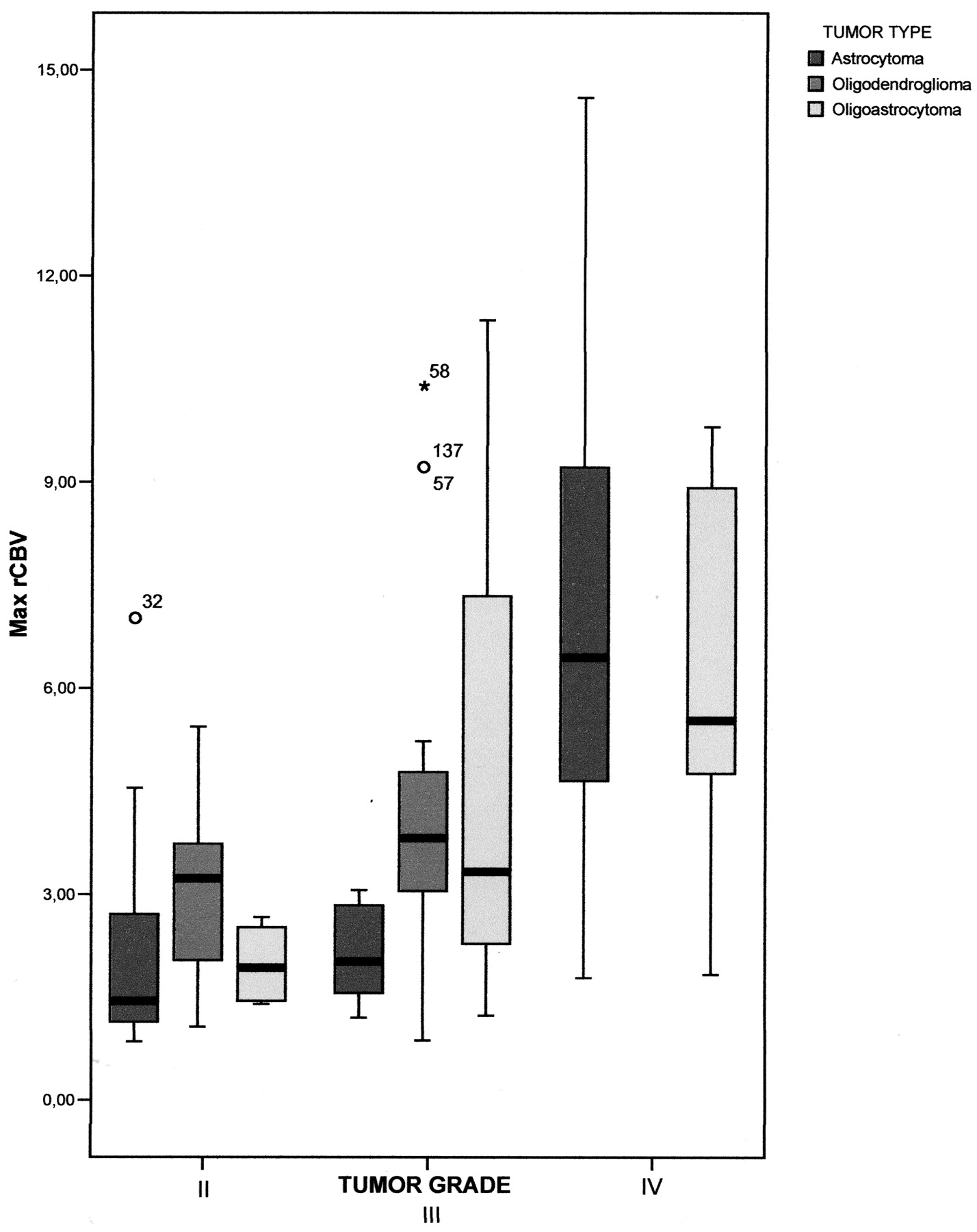
  • Fig 2.
    • Download figure
    • Open in new tab
    • Download powerpoint
    Fig 2.

    Boxplots show the range of maximum rCBV values determined by using DSC perfusion imaging in 162 patients with diffuse gliomas distributed by tumor histology and grade. The box represents the interquartile range (ie, 25%–75%); and black bar, the median. Th perfusion difference between grade II and III tumors was not statistically significant.

  • Fig 3.
    • Download figure
    • Open in new tab
    • Download powerpoint
    Fig 3.

    Boxplots show the range of minimum ADC values of 162 diffuse gliomas distributed by tumor histology and grade. The box represents the interquartile range (ie, 25%–75%); and the black bar, the median. Differences in minimum ADC values were statistically significant among grade II, III, and IV gliomas (P < .001). However, if we consider astrocytomas, oligodendrogliomas, and oligoastrocytomas separately, grades II and III oligodendrogliomas and grades III and IV oligoastrocytomas were not significantly different in ADC values.

  • Fig 4.
    • Download figure
    • Open in new tab
    • Download powerpoint
    Fig 4.

    ROC curve analysis showing the effect of using rCBV (A) and ADC (B) as individual or combined (C) variables in differentiating high- and low-grade gliomas. The area under the ROC curve for the maximum rCBV, minimum ADC, and a combination of both parameters is 0.72; 0.75; and 0.83, respectively.

Tables

  • Figures
    • View popup
    Table 1:

    Histopathologic diagnosis distributed by tumor grade

    HistologyGrade IIGrade IIIGrade IVTotal
    Astrocytomas18 (15.1%)9 (7.6%)92 (77.3%) (GBM)119 (73.5%)
    Oligodendrogliomas10 (37.03%)17 (62.97%)27 (16.7%)
    Oligoastrocytomas4 (25%)3 (18.8%)9 (56.3%) (GBM-O)16 (9.9%)
    Total32 (19.8%)29 (17.9%)101 (62.3%)162 (100%)
    • View popup
    Table 2:

    Minimum ADC and maximum rCBV values of different tumor types and grades

    Type and GradeMax rCBV (Mean)Min ADC (Mean)(× 10−3 mm2/s)
    Astrocytomas
        Grade II2.11 ± 1.581.2731 ± 0.293
        Grade III2.15 ± 0.711.0673 ± 0.276a
        Grade IV6.92 ± 3.12a,b0.7456 ± 0.135a,b
    Oligodendrogliomas
        Grade II2.98 ± 1.301.0827 ± 0.195
        Grade III4.36 ± 2.800.9326 ± 0.199
    Oligoastrocytomas
        Grade II1.98 ± 0.631.2925 ± 0.300
        Grade III5.30 ± 4.340.8410 ± 0.367a
        Grade IV6.30 ± 2.970.7651 ± 0.768a
    • Note:—Max indicates maximum; Min, minimum.

    • ↵a Significantly different from grade II (P < .01).

    • ↵b Significantly different from grade III (P < .01).

    • View popup
    Table 3:

    Sensitivity, specificity, PPV, and NPV for rCBV, ADC, and their combination in the differentiation of high-grade from low-grade gliomas

    SensitivitySpecificityPPVNPV
    Max rCBV ≥ 1.7494.4%50.0%88%69.6%
    Min ADC ≤ 1.18597.6%53.1%89%85%
    Max rCBV ≥ 1.74 OR Min ADC ≤ 1.18598.4%31.3%84.7%83.3%
    Max rCBV ≥ 1.74 AND Min ADC ≤ 1.18593.5%71.9%92.8%74.2%
    • Note:—PPV indicates positive predictive value; NPV, negative predictive value; Max, maximum; Min, minimum.

PreviousNext
Back to top

In this issue

American Journal of Neuroradiology: 33 (4)
American Journal of Neuroradiology
Vol. 33, Issue 4
1 Apr 2012
  • Table of Contents
  • Index by author
Advertisement
Print
Download PDF
Email Article

Thank you for your interest in spreading the word on American Journal of Neuroradiology.

NOTE: We only request your email address so that the person you are recommending the page to knows that you wanted them to see it, and that it is not junk mail. We do not capture any email address.

Enter multiple addresses on separate lines or separate them with commas.
The Added Value of Apparent Diffusion Coefficient to Cerebral Blood Volume in the Preoperative Grading of Diffuse Gliomas
(Your Name) has sent you a message from American Journal of Neuroradiology
(Your Name) thought you would like to see the American Journal of Neuroradiology web site.
CAPTCHA
This question is for testing whether or not you are a human visitor and to prevent automated spam submissions.
Cite this article
A. Hilario, A. Ramos, A. Perez-Nuñez, E. Salvador, J.M. Millan, A. Lagares, J.M. Sepulveda, P. Gonzalez-Leon, A. Hernandez-Lain, J.R. Ricoy
The Added Value of Apparent Diffusion Coefficient to Cerebral Blood Volume in the Preoperative Grading of Diffuse Gliomas
American Journal of Neuroradiology Apr 2012, 33 (4) 701-707; DOI: 10.3174/ajnr.A2846

Citation Manager Formats

  • BibTeX
  • Bookends
  • EasyBib
  • EndNote (tagged)
  • EndNote 8 (xml)
  • Medlars
  • Mendeley
  • Papers
  • RefWorks Tagged
  • Ref Manager
  • RIS
  • Zotero
0 Responses
Respond to this article
Share
Bookmark this article
The Added Value of Apparent Diffusion Coefficient to Cerebral Blood Volume in the Preoperative Grading of Diffuse Gliomas
A. Hilario, A. Ramos, A. Perez-Nuñez, E. Salvador, J.M. Millan, A. Lagares, J.M. Sepulveda, P. Gonzalez-Leon, A. Hernandez-Lain, J.R. Ricoy
American Journal of Neuroradiology Apr 2012, 33 (4) 701-707; DOI: 10.3174/ajnr.A2846
del.icio.us logo Twitter logo Facebook logo Mendeley logo
  • Tweet Widget
  • Facebook Like
  • Google Plus One
Purchase

Jump to section

  • Article
    • Abstract
    • ABBREVIATIONS:
    • Materials and Methods
    • Results
    • Discussion
    • Conclusion
    • Footnotes
    • References
  • Figures & Data
  • Info & Metrics
  • Responses
  • References
  • PDF

Related Articles

  • No related articles found.
  • PubMed
  • Google Scholar

Cited By...

  • Utility of Early Postoperative DWI to Assess the Extent of Resection of Adult-Type World Health Organization Grade 2 and 3 Diffuse Gliomas
  • Differentiating Low-Grade from High-Grade Intracranial Ependymomas: Comparison of Dynamic Contrast-Enhanced MRI and Diffusion-Weighted Imaging
  • Utilizing the Amide Proton Transfer Technique to Characterize Diffuse Gliomas Based on the WHO 2021 Classification of CNS Tumors
  • Regional and Volumetric Parameters for Diffusion-Weighted WHO Grade II and III Glioma Genotyping: A Method Comparison
  • High-Grade Gliomas in Children with Neurofibromatosis Type 1: Literature Review and Illustrative Cases
  • Predicting Genotype and Survival in Glioma Using Standard Clinical MR Imaging Apparent Diffusion Coefficient Images: A Pilot Study from The Cancer Genome Atlas
  • 3D Pseudocontinuous Arterial Spin-Labeling MR Imaging in the Preoperative Evaluation of Gliomas
  • Discrimination between Glioma Grades II and III Using Dynamic Susceptibility Perfusion MRI: A Meta-Analysis
  • Comparison of the Effect of Vessel Size Imaging and Cerebral Blood Volume Derived from Perfusion MR Imaging on Glioma Grading
  • Mean Diffusional Kurtosis in Patients with Glioma: Initial Results with a Fast Imaging Method in a Clinical Setting
  • Diffusion-Weighted Imaging in Cancer: Physical Foundations and Applications of Restriction Spectrum Imaging
  • A Prognostic Model Based on Preoperative MRI Predicts Overall Survival in Patients with Diffuse Gliomas
  • Multimodal MR Imaging (Diffusion, Perfusion, and Spectroscopy): Is It Possible to Distinguish Oligodendroglial Tumor Grade and 1p/19q Codeletion in the Pretherapeutic Diagnosis?
  • Crossref
  • Google Scholar

This article has not yet been cited by articles in journals that are participating in Crossref Cited-by Linking.

More in this TOC Section

  • Predictors of Reperfusion in Patients with Acute Ischemic Stroke
  • Enhanced Axonal Metabolism during Early Natalizumab Treatment in Relapsing-Remitting Multiple Sclerosis
  • Progression of Microstructural Damage in Spinocerebellar Ataxia Type 2: A Longitudinal DTI Study
Show more Brain

Similar Articles

Advertisement

Indexed Content

  • Current Issue
  • Accepted Manuscripts
  • Article Preview
  • Past Issues
  • Editorials
  • Editor's Choice
  • Fellows' Journal Club
  • Letters to the Editor
  • Video Articles

Cases

  • Case Collection
  • Archive - Case of the Week
  • Archive - Case of the Month
  • Archive - Classic Case

More from AJNR

  • Trainee Corner
  • Imaging Protocols
  • MRI Safety Corner
  • Book Reviews

Multimedia

  • AJNR Podcasts
  • AJNR Scantastics

Resources

  • Turnaround Time
  • Submit a Manuscript
  • Submit a Video Article
  • Submit an eLetter to the Editor/Response
  • Manuscript Submission Guidelines
  • Statistical Tips
  • Fast Publishing of Accepted Manuscripts
  • Graphical Abstract Preparation
  • Imaging Protocol Submission
  • Evidence-Based Medicine Level Guide
  • Publishing Checklists
  • Author Policies
  • Become a Reviewer/Academy of Reviewers
  • News and Updates

About Us

  • About AJNR
  • Editorial Board
  • Editorial Board Alumni
  • Alerts
  • Permissions
  • Not an AJNR Subscriber? Join Now
  • Advertise with Us
  • Librarian Resources
  • Feedback
  • Terms and Conditions
  • AJNR Editorial Board Alumni

American Society of Neuroradiology

  • Not an ASNR Member? Join Now

© 2025 by the American Society of Neuroradiology All rights, including for text and data mining, AI training, and similar technologies, are reserved.
Print ISSN: 0195-6108 Online ISSN: 1936-959X

Powered by HighWire