
Article Figures & Data
Figures
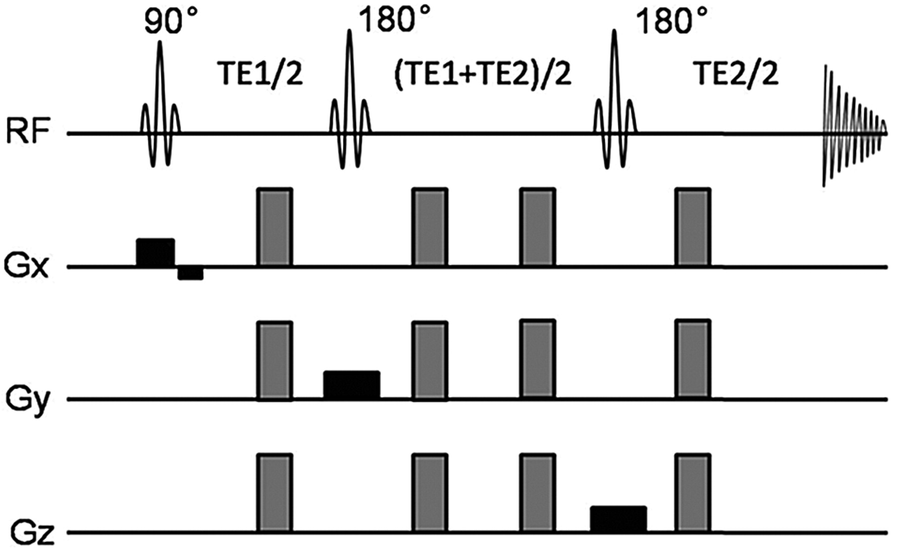
- Fig 1.
Schematic radio-frequency pulse and diffusion gradients for diffusion-weighted PRESS. The diffusion-weighted gradients are present around the second and third 180° pulses. The amplitudes are fixed at 80% of the maximum amplitude of the system, and the gradients of 3 directions are used together to increase the b-value.
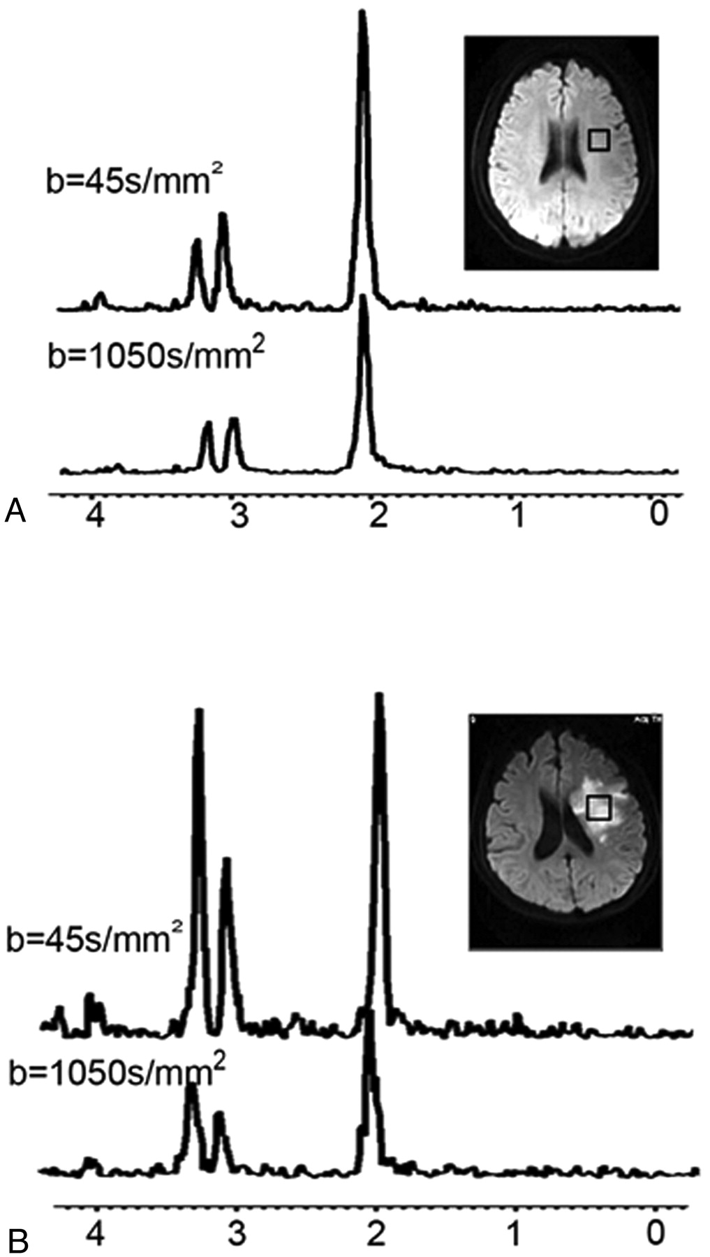
- Fig 2.
Brain regions. A, Brain regions of volunteers. The volume of interest (2.0 × 2.0 × 2.0 = 8 cm3) is located in the left centrum semiovale, and the spectrum is taken at low (45 s/mm2) and high (1050 s/mm2) b-values. B, Brain regions of patients. The volume of interest (2.0 × 2.0 × 2.0 = 8 cm3) is located in the region of infarct, and the spectrum is taken at low (45 s/mm2) and high (1050 s/mm2) b-values.
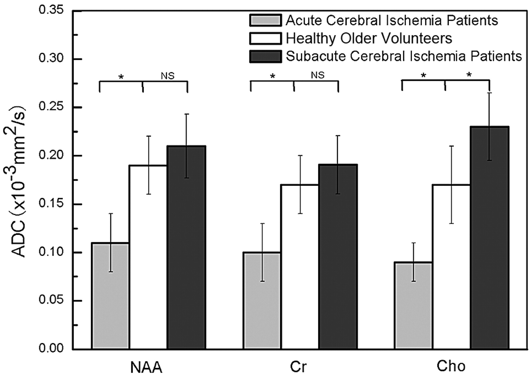
- Fig 3.
Mean metabolite ADCs for regions of interest across subjects with acute cerebral ischemia and subacute cerebral ischemia and the healthy older volunteer group, respectively. Gray bars show ADC values for the acute cerebral ischemia group, white bars show ADC values for the healthy older volunteer group, and black bars are used for the subacute cerebral ischemia group. Standard error bars and significance are also shown. Statistically significant differences relative to controls: the asterisk indicates P < .05; NS, no significance.


Tables
- Table 1:
Healthy young, healthy older, acute ischemia, and subacute ischemia group demographics and stroke characteristics
Healthy Young Healthy Older Acute Ischemia Subacute Ischemia No. 26 17 7 12 Age (yr, mean) 24 ± 2.2 63 ± 7.0 57 ± 4.0 62 ± 7.8 Age range (yr) 21–31 54–78 52–77 54–74 Sex (male/female) 13/13 6/11 5/2 10/2 Body mass (kg, mean) 65 ± 14.0 68 ± 5.7 63 ± 4.3 64 ± 6.3 Onset time of ischemia (hr) – – 24–48 ≥72 -
Note:—indicates no onset time of ischemia for healthy group.
-
- Table 2:
ADCs of metabolites and water in the left centrum semiovale of healthy volunteers and patients with ischemia at different ages
Subject Age (yr) No. ADC (mean ± SD, ×10−3 mm2/s) NAA Cr Cho Water Healthy young volunteers 21–31 26 0.26 ± 0.06a 0.25 ± 0.06a 0.23 ± 0.05a 0.78 ± 0.02 Healthy older volunteers 54–78 17 0.19 ± 0.03 0.17 ± 0.03 0.17 ± 0.04 0.79 ± 0.04 Patients with acute cerebral ischemia 52–77 7 0.11 ± 0.03b 0.10 ± 0.03b 0.09 ± 0.02b 0.54 ± 0.10 Patients with subacute cerebral ischemia 53–74 12 0.21 ± 0.03 0.19 ± 0.03 0.23 ± 0.04c 0.62 ± 0.08 -
↵a Significantly higher in the healthy young volunteer group compared with the healthy older volunteer group (P < .01).
-
↵b Significantly lower in the acute cerebral ischemia patient group compared with the healthy older volunteer group (P < .05).
-
↵c Significantly higher in the subacute cerebral ischemia patient group compared with the healthy older volunteer group (P < .05).
-
In this issue


Jump to section
Related Articles
Cited By...
- No citing articles found.