
Article Figures & Data
Figures
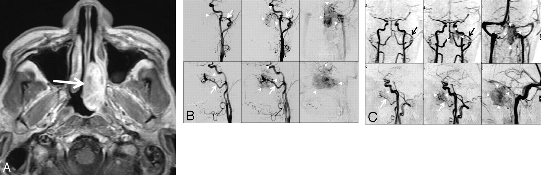
- Fig 1.
A 44-year-old man with juvenile angiofibroma. A, Axial contrast-enhanced T1-weighted MR image shows an enhanced mass in the left nasopharyngeal space (arrow). B, Anteroposterior (upper) and lateral (lower) DSA projections from the left common carotid artery during the early arterial (left), late arterial (middle), and venous (right) phase. DSA shows a tumor stain (arrowheads) at the early-arterial-to-venous phase; supply is from branches of the left internal maxillary artery (arrow). C, Anteroposterior (upper) and lateral (lower) projections of maximum-intensity 4D-CE-MRA images (2.9/1.4, 20° flip angle). Tumor stain (arrowheads) is seen from the early arterial phase (left); it is apparent in the late arterial (middle) and venous phases (right). The tumor is mainly supplied by branches from the left internal maxillary artery (arrow). Both readers judged that the internal maxillary artery was the main arterial feeder and that the tumor stain was grade 2.
- Fig 2.
A 54-year-old man with cerebellar hemangioblastoma. A, Contrast-enhanced MR image shows a ring-enhanced mass in the left cerebellar hemisphere (arrow). B, Lateral DSA projections from the left vertebral (left, middle) and the external carotid (right) artery at the early arterial (left, right) and venous (middle) phases. DSA shows a tumor mainly supplied by branches from the posterior meningeal artery (white arrow). The other feeders were the posterior inferior cerebellar (small white arrow) and occipital (black arrow) arteries. Tumor stain is seen at the early arterial and venous phases (arrowheads). C, Lateral projections of maximum-intensity 4D-CE-MRA images (2.9/1.4, 20° flip angle). Tumor stain (arrowheads) is seen from the early arterial (left) to the venous phase (right). Both readers judged that the occipital artery (arrow) was the main arterial feeder and that the tumor stain was grade 2.


Tables
4D-CE-MRA Interobserver Agreement DSA 4D-CE-MRAa Intermodality Agreement Observer 1 Observer 2 Main feeders IMA 3 3 4 3 Oph A 2 3 3 2 Facial A 2 1 2 1 Occipital A 1 1 0 1 PCA 0 1 6/15 (40%) 1 1 8/15 (53%) ACA 0 2 2 1 Cerebellar A 0 0 1 0 ICA 0 1 1 0 Other feeders 7 3 1 6 Tumor stainb Grade 0 0 0 0 0 Grade 1 8 7 14/15 (93%) 6 8 13/15 (87%) Grade 2 7 8 9 7 -
Note:—IMA indicates internal maxillary artery; Oph A, ophthalmic artery; Facial A, facial artery; Occipital A, occipital artery; PCA, posterior cerebral artery; ACA, anterior cerebral artery; Cerebellar A, cerebellar artery; ICA, internal carotid artery.
-
↵a Consensus reading of the 2 observers.
-
↵b The degree of tumor stain was classified as grade 0, not visible; grade 1, visible only after the late arterial phase; and grade 2, visible from the early arterial phase.
-
Main Feeders Tumor Stain Interobserver agreement 0.283 (0.095–0.662) 0.867 (0.618–1.000) Intermodality agreement 0.454 (0.165–0.743) 0.737 (0.409–1.000) -
↵a Data are κ statistics, with 95% CIs in parentheses.
-
In this issue


Jump to section
Related Articles
Cited By...
- No citing articles found.