Advertisement

Main menu

  • Home
  • Content
    • Current Issue
    • Accepted Manuscripts
    • Article Preview
    • Past Issue Archive
    • Video Articles
    • AJNR Case Collection
    • Case of the Week Archive
    • Case of the Month Archive
    • Classic Case Archive
  • Special Collections
    • Advancing NeuroMRI with High-Relaxivity Contrast Agents
    • AJNR Awards
    • Low-Field MRI
    • Alzheimer Disease
    • ASNR Foundation Special Collection
    • Photon-Counting CT
    • View All
  • Multimedia
    • AJNR Podcasts
    • AJNR SCANtastic
    • Trainee Corner
    • MRI Safety Corner
    • Imaging Protocols
  • For Authors
    • Submit a Manuscript
    • Submit a Video Article
    • Submit an eLetter to the Editor/Response
    • Manuscript Submission Guidelines
    • Statistical Tips
    • Fast Publishing of Accepted Manuscripts
    • Graphical Abstract Preparation
    • Imaging Protocol Submission
    • Author Policies
  • About Us
    • About AJNR
    • Editorial Board
    • Editorial Board Alumni
  • More
    • Become a Reviewer/Academy of Reviewers
    • Subscribers
    • Permissions
    • Alerts
    • Feedback
    • Advertisers
    • ASNR Home

User menu

  • Alerts
  • Log in

Search

  • Advanced search
American Journal of Neuroradiology

American Journal of Neuroradiology

ASHNR American Society of Functional Neuroradiology ASHNR American Society of Pediatric Neuroradiology ASSR
  • Alerts
  • Log in

Advanced Search

  • Home
  • Content
    • Current Issue
    • Accepted Manuscripts
    • Article Preview
    • Past Issue Archive
    • Video Articles
    • AJNR Case Collection
    • Case of the Week Archive
    • Case of the Month Archive
    • Classic Case Archive
  • Special Collections
    • Advancing NeuroMRI with High-Relaxivity Contrast Agents
    • AJNR Awards
    • Low-Field MRI
    • Alzheimer Disease
    • ASNR Foundation Special Collection
    • Photon-Counting CT
    • View All
  • Multimedia
    • AJNR Podcasts
    • AJNR SCANtastic
    • Trainee Corner
    • MRI Safety Corner
    • Imaging Protocols
  • For Authors
    • Submit a Manuscript
    • Submit a Video Article
    • Submit an eLetter to the Editor/Response
    • Manuscript Submission Guidelines
    • Statistical Tips
    • Fast Publishing of Accepted Manuscripts
    • Graphical Abstract Preparation
    • Imaging Protocol Submission
    • Author Policies
  • About Us
    • About AJNR
    • Editorial Board
    • Editorial Board Alumni
  • More
    • Become a Reviewer/Academy of Reviewers
    • Subscribers
    • Permissions
    • Alerts
    • Feedback
    • Advertisers
    • ASNR Home
  • Follow AJNR on Twitter
  • Visit AJNR on Facebook
  • Follow AJNR on Instagram
  • Join AJNR on LinkedIn
  • RSS Feeds

AJNR Awards, New Junior Editors, and more. Read the latest AJNR updates


Improved Turnaround Times | Median time to first decision: 12 days

Research ArticleHead and Neck Imaging

Diagnostic Accuracy of 4D-CT for Parathyroid Adenomas and Hyperplasia

J.L. Chazen, A. Gupta, A. Dunning and C.D. Phillips
American Journal of Neuroradiology March 2012, 33 (3) 429-433; DOI: https://doi.org/10.3174/ajnr.A2805
J.L. Chazen
  • Find this author on Google Scholar
  • Find this author on PubMed
  • Search for this author on this site
A. Gupta
  • Find this author on Google Scholar
  • Find this author on PubMed
  • Search for this author on this site
A. Dunning
  • Find this author on Google Scholar
  • Find this author on PubMed
  • Search for this author on this site
C.D. Phillips
  • Find this author on Google Scholar
  • Find this author on PubMed
  • Search for this author on this site
  • Article
  • Figures & Data
  • Info & Metrics
  • Responses
  • References
  • PDF
Loading

Article Figures & Data

Figures

  • Tables
  • Fig 1.
    • Download figure
    • Open in new tab
    • Download powerpoint
    Fig 1.

    Axial noncontrast (A), axial early phase postcontrast (B), and axial delayed phase postcontrast (C) images show a hypoattenuated hypodense nodule contiguous with the left posterior thyroid gland, which demonstrates avid early contrast enhancement and washout. Pathology revealed a 600-mg parathyroid adenoma.
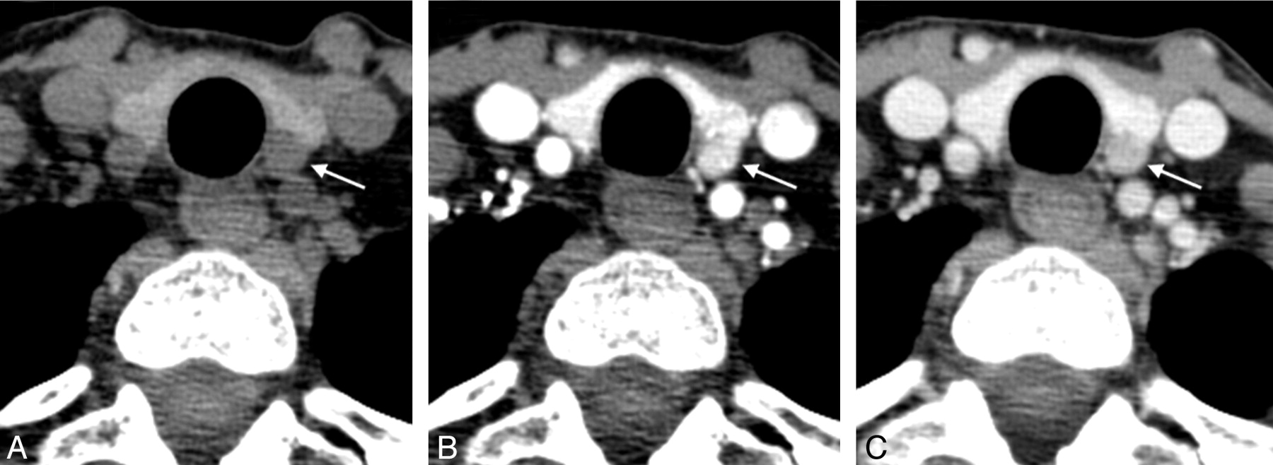
  • Fig 2.
    • Download figure
    • Open in new tab
    • Download powerpoint
    Fig 2.

    Coronal early phase postcontrast (A), axial early phase postcontrast (B), and axial delayed phase postcontrast (C) images showing an early enhancing 1.3-cm nodule in the right tracheoesophageal groove, with modest contrast washout representing a right inferior parathyroid gland in an orthotopic location. Pathology demonstrated a 3-g parathyroid adenoma.

  • Fig 3.
    • Download figure
    • Open in new tab
    • Download powerpoint
    Fig 3.

    Axial (A) and coronal (B) postcontrast images showing an enhancing retrosternal ectopic parathyroid adenoma.

Tables

  • Figures
    • View popup
    Table 1:

    Descriptives

    VariableMean/FreqSD/Percent
    Age (yr)59.39.9
    Female gender (n)2674%
    BMI (kg/m2)26.85.6
    Weight (kg)73.216.9
    Lesion weight (mg)566.7611.0
    Prior neck surgery (n)26%
    Single-gland disease (n)2880%
    Multigland disease (n)720%
    Peak preoperative parathyroid hormone (pg/mL)190.3288.6
    Postoperative parathyroid hormone (pg/mL)29.527.0
    • Note:—Freq indicates frequency.

    • View popup
    Table 2:

    Distribution of findings

    VariablePathology Findings4D-CT Findings
    Mean/FreqSD/PercentMean/FreqSD/Percent
    Lesion detection
        Positive27100.0%27100.0%
        Negative00.0%00.0%
    Lesion location
        Right superior622.2%414.8%
        Right inferior622.2%829.6%
        Left superior933.3%829.6%
        Left inferior622.2%725.9%
    Lesion side
        Right1244.4%1244.4%
        Left1555.6%1555.6%
    Lesion z-axis
        Superior1555.6%1244.4%
        Inferior1244.4%1555.6%
    • Note:—Freq indicates frequency.

    • View popup
    Table 3:

    Single-gland lateralization

    Measure%RateExact Binomial 95% CINumeratorDenominator
    LowerUpper
    Sensitivity91.761.599.81112
    Specificity93.368.199.81415
    PPV91.761.599.81112
    PNV93.368.199.81415
    Accuracy92.675.799.12527
    CT SidePathology Side
    LeftRightTotal
    Left14115
    Right11112
    Total151227
    • View popup
    Table 4:

    Single-gland quadrant localization

    Measure%RateExact Binomial 95% CINumeratorDenominator
    LowerUpper
    Sensitivity85.757.298.21214
    Specificity10071.51001111
    PPV10073.51001212
    PNV84.654.698.11113
    Accuracy92.074.099.02325
    CT QuadrantPathology Quadrant
    InferiorSuperiorTotal
    Inferior11213
    Superior01212
    Total111425
    • View popup
    Table 5:

    Multigland disease

    Measure%RateExact Binomial 95% CINumeratorDenominator
    LowerUpper
    Sensitivity42.99.981.637
    Specificity10087.71002828
    PPV10029.210033
    PNV87.571.096.52832
    Accuracy88.673.396.83135
    CT MultiglandPathology Multigland
    NoYesTotal
    No28432
    Yes033
    Total28735
PreviousNext
Back to top

In this issue

American Journal of Neuroradiology: 33 (3)
American Journal of Neuroradiology
Vol. 33, Issue 3
1 Mar 2012
  • Table of Contents
  • Index by author
Advertisement
Print
Download PDF
Email Article

Thank you for your interest in spreading the word on American Journal of Neuroradiology.

NOTE: We only request your email address so that the person you are recommending the page to knows that you wanted them to see it, and that it is not junk mail. We do not capture any email address.

Enter multiple addresses on separate lines or separate them with commas.
Diagnostic Accuracy of 4D-CT for Parathyroid Adenomas and Hyperplasia
(Your Name) has sent you a message from American Journal of Neuroradiology
(Your Name) thought you would like to see the American Journal of Neuroradiology web site.
CAPTCHA
This question is for testing whether or not you are a human visitor and to prevent automated spam submissions.
Cite this article
J.L. Chazen, A. Gupta, A. Dunning, C.D. Phillips
Diagnostic Accuracy of 4D-CT for Parathyroid Adenomas and Hyperplasia
American Journal of Neuroradiology Mar 2012, 33 (3) 429-433; DOI: 10.3174/ajnr.A2805

Citation Manager Formats

  • BibTeX
  • Bookends
  • EasyBib
  • EndNote (tagged)
  • EndNote 8 (xml)
  • Medlars
  • Mendeley
  • Papers
  • RefWorks Tagged
  • Ref Manager
  • RIS
  • Zotero
0 Responses
Respond to this article
Share
Bookmark this article
Diagnostic Accuracy of 4D-CT for Parathyroid Adenomas and Hyperplasia
J.L. Chazen, A. Gupta, A. Dunning, C.D. Phillips
American Journal of Neuroradiology Mar 2012, 33 (3) 429-433; DOI: 10.3174/ajnr.A2805
del.icio.us logo Twitter logo Facebook logo Mendeley logo
  • Tweet Widget
  • Facebook Like
  • Google Plus One
Purchase

Jump to section

  • Article
    • Abstract
    • ABBREVIATIONS:
    • Materials and Methods
    • Results
    • Discussion
    • Conclusions
    • Footnotes
    • References
  • Figures & Data
  • Info & Metrics
  • Responses
  • References
  • PDF

Related Articles

  • No related articles found.
  • PubMed
  • Google Scholar

Cited By...

  • Distinguishing Recurrent Thyroid Cancer from Residual Nonmalignant Thyroid Tissue Using Multiphasic Multidetector CT
  • 4D-Dynamic Contrast-Enhanced MRI for Preoperative Localization in Patients with Primary Hyperparathyroidism
  • Clinical Images: Four-Dimensional Computed Tomography--Future of Preoperative Parathyroid Adenoma Imaging
  • Accuracy of 2-Phase Parathyroid CT for the Preoperative Localization of Parathyroid Adenomas in Primary Hyperparathyroidism
  • Dynamic 4D MRI for Characterization of Parathyroid Adenomas: Multiparametric Analysis
  • Dynamic CT for Parathyroid Disease: Are Multiple Phases Necessary?
  • 4D-CT for Preoperative Localization of Abnormal Parathyroid Glands in Patients with Hyperparathyroidism: Accuracy and Ability to Stratify Patients by Unilateral versus Bilateral Disease in Surgery-Naive and Re-Exploration Patients
  • Dual-Energy 4-Phase CT Scan in Primary Hyperparathyroidism
  • Crossref
  • Google Scholar

This article has not yet been cited by articles in journals that are participating in Crossref Cited-by Linking.

More in this TOC Section

  • Diagnostic Performance of US in Neck Node NIRADS 2
  • Hydrops Herniation into the Semicircular Canals
  • DIVr of NPC Predicted Outcome
Show more HEAD AND NECK IMAGING

Similar Articles

Advertisement

Indexed Content

  • Current Issue
  • Accepted Manuscripts
  • Article Preview
  • Past Issues
  • Editorials
  • Editor's Choice
  • Fellows' Journal Club
  • Letters to the Editor
  • Video Articles

Cases

  • Case Collection
  • Archive - Case of the Week
  • Archive - Case of the Month
  • Archive - Classic Case

More from AJNR

  • Trainee Corner
  • Imaging Protocols
  • MRI Safety Corner
  • Book Reviews

Multimedia

  • AJNR Podcasts
  • AJNR Scantastics

Resources

  • Turnaround Time
  • Submit a Manuscript
  • Submit a Video Article
  • Submit an eLetter to the Editor/Response
  • Manuscript Submission Guidelines
  • Statistical Tips
  • Fast Publishing of Accepted Manuscripts
  • Graphical Abstract Preparation
  • Imaging Protocol Submission
  • Evidence-Based Medicine Level Guide
  • Publishing Checklists
  • Author Policies
  • Become a Reviewer/Academy of Reviewers
  • News and Updates

About Us

  • About AJNR
  • Editorial Board
  • Editorial Board Alumni
  • Alerts
  • Permissions
  • Not an AJNR Subscriber? Join Now
  • Advertise with Us
  • Librarian Resources
  • Feedback
  • Terms and Conditions
  • AJNR Editorial Board Alumni

American Society of Neuroradiology

  • Not an ASNR Member? Join Now

© 2025 by the American Society of Neuroradiology All rights, including for text and data mining, AI training, and similar technologies, are reserved.
Print ISSN: 0195-6108 Online ISSN: 1936-959X

Powered by HighWire