Advertisement

Main menu

  • Home
  • Content
    • Current Issue
    • Accepted Manuscripts
    • Article Preview
    • Past Issue Archive
    • Video Articles
    • AJNR Case Collection
    • Case of the Week Archive
    • Case of the Month Archive
    • Classic Case Archive
  • Special Collections
    • AJNR Awards
    • Low-Field MRI
    • Alzheimer Disease
    • ASNR Foundation Special Collection
    • Photon-Counting CT
    • View All
  • Multimedia
    • AJNR Podcasts
    • AJNR SCANtastic
    • Trainee Corner
    • MRI Safety Corner
    • Imaging Protocols
  • For Authors
    • Submit a Manuscript
    • Submit a Video Article
    • Submit an eLetter to the Editor/Response
    • Manuscript Submission Guidelines
    • Statistical Tips
    • Fast Publishing of Accepted Manuscripts
    • Graphical Abstract Preparation
    • Imaging Protocol Submission
    • Author Policies
  • About Us
    • About AJNR
    • Editorial Board
    • Editorial Board Alumni
  • More
    • Become a Reviewer/Academy of Reviewers
    • Subscribers
    • Permissions
    • Alerts
    • Feedback
    • Advertisers
    • ASNR Home

User menu

  • Alerts
  • Log in

Search

  • Advanced search
American Journal of Neuroradiology

American Journal of Neuroradiology

ASHNR American Society of Functional Neuroradiology ASHNR American Society of Pediatric Neuroradiology ASSR
  • Alerts
  • Log in

Advanced Search

  • Home
  • Content
    • Current Issue
    • Accepted Manuscripts
    • Article Preview
    • Past Issue Archive
    • Video Articles
    • AJNR Case Collection
    • Case of the Week Archive
    • Case of the Month Archive
    • Classic Case Archive
  • Special Collections
    • AJNR Awards
    • Low-Field MRI
    • Alzheimer Disease
    • ASNR Foundation Special Collection
    • Photon-Counting CT
    • View All
  • Multimedia
    • AJNR Podcasts
    • AJNR SCANtastic
    • Trainee Corner
    • MRI Safety Corner
    • Imaging Protocols
  • For Authors
    • Submit a Manuscript
    • Submit a Video Article
    • Submit an eLetter to the Editor/Response
    • Manuscript Submission Guidelines
    • Statistical Tips
    • Fast Publishing of Accepted Manuscripts
    • Graphical Abstract Preparation
    • Imaging Protocol Submission
    • Author Policies
  • About Us
    • About AJNR
    • Editorial Board
    • Editorial Board Alumni
  • More
    • Become a Reviewer/Academy of Reviewers
    • Subscribers
    • Permissions
    • Alerts
    • Feedback
    • Advertisers
    • ASNR Home
  • Follow AJNR on Twitter
  • Visit AJNR on Facebook
  • Follow AJNR on Instagram
  • Join AJNR on LinkedIn
  • RSS Feeds

AJNR Awards, New Junior Editors, and more. Read the latest AJNR updates

Research ArticlePediatrics

Optic Nerve Sheath Diameter on MR Imaging: Establishment of Norms and Comparison of Pediatric Patients with Idiopathic Intracranial Hypertension with Healthy Controls

B. Shofty, L. Ben-Sira, S. Constantini, S. Freedman and A. Kesler
American Journal of Neuroradiology February 2012, 33 (2) 366-369; DOI: https://doi.org/10.3174/ajnr.A2779
B. Shofty
  • Find this author on Google Scholar
  • Find this author on PubMed
  • Search for this author on this site
L. Ben-Sira
  • Find this author on Google Scholar
  • Find this author on PubMed
  • Search for this author on this site
S. Constantini
  • Find this author on Google Scholar
  • Find this author on PubMed
  • Search for this author on this site
S. Freedman
  • Find this author on Google Scholar
  • Find this author on PubMed
  • Search for this author on this site
A. Kesler
  • Find this author on Google Scholar
  • Find this author on PubMed
  • Search for this author on this site
  • Article
  • Figures & Data
  • Info & Metrics
  • Responses
  • References
  • PDF
Loading

Article Figures & Data

Figures

  • Tables
  • Fig 1.
    • Download figure
    • Open in new tab
    • Download powerpoint
    Fig 1.

    Example of our measurement method. ONSD is measured 10 mm anterior to the optic foramen. In this example, a distended nerve is measured.
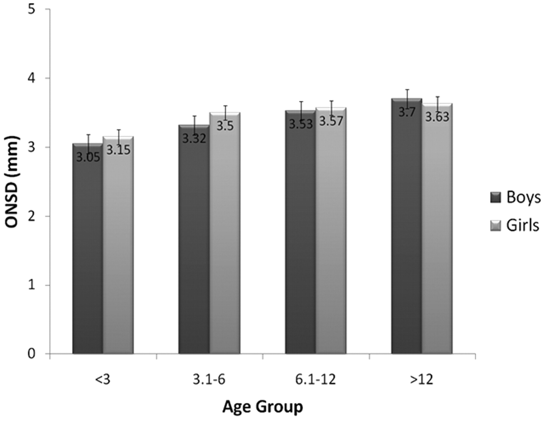
  • Fig 2.
    • Download figure
    • Open in new tab
    • Download powerpoint
    Fig 2.

    Mean ONSD of each age group in our control cohort, stratified by sex. Error bars at the top of each column represent the standard error for that column, ranging from 0.07 to 0.16 mm.

  • Fig 3.
    • Download figure
    • Open in new tab
    • Download powerpoint
    Fig 3.

    Mean ONSD width by age group in controls versus patients with IIH.
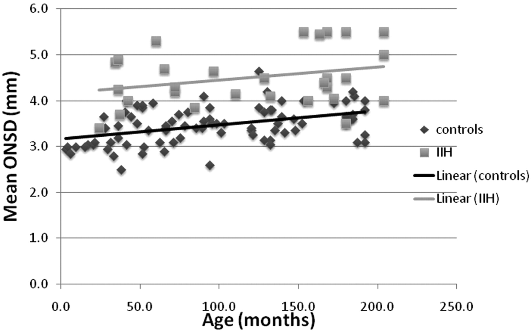
  • Fig 4.
    • Download figure
    • Open in new tab
    • Download powerpoint
    Fig 4.

    Scattergraph and trend lines of the mean ONSD of both controls and patients with IIH.

Tables

  • Figures
    • View popup
    Table 1:

    Revised modified Dandy criteriaa

    Criteria
    1) If symptoms or signs are present, they may only reflect those of generalized intracranial hypertension or papilledema
    2) Documented elevated intracranial pressure measured in the lateral decubitus position
    3) Normal CSF composition
    4) No evidence of hydrocephalus, mass, structural, or vascular lesion on MRI or contrast-enhanced CT for typical patients, and MRI and MR venography for all others
    5) No other cause of intracranial hypertension identified
    • ↵a All of the above criteria should be met in order to make the diagnosis of IIH.

    • View popup
    Table 2:

    Study participant data organized by age group

    ControlIIH
    Total No.8629
    SexM: 41; F: 45M: 11; F: 28
    Age range4 mo-16 yr24 mo-17 yr
    (mean, 8.1 yr; median, 7.7 yr)(mean, 10.1 yr; median, 11.5 yr)
    Group I: 0–3 yr
        No.154
        ONSD (avg)3.1 mm4.35 mm
        SD0.23 mm0.7 mm
        CI (95%)0.12 mm0.69 mm
    Group II: 3–6 yr
        No.206
        ONSD (avg)3.41 mm4.37 mm
        SD0.42 mm0.57 mm
        CI (95%)0.18 mm0.46 mm
    Group III: 6–12 yr
        No.325
        ONSD (avg)3.55 mm4.25 mm
        SD0.38 mm0.32 mm
        CI (95%)0.13 mm0.28 mm
    Group IV: 12–18 yr
        No.1914
        ONSD (avg)3.56 mm4.69 mm
        SD0.32 mm0.7 mm
        CI (95%)0.14 mm0.37 mm
    • Note:—avg indicates average; CI, confidence interval.

PreviousNext
Back to top

In this issue

American Journal of Neuroradiology: 33 (2)
American Journal of Neuroradiology
Vol. 33, Issue 2
1 Feb 2012
  • Table of Contents
  • Index by author
Advertisement
Print
Download PDF
Email Article

Thank you for your interest in spreading the word on American Journal of Neuroradiology.

NOTE: We only request your email address so that the person you are recommending the page to knows that you wanted them to see it, and that it is not junk mail. We do not capture any email address.

Enter multiple addresses on separate lines or separate them with commas.
Optic Nerve Sheath Diameter on MR Imaging: Establishment of Norms and Comparison of Pediatric Patients with Idiopathic Intracranial Hypertension with Healthy Controls
(Your Name) has sent you a message from American Journal of Neuroradiology
(Your Name) thought you would like to see the American Journal of Neuroradiology web site.
CAPTCHA
This question is for testing whether or not you are a human visitor and to prevent automated spam submissions.
Cite this article
B. Shofty, L. Ben-Sira, S. Constantini, S. Freedman, A. Kesler
Optic Nerve Sheath Diameter on MR Imaging: Establishment of Norms and Comparison of Pediatric Patients with Idiopathic Intracranial Hypertension with Healthy Controls
American Journal of Neuroradiology Feb 2012, 33 (2) 366-369; DOI: 10.3174/ajnr.A2779

Citation Manager Formats

  • BibTeX
  • Bookends
  • EasyBib
  • EndNote (tagged)
  • EndNote 8 (xml)
  • Medlars
  • Mendeley
  • Papers
  • RefWorks Tagged
  • Ref Manager
  • RIS
  • Zotero
0 Responses
Respond to this article
Share
Bookmark this article
Optic Nerve Sheath Diameter on MR Imaging: Establishment of Norms and Comparison of Pediatric Patients with Idiopathic Intracranial Hypertension with Healthy Controls
B. Shofty, L. Ben-Sira, S. Constantini, S. Freedman, A. Kesler
American Journal of Neuroradiology Feb 2012, 33 (2) 366-369; DOI: 10.3174/ajnr.A2779
del.icio.us logo Twitter logo Facebook logo Mendeley logo
  • Tweet Widget
  • Facebook Like
  • Google Plus One
Purchase

Jump to section

  • Article
    • Abstract
    • ABBREVIATIONS:
    • Materials and Methods
    • Results
    • Discussion
    • Conclusions
    • Acknowledgments
    • Footnotes
    • References
  • Figures & Data
  • Info & Metrics
  • Responses
  • References
  • PDF

Related Articles

  • No related articles found.
  • PubMed
  • Google Scholar

Cited By...

  • The utility of MRI radiological biomarkers in determining intracranial pressure
  • Dilated Optic Nerve Sheath in Mucopolysaccharidosis I: Common and Not Necessarily High Intracranial Pressure
  • Brain MRI and Ophthalmic Biomarkers of Intracranial Pressure
  • Review of non-invasive intracranial pressure measurement techniques for ophthalmology applications
  • Optic Nerve Measurement on MRI in the Pediatric Population: Normative Values and Correlations
  • Fifteen-minute consultation: the child with idiopathic intracranial hypertension
  • Update on the pathophysiology and management of idiopathic intracranial hypertension
  • Crossref (84)
  • Google Scholar

This article has been cited by the following articles in journals that are participating in Crossref Cited-by Linking.

  • Brain Imaging in Idiopathic Intracranial Hypertension
    Samuel Bidot, Amit M. Saindane, Jason H. Peragallo, Beau B. Bruce, Nancy J. Newman, Valérie Biousse
    Journal of Neuro-Ophthalmology 2015 35 4
  • Systematic review and meta-analysis of MRI signs for diagnosis of idiopathic intracranial hypertension
    Robert M. Kwee, Thomas C. Kwee
    European Journal of Radiology 2019 116
  • Noninvasive methods of detecting increased intracranial pressure
    Wen Xu, Patrick Gerety, Tomas Aleman, Jordan Swanson, Jesse Taylor
    Child's Nervous System 2016 32 8
  • MR imaging findings in children with pseudotumor cerebri and comparison with healthy controls
    Süreyya Burcu Görkem, Selim Doğanay, Mehmet Canpolat, Gonca Koc, Mehmet S. Dogan, Hüseyin Per, Abdülhakim Coşkun
    Child's Nervous System 2015 31 3
  • Measurement of the Optic Nerve Sheath Diameter with Magnetic Resonance Imaging and Its Association with Eyeball Diameter in Healthy Adults
    Dong Hwan Kim, Jin-Sun Jun, Ryul Kim
    Journal of Clinical Neurology 2018 14 3
  • High resolution transbulbar sonography in children with suspicion of increased intracranial pressure
    Marc Steinborn, Melanie Friedmann, Christine Makowski, Helmut Hahn, Alexander Hapfelmeier, Hendrik Juenger
    Child's Nervous System 2016 32 4
  • Magnetic Resonance Imaging Findings of Elevated Intracranial Pressure in Cerebral Venous Thrombosis Versus Idiopathic Intracranial Hypertension with Transverse Sinus Stenosis
    Maysa A. Ridha, Amit M. Saindane, Beau B. Bruce, Bryan D. Riggeal, Linda P. Kelly, Nancy J. Newman, Valerie Biousse
    Neuro-Ophthalmology 2013 37 1
  • Ultrasonographic optic nerve sheath diameter to detect increased intracranial pressure in adults: a meta-analysis
    Sung-Eun Kim, Eun Pyo Hong, Heung Cheol Kim, Si Un Lee, Jin Pyeong Jeon
    Acta Radiologica 2019 60 2
  • Pediatric Pseudotumor Cerebri Syndrome
    Paul H. Phillips, Claire A. Sheldon
    Journal of Neuro-Ophthalmology 2017 37 1
  • Bedside Optic Nerve Sheath Diameter Assessment in the Identification of Increased Intracranial Pressure in Suspected Idiopathic Intracranial Hypertension
    Jose E. Irazuzta, Martha E. Brown, Javed Akhtar
    Pediatric Neurology 2016 54

More in this TOC Section

  • SyMRI & MR Fingerprinting in Brainstem Myelination
  • Comparison of Image Quality and Radiation Dose in Pediatric Temporal Bone CT Using Photon-Counting Detector CT and Energy-Integrating Detector CT
  • Dandy-Walker Phenotype with Brainstem Involvement
Show more Pediatrics

Similar Articles

Advertisement

Indexed Content

  • Current Issue
  • Accepted Manuscripts
  • Article Preview
  • Past Issues
  • Editorials
  • Editor's Choice
  • Fellows' Journal Club
  • Letters to the Editor
  • Video Articles

Cases

  • Case Collection
  • Archive - Case of the Week
  • Archive - Case of the Month
  • Archive - Classic Case

More from AJNR

  • Trainee Corner
  • Imaging Protocols
  • MRI Safety Corner
  • Book Reviews

Multimedia

  • AJNR Podcasts
  • AJNR Scantastics

Resources

  • Turnaround Time
  • Submit a Manuscript
  • Submit a Video Article
  • Submit an eLetter to the Editor/Response
  • Manuscript Submission Guidelines
  • Statistical Tips
  • Fast Publishing of Accepted Manuscripts
  • Graphical Abstract Preparation
  • Imaging Protocol Submission
  • Evidence-Based Medicine Level Guide
  • Publishing Checklists
  • Author Policies
  • Become a Reviewer/Academy of Reviewers
  • News and Updates

About Us

  • About AJNR
  • Editorial Board
  • Editorial Board Alumni
  • Alerts
  • Permissions
  • Not an AJNR Subscriber? Join Now
  • Advertise with Us
  • Librarian Resources
  • Feedback
  • Terms and Conditions
  • AJNR Editorial Board Alumni

American Society of Neuroradiology

  • Not an ASNR Member? Join Now

© 2025 by the American Society of Neuroradiology All rights, including for text and data mining, AI training, and similar technologies, are reserved.
Print ISSN: 0195-6108 Online ISSN: 1936-959X

Powered by HighWire