Advertisement

Main menu

  • Home
  • Content
    • Current Issue
    • Accepted Manuscripts
    • Article Preview
    • Past Issue Archive
    • Video Articles
    • AJNR Case Collection
    • Case of the Week Archive
    • Case of the Month Archive
    • Classic Case Archive
  • Special Collections
    • AJNR Awards
    • Low-Field MRI
    • Alzheimer Disease
    • ASNR Foundation Special Collection
    • Photon-Counting CT
    • View All
  • Multimedia
    • AJNR Podcasts
    • AJNR SCANtastic
    • Trainee Corner
    • MRI Safety Corner
    • Imaging Protocols
  • For Authors
    • Submit a Manuscript
    • Submit a Video Article
    • Submit an eLetter to the Editor/Response
    • Manuscript Submission Guidelines
    • Statistical Tips
    • Fast Publishing of Accepted Manuscripts
    • Graphical Abstract Preparation
    • Imaging Protocol Submission
    • Author Policies
  • About Us
    • About AJNR
    • Editorial Board
    • Editorial Board Alumni
  • More
    • Become a Reviewer/Academy of Reviewers
    • Subscribers
    • Permissions
    • Alerts
    • Feedback
    • Advertisers
    • ASNR Home

User menu

  • Alerts
  • Log in

Search

  • Advanced search
American Journal of Neuroradiology

American Journal of Neuroradiology

ASHNR American Society of Functional Neuroradiology ASHNR American Society of Pediatric Neuroradiology ASSR
  • Alerts
  • Log in

Advanced Search

  • Home
  • Content
    • Current Issue
    • Accepted Manuscripts
    • Article Preview
    • Past Issue Archive
    • Video Articles
    • AJNR Case Collection
    • Case of the Week Archive
    • Case of the Month Archive
    • Classic Case Archive
  • Special Collections
    • AJNR Awards
    • Low-Field MRI
    • Alzheimer Disease
    • ASNR Foundation Special Collection
    • Photon-Counting CT
    • View All
  • Multimedia
    • AJNR Podcasts
    • AJNR SCANtastic
    • Trainee Corner
    • MRI Safety Corner
    • Imaging Protocols
  • For Authors
    • Submit a Manuscript
    • Submit a Video Article
    • Submit an eLetter to the Editor/Response
    • Manuscript Submission Guidelines
    • Statistical Tips
    • Fast Publishing of Accepted Manuscripts
    • Graphical Abstract Preparation
    • Imaging Protocol Submission
    • Author Policies
  • About Us
    • About AJNR
    • Editorial Board
    • Editorial Board Alumni
  • More
    • Become a Reviewer/Academy of Reviewers
    • Subscribers
    • Permissions
    • Alerts
    • Feedback
    • Advertisers
    • ASNR Home
  • Follow AJNR on Twitter
  • Visit AJNR on Facebook
  • Follow AJNR on Instagram
  • Join AJNR on LinkedIn
  • RSS Feeds

AJNR Awards, New Junior Editors, and more. Read the latest AJNR updates

Research ArticleBrain
Open Access

Significant Volume Reduction and Shape Abnormalities of the Basal Ganglia in Cases of Chronic Liver Cirrhosis

W.-C. Lin, K.-H. Chou, C.-L. Chen, C.-H. Chen, H.-L. Chen, J.A. Feekes, N.-W. Hsu, S.-H. Li, Y.-F. Cheng and C.-P. Lin
American Journal of Neuroradiology February 2012, 33 (2) 239-245; DOI: https://doi.org/10.3174/ajnr.A2769
W.-C. Lin
  • Find this author on Google Scholar
  • Find this author on PubMed
  • Search for this author on this site
K.-H. Chou
  • Find this author on Google Scholar
  • Find this author on PubMed
  • Search for this author on this site
C.-L. Chen
  • Find this author on Google Scholar
  • Find this author on PubMed
  • Search for this author on this site
C.-H. Chen
  • Find this author on Google Scholar
  • Find this author on PubMed
  • Search for this author on this site
H.-L. Chen
  • Find this author on Google Scholar
  • Find this author on PubMed
  • Search for this author on this site
J.A. Feekes
  • Find this author on Google Scholar
  • Find this author on PubMed
  • Search for this author on this site
N.-W. Hsu
  • Find this author on Google Scholar
  • Find this author on PubMed
  • Search for this author on this site
S.-H. Li
  • Find this author on Google Scholar
  • Find this author on PubMed
  • Search for this author on this site
Y.-F. Cheng
  • Find this author on Google Scholar
  • Find this author on PubMed
  • Search for this author on this site
C.-P. Lin
  • Find this author on Google Scholar
  • Find this author on PubMed
  • Search for this author on this site
  • Article
  • Figures & Data
  • Supplemental
  • Info & Metrics
  • Responses
  • References
  • PDF
Loading

Article Figures & Data

Figures

  • Tables
  • Fig 1.
    • Download figure
    • Open in new tab
    • Download powerpoint
    Fig 1.

    FIRST segmentation of the subcortical nuclei in a patient with liver cirrhosis. Volume estimation for 7 bilateral subcortical nuclei, including the nucleus accumbens, amygdala, caudate, hippocampus, pallidum, putamen, and thalamus.

  • Fig 2.
    • Download figure
    • Open in new tab
    • Download powerpoint
    Fig 2.

    Statistical 3D maps revealing putaminal volume reduction in Child B and C groups versus controls, with P values color-coded at each surface point (FDR, P < .05 corrected for multiple comparisons). Red areas indicate significant volume reduction (P < .005); blue areas indicate little or no volume reduction (P > .05). R indicates right; L, left; A, anterior.

  • Fig 3.
    • Download figure
    • Open in new tab
    • Download powerpoint
    Fig 3.

    Statistical 3D maps revealing significant caudate nucleus volume reduction in the left lateral aspect in the Child C group versus controls. R indicates right; L, left; A, anterior.

  • Fig 4.
    • Download figure
    • Open in new tab
    • Download powerpoint
    Fig 4.

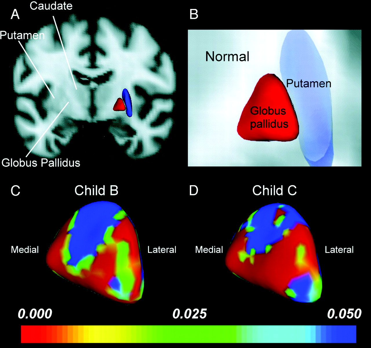
    Shape analysis of the GP. A and B, Anterior coronal views display the relative anatomic location of the putamen and GP in a healthy control. C and D, Statistical 3D maps display right pallidal volume changes with P values color-coded at each surface point in the Child B (C) and Child C groups (D). Note that the GP volume decrease in patients with cirrhosis, while not directly measured, is indicated by the location of the shape change. Medial expansion and lateral compression of the GP was found to be proportional to the severity of liver cirrhosis and further suggested counterbalancing roles between the medial and lateral GP physiologically.

  • Fig 5.
    • Download figure
    • Open in new tab
    • Download powerpoint
    Fig 5.

    The relationship between total CASI scores and volume of the bilateral putamina.

Tables

  • Figures
    • View popup
    Table 1:

    Demographic and clinical characteristics among patients with Child B and Child C liver cirrhosis and controls

    GroupControlChild BChild CF or χ2P Value
    No. of subjects281612
    Age (yr)54.21 ± 10.8552.75 ± 7.8056.17 ± 11.470.383.684
    Sex7 F/21 M3 F/13 M4 F/8 M0.778.678
    Education (yr)11.75 ± 4.3410.75 ± 3.538.83 ± 3.192.350.105
    Type of hepatitis12 (B)/4 (C)9 (B)/3 (C)0.761.672
    TIV (cm3)1521.86 ± 129.581528.18 ± 164.671461.36 ± 182.170.272.763
    GM (cm3)658.27 ± 60.28652.71 ± 55.62626.14 ± 85.140.310.735
    WM (cm3)467.61 ± 52.68471.96 ± 73.61426.86 ± 57.581.222.303
    Creatinine (mg/dL)–0.82 ± 0.300.70 ± 0.271.148.294
    Aspartate aminotransferase (IU/L)–88.70 ± 32.00105.57 ± 31.460.491.490
    Bilirubin (mg/dL)–1.34 ± 0.874.63 ± 2.0128.133<.001a
    Albumin (mg/dL)–3.56 ± 0.572.75 ± 0.3813.045.001a
    INR–1.14 ± 0.131.37 ± 0.2112.408.002a
    Venous ammonia (mg/dL)–103.25 ± 37.51147.08 ± 102.022.526.124
    Pallidal index0.90 ± 0.051.03 ± 0.961.04 ± 0.8224.406<.001a
    CASI score (0–100)97.39 ± 4.9982.27 ± 25.0971.00 ± 23.867.268.002a
    • Note:–indicates not available.

    • ↵a Indicates threshold for statistical significance was set at P < 0.05.

    • View popup
    Table 2:

    Mean subcortical nuclei volume in cubic centimeters per diagnosis: MANCOVA analysis applied for estimating group differencesa

    AnatomyABCMANCOVAPost Hoc ComparisonCohen D
    ControlChild BChild C
    Mean Volume (SD) (cm3)FP ValueA vs BB vs CA vs C
    L accumbens0.39(0.11)0.35(0.09)0.30(0.07)2.64.080.420.691.01
    L amygdala1.12(0.19)1.02(0.20)0.94(0.23)3.12.05bA > C; A, B (NS); B, C (NS)0.510.390.87
    L caudate3.09(0.35)2.92(0.48)2.70(0.41)3.53.04bA > C; A, B (NS); B, C (NS)0.390.491.01
    L hippocampus3.61(0.44)3.66(0.41)3.51(0.72)0.11.90−0.130.270.17
    L pallidus1.78(0.28)1.60(0.48)1.57(0.56)1.21.310.470.040.47
    L putamen4.77(0.54)4.33(0.87)3.81(0.75)7.17<.01bA > B; A > C; B, C (NS)0.600.641.47
    L thalamus7.24(0.62)7.25(0.56)6.84(0.63)0.78.46−0.020.690.64
    R accumbens0.29(0.09)0.26(0.12)0.22(0.09)1.03.360.300.320.71
    R amygdala1.08(0.22)0.95(0.25)0.93(0.20)2.59.090.530.100.70
    R caudate3.22(0.40)3.20(0.52)2.97(0.42)0.76.470.040.490.61
    R hippocampus3.77(0.53)3.85(0.58)3.75(0.38)0.06.94−0.140.200.04
    R pallidus1.78(0.30)1.63(0.44)1.56(0.46)1.13.330.380.170.57
    R putamen4.58(0.51)4.24(0.81)3.96(0.73)3.71.03bA > B; A > C; B, C (NS)0.510.370.99
    R thalamus6.99(0.61)7.07(0.54)6.74(0.70)0.12.89−0.140.530.38
    • Note:—L indicates left; R, right; NS, no significance.

    • ↵a Adjusted for age, sex, education years, and TIV.

    • ↵b Indicates threshold for statistical significance was set at P < 0.05.

PreviousNext
Back to top

In this issue

American Journal of Neuroradiology: 33 (2)
American Journal of Neuroradiology
Vol. 33, Issue 2
1 Feb 2012
  • Table of Contents
  • Index by author
Advertisement
Print
Download PDF
Email Article

Thank you for your interest in spreading the word on American Journal of Neuroradiology.

NOTE: We only request your email address so that the person you are recommending the page to knows that you wanted them to see it, and that it is not junk mail. We do not capture any email address.

Enter multiple addresses on separate lines or separate them with commas.
Significant Volume Reduction and Shape Abnormalities of the Basal Ganglia in Cases of Chronic Liver Cirrhosis
(Your Name) has sent you a message from American Journal of Neuroradiology
(Your Name) thought you would like to see the American Journal of Neuroradiology web site.
CAPTCHA
This question is for testing whether or not you are a human visitor and to prevent automated spam submissions.
Cite this article
W.-C. Lin, K.-H. Chou, C.-L. Chen, C.-H. Chen, H.-L. Chen, J.A. Feekes, N.-W. Hsu, S.-H. Li, Y.-F. Cheng, C.-P. Lin
Significant Volume Reduction and Shape Abnormalities of the Basal Ganglia in Cases of Chronic Liver Cirrhosis
American Journal of Neuroradiology Feb 2012, 33 (2) 239-245; DOI: 10.3174/ajnr.A2769

Citation Manager Formats

  • BibTeX
  • Bookends
  • EasyBib
  • EndNote (tagged)
  • EndNote 8 (xml)
  • Medlars
  • Mendeley
  • Papers
  • RefWorks Tagged
  • Ref Manager
  • RIS
  • Zotero
0 Responses
Respond to this article
Share
Bookmark this article
Significant Volume Reduction and Shape Abnormalities of the Basal Ganglia in Cases of Chronic Liver Cirrhosis
W.-C. Lin, K.-H. Chou, C.-L. Chen, C.-H. Chen, H.-L. Chen, J.A. Feekes, N.-W. Hsu, S.-H. Li, Y.-F. Cheng, C.-P. Lin
American Journal of Neuroradiology Feb 2012, 33 (2) 239-245; DOI: 10.3174/ajnr.A2769
del.icio.us logo Twitter logo Facebook logo Mendeley logo
  • Tweet Widget
  • Facebook Like
  • Google Plus One
Purchase

Jump to section

  • Article
    • Abstract
    • ABBREVIATIONS:
    • Materials and Methods
    • Results
    • Discussion
    • Conclusions
    • Acknowledgments
    • Footnotes
    • References
  • Figures & Data
  • Supplemental
  • Info & Metrics
  • Responses
  • References
  • PDF

Related Articles

  • PubMed
  • Google Scholar

Cited By...

  • No citing articles found.
  • Crossref (14)
  • Google Scholar

This article has been cited by the following articles in journals that are participating in Crossref Cited-by Linking.

  • Brain Edema in Chronic Hepatic Encephalopathy
    Cristina Cudalbu, Simon D. Taylor-Robinson
    Journal of Clinical and Experimental Hepatology 2019 9 3
  • Connectivity of Default-Mode Network Is Associated with Cerebral Edema in Hepatic Encephalopathy
    Wei-Che Lin, Tun-Wei Hsu, Chao-Long Chen, Changwei W. Wu, Cheng-Hsien Lu, Hsiu-Ling Chen, Shau-Hsuan Li, Pin-Yang Yeh, Yu-Fan Cheng, Ching-Po Lin, Christopher P. Hess
    PLoS ONE 2012 7 5
  • Longitudinal Brain White Matter Alterations in Minimal Hepatic Encephalopathy before and after Liver Transplantation
    Wei-Che Lin, Kun-Hsien Chou, Chao-Long Chen, Hsiu-Ling Chen, Cheng-Hsien Lu, Shau-Hsuan Li, Chu-Chung Huang, Ching-Po Lin, Yu-Fan Cheng, Elizabeth J. Coulson
    PLoS ONE 2014 9 8
  • Brain Microstructural Abnormalities in Patients With Cirrhosis Without Overt Hepatic Encephalopathy: A Voxel-Based Diffusion Kurtosis Imaging Study
    Hua-Jun Chen, Peng-Fei Liu, Qiu-Feng Chen, Hai-Bin Shi
    American Journal of Roentgenology 2017 209 5
  • Reestablishing Brain Networks in Patients without Overt Hepatic Encephalopathy after Liver Transplantation
    Wei-Che Lin, Tun-Wei Hsu, Chao-Long Chen, Cheng-Hsien Lu, Hsiu-Ling Chen, Yu-Fan Cheng, Ching-Po Lin
    Journal of Cerebral Blood Flow & Metabolism 2014 34 12
  • Role of inflammation in alcohol-related brain abnormalities: a translational study
    Anastasia Lanquetin, Sophie Leclercq, Philippe de Timary, Shailendra Segobin, Mikaël Naveau, Laurent Coulbault, Paola Maccioni, Irene Lorrai, Giancarlo Colombo, Denis Vivien, Marina Rubio, Anne-Lise Pitel
    Brain Communications 2021 3 3
  • Altered metal metabolism in patients with HCV-related cirrhosis and hepatic encephalopathy
    Massimo Marano, Umberto Vespasiani Gentilucci, Claudia Altamura, Mariacristina Siotto, Rosanna Squitti, Serena Bucossi, Livia Quintiliani, Simone Migliore, Federico Greco, Laura Scarciolla, Carlo Cosimo Quattrocchi, Antonio Picardi, Fabrizio Vernieri
    Metabolic Brain Disease 2015 30 6
  • Reduced Cortical Complexity in Cirrhotic Patients with Minimal Hepatic Encephalopathy
    Qiu-Feng Chen, Xiao-Hong Zhang, Tian-Xiu Zou, Nao-Xin Huang, Hua-Jun Chen
    Neural Plasticity 2020 2020
  • Diabetes Mellitus is Associated with More Severe Brain Spontaneous Activity Impairment and Gray Matter Loss in Patients with Cirrhosis
    Yun Fei Wang, Xiang Kong, Guang Ming Lu, Long Jiang Zhang
    Scientific Reports 2017 7 1
  • Predictive models of minimal hepatic encephalopathy for cirrhotic patients based on large-scale brain intrinsic connectivity networks
    Yun Jiao, Xun-Heng Wang, Rong Chen, Tian-Yu Tang, Xi-Qi Zhu, Gao-Jun Teng
    Scientific Reports 2017 7 1

More in this TOC Section

  • Predictors of Reperfusion in Patients with Acute Ischemic Stroke
  • Enhanced Axonal Metabolism during Early Natalizumab Treatment in Relapsing-Remitting Multiple Sclerosis
  • Progression of Microstructural Damage in Spinocerebellar Ataxia Type 2: A Longitudinal DTI Study
Show more BRAIN

Similar Articles

Advertisement

Indexed Content

  • Current Issue
  • Accepted Manuscripts
  • Article Preview
  • Past Issues
  • Editorials
  • Editor's Choice
  • Fellows' Journal Club
  • Letters to the Editor
  • Video Articles

Cases

  • Case Collection
  • Archive - Case of the Week
  • Archive - Case of the Month
  • Archive - Classic Case

More from AJNR

  • Trainee Corner
  • Imaging Protocols
  • MRI Safety Corner
  • Book Reviews

Multimedia

  • AJNR Podcasts
  • AJNR Scantastics

Resources

  • Turnaround Time
  • Submit a Manuscript
  • Submit a Video Article
  • Submit an eLetter to the Editor/Response
  • Manuscript Submission Guidelines
  • Statistical Tips
  • Fast Publishing of Accepted Manuscripts
  • Graphical Abstract Preparation
  • Imaging Protocol Submission
  • Evidence-Based Medicine Level Guide
  • Publishing Checklists
  • Author Policies
  • Become a Reviewer/Academy of Reviewers
  • News and Updates

About Us

  • About AJNR
  • Editorial Board
  • Editorial Board Alumni
  • Alerts
  • Permissions
  • Not an AJNR Subscriber? Join Now
  • Advertise with Us
  • Librarian Resources
  • Feedback
  • Terms and Conditions
  • AJNR Editorial Board Alumni

American Society of Neuroradiology

  • Not an ASNR Member? Join Now

© 2025 by the American Society of Neuroradiology All rights, including for text and data mining, AI training, and similar technologies, are reserved.
Print ISSN: 0195-6108 Online ISSN: 1936-959X

Powered by HighWire