Advertisement

Main menu

  • Home
  • Content
    • Current Issue
    • Accepted Manuscripts
    • Article Preview
    • Past Issue Archive
    • Video Articles
    • AJNR Case Collection
    • Case of the Week Archive
    • Case of the Month Archive
    • Classic Case Archive
  • Special Collections
    • AJNR Awards
    • Low-Field MRI
    • Alzheimer Disease
    • ASNR Foundation Special Collection
    • Photon-Counting CT
    • View All
  • Multimedia
    • AJNR Podcasts
    • AJNR SCANtastic
    • Trainee Corner
    • MRI Safety Corner
    • Imaging Protocols
  • For Authors
    • Submit a Manuscript
    • Submit a Video Article
    • Submit an eLetter to the Editor/Response
    • Manuscript Submission Guidelines
    • Statistical Tips
    • Fast Publishing of Accepted Manuscripts
    • Graphical Abstract Preparation
    • Imaging Protocol Submission
    • Author Policies
  • About Us
    • About AJNR
    • Editorial Board
    • Editorial Board Alumni
  • More
    • Become a Reviewer/Academy of Reviewers
    • Subscribers
    • Permissions
    • Alerts
    • Feedback
    • Advertisers
    • ASNR Home

User menu

  • Alerts
  • Log in

Search

  • Advanced search
American Journal of Neuroradiology

American Journal of Neuroradiology

ASHNR American Society of Functional Neuroradiology ASHNR American Society of Pediatric Neuroradiology ASSR
  • Alerts
  • Log in

Advanced Search

  • Home
  • Content
    • Current Issue
    • Accepted Manuscripts
    • Article Preview
    • Past Issue Archive
    • Video Articles
    • AJNR Case Collection
    • Case of the Week Archive
    • Case of the Month Archive
    • Classic Case Archive
  • Special Collections
    • AJNR Awards
    • Low-Field MRI
    • Alzheimer Disease
    • ASNR Foundation Special Collection
    • Photon-Counting CT
    • View All
  • Multimedia
    • AJNR Podcasts
    • AJNR SCANtastic
    • Trainee Corner
    • MRI Safety Corner
    • Imaging Protocols
  • For Authors
    • Submit a Manuscript
    • Submit a Video Article
    • Submit an eLetter to the Editor/Response
    • Manuscript Submission Guidelines
    • Statistical Tips
    • Fast Publishing of Accepted Manuscripts
    • Graphical Abstract Preparation
    • Imaging Protocol Submission
    • Author Policies
  • About Us
    • About AJNR
    • Editorial Board
    • Editorial Board Alumni
  • More
    • Become a Reviewer/Academy of Reviewers
    • Subscribers
    • Permissions
    • Alerts
    • Feedback
    • Advertisers
    • ASNR Home
  • Follow AJNR on Twitter
  • Visit AJNR on Facebook
  • Follow AJNR on Instagram
  • Join AJNR on LinkedIn
  • RSS Feeds

AJNR Awards, New Junior Editors, and more. Read the latest AJNR updates

Research ArticlePatient Safety

Iterative Reconstruction in Head CT: Image Quality of Routine and Low-Dose Protocols in Comparison with Standard Filtered Back-Projection

A. Korn, M. Fenchel, B. Bender, S. Danz, T.K. Hauser, D. Ketelsen, T. Flohr, C.D. Claussen, M. Heuschmid, U. Ernemann and H. Brodoefel
American Journal of Neuroradiology February 2012, 33 (2) 218-224; DOI: https://doi.org/10.3174/ajnr.A2749
A. Korn
  • Find this author on Google Scholar
  • Find this author on PubMed
  • Search for this author on this site
M. Fenchel
  • Find this author on Google Scholar
  • Find this author on PubMed
  • Search for this author on this site
B. Bender
  • Find this author on Google Scholar
  • Find this author on PubMed
  • Search for this author on this site
S. Danz
  • Find this author on Google Scholar
  • Find this author on PubMed
  • Search for this author on this site
T.K. Hauser
  • Find this author on Google Scholar
  • Find this author on PubMed
  • Search for this author on this site
D. Ketelsen
  • Find this author on Google Scholar
  • Find this author on PubMed
  • Search for this author on this site
T. Flohr
  • Find this author on Google Scholar
  • Find this author on PubMed
  • Search for this author on this site
C.D. Claussen
  • Find this author on Google Scholar
  • Find this author on PubMed
  • Search for this author on this site
M. Heuschmid
  • Find this author on Google Scholar
  • Find this author on PubMed
  • Search for this author on this site
U. Ernemann
  • Find this author on Google Scholar
  • Find this author on PubMed
  • Search for this author on this site
H. Brodoefel
  • Find this author on Google Scholar
  • Find this author on PubMed
  • Search for this author on this site
  • Article
  • Figures & Data
  • Info & Metrics
  • Responses
  • References
  • PDF
Loading

Article Figures & Data

Figures

  • Tables
  • Fig 1.
    • Download figure
    • Open in new tab
    • Download powerpoint
    Fig 1.

    CNR in nonenhanced (A) and contrast-enhanced (B) head CT with the use of various tube currents and reconstruction of data by FBP or IR. In the boxplot diagrams, the line across the middle of the box identifies the median sample value; boxes extend from the 25th to the 75th quartile, and whiskers, down to the lowest and highest values.
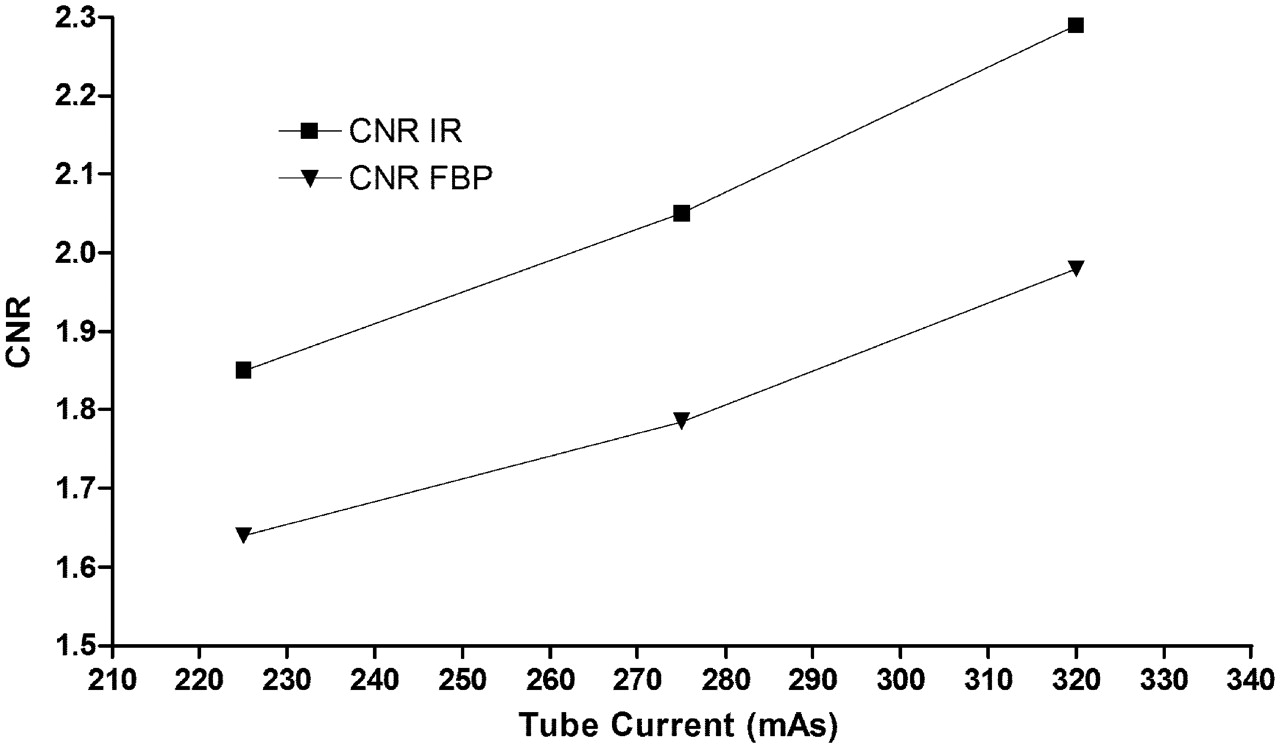
  • Fig 2.
    • Download figure
    • Open in new tab
    • Download powerpoint
    Fig 2.

    Regression plot of CNR against tube current with IR and FBP. According to linear regression equation, the x-intercept is at a tube current of 255 mAs when y is at a standard 1.98 CNR (320-mAs FBP).
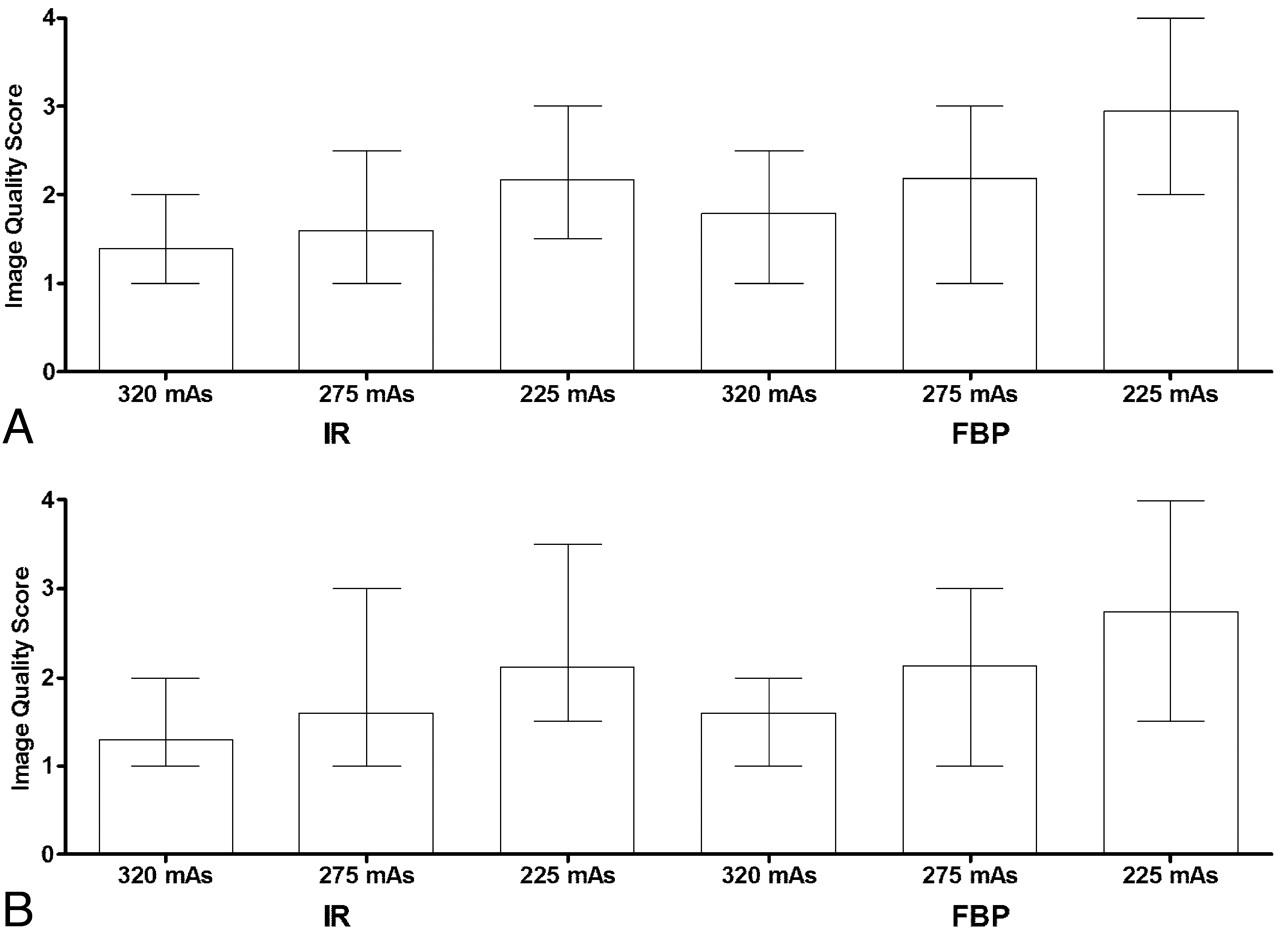
  • Fig 3.
    • Download figure
    • Open in new tab
    • Download powerpoint
    Fig 3.

    Subjective grading of WM-GM differentiation in nonenhanced (A) and contrast-enhanced (B) head CT with use of various tube currents and reconstruction of data by FBP or IR. Data are presented as means and ranges.
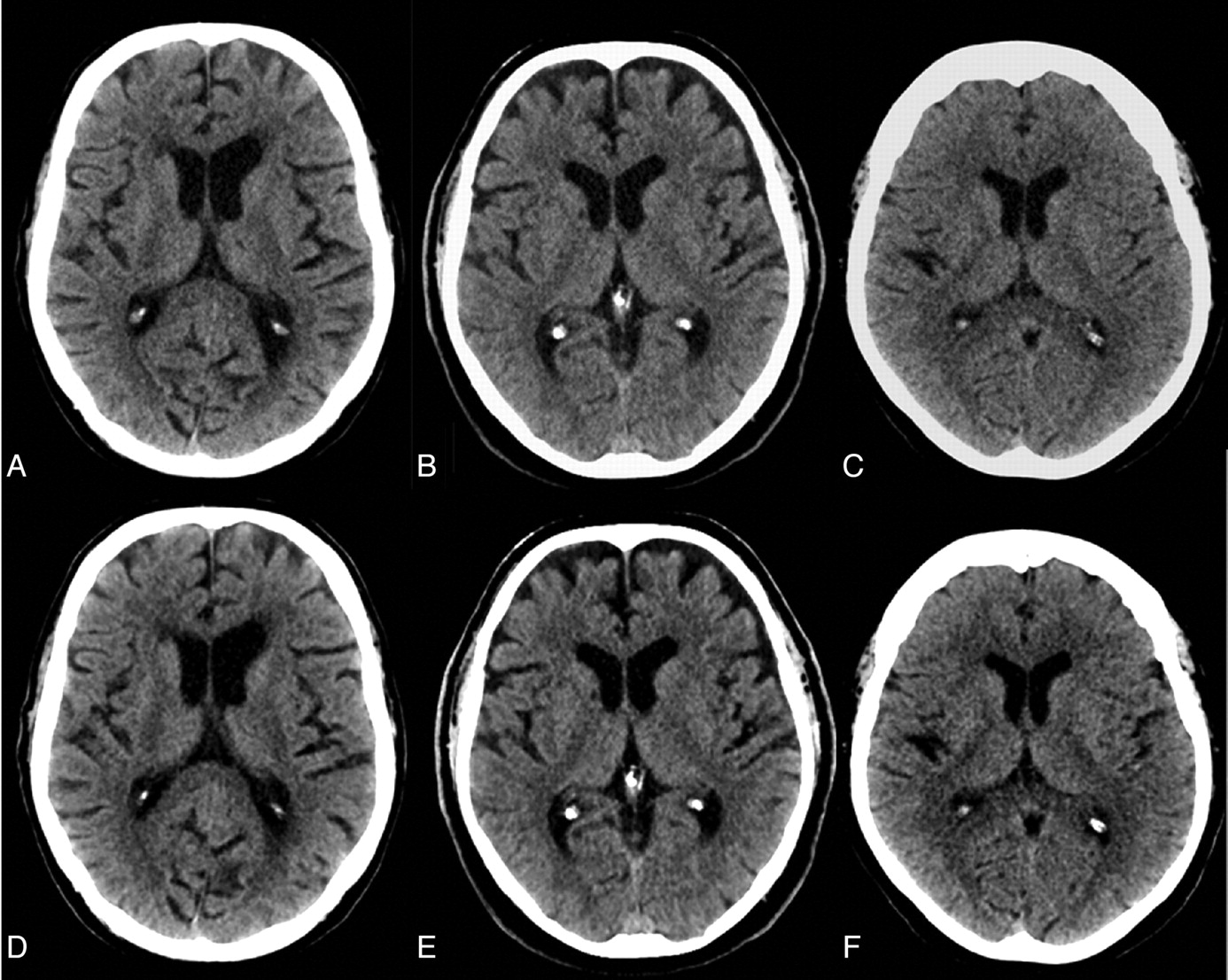
  • Fig 4.
    • Download figure
    • Open in new tab
    • Download powerpoint
    Fig 4.

    Example of image quality at 320 (A and D), 275 (B and E), and 225 (C and F) mAs. At all dose levels, use of IR (D−F) is associated with a considerable reduction of noise and enhancement of image quality. As demonstrated in our study, this is achieved without significant loss of image sharpness. While image quality at 85% dose and IR (E) is similar to the standard of reference (A), a 30% dose reduction results in substantial increase of noise despite using IR (F).

Tables

  • Figures
    • View popup
    Table 1:

    Patient characteristics and radiation dose in CT protocols with various tube currents

    Characteristic320 mAs275 mAs225 mAsP
    Age63 ± 1467 ± 1366 ± 12.54
    Sex (male/female)12/1814/1619/11.17
    CTDIvol (mGy)60.151.842.3—
    DLP (mGy.cm)1043 ± 53890 ± 34733 ± 52<.0001
    Effective dose (mSv)2.2 ± 0.11.8 ± 0.071.5 ± 1.0<.0001
    • View popup
    Table 2:

    Mean and SD of SNR in various CT protocols with either IR or FBPa

    Tube Current 320 mAsTube Current 275 mAsTube Current 225 mAs
    IRFBPIRFBPIRFBP
    Non-Enhanced
        WM8.8 ± 2.17.9 ± 1, P = 0.018.0 ± 1.77.0 ± 1.3, P < .00017.6 ± 1.56.7 ± 1.4, P = .006
        GM10.9 ± 29.4 ± 1.4, P < .00019.8 ± 1.48.3 ± 1.3, P < .00019.7 ± 1.88.4 ± 1.6, P < .0001
        LQ1.4 ± 0.51.2 ± 0.4, P < .00011.3 ± 0.41.0 ± 0.3, P < .00011.2 ± 0.31.0 ± 0.3, P < .0006
        BG−448−302, P < .0001−385−210, P < .0001−295−244, P < .0001
    Contrast-Enhanced
        WM8.9 ± 1.87.6 ± 1.4, P < .00017.9 ± 1.56.8 ± 1.1, P < .00017.8 ± 1.46.5 ± 1.0, P < .0001
        GM11.2 ± 1.710.2 ± 1.6, P = .00110.2 ± 1.79.0 ± 1.4, P = .00089.4 ± 2.18.6 ± 1.7, P = .02
        LQ1.3 ± 0.61.0 ± 0.4, P < .00011.2 ± 0.51.0 ± 0.4, P = 0.0041.1 ± 0.30.9 ± 0.4, P < .001
        BG−385−328, P < .0001−222−192, P < .008−221−191, P < .0001
    • Note: —LQ indicates liquor; BG, background.

    • ↵a Data are shown for WM, GM, LQ, and BG measurements in air outside the skull. P values refer to differences between IR and FBP.

    • View popup
    Table 3:

    Mean and median of qualitative image scores in various CT protocols with either IR or FBPa

    Tube Current 320 mAsTube Current 275 mAsTube Current 225 mAs
    IRFBPIRFBPIRFBP
    Non-Enhanced
        Noise1.2 (1)1.6 (1.5), P = .0031.6 (2)1.9 (1.75), P = .0012.0 (2)2.5 (2.5), P < .0001
        GM/WM1.4 (1)1.8 (2), P = .0021.6 (1.5)2.2 (2.25), P = .0092.2 (2)2.9 (3.0), P < .0001
        SS1.4 (1)1.7 (2), P = .0071.6 (2)2.0 (2), P = .012.1 (2)2.7 (3), P = .0006
        PF1.6 (2)2.0 (2), P = .12.0 (2)2.7 (2.5), P = .0012.8 (3)3.4 (3), P = .0002
        DA1.3 (1)1.7 (2), P = .0021.7 (2)2.2 (2), P = .00052.2 (2)2.8 (2.5), P < .0001
    Contrast-Enhanced
        Noise1.2 (1)1.4 (1.5), P = .021.4 (1)1.8 (1.75), P = .0032.1 (2)2.5 (2.5), P = .0002
        GM/WM1.3 (1)1.6 (2), P = .0021.6 (1.5)2.1 (2), P = .0012.1 (2)2.7 (3), P = .0004
        SS1.3 (1)1.6 (2), P = .0041.5 (1.75)1.9 (2), P = .0082.0 (2)2.6 (3), P < .0001
        PF1.6 (2)2.0 (2), P = .012.2 (2)2.5 (2.5), P = .092.8 (3)3.0 (3), P = .2
        DA1.2 (1)1.8 (2), P = .0071.6 (2)2.0 (2), P = .0082.1 (2)2.6 (2.5), P = .0003
    • Note:—SS indicates subarachnoid space margins; PF, distinctness of posterior fossa contents; DA, overall diagnostic acceptability.

    • ↵a Image-quality grading is provided for noise, GM-WM matter differentiation, sharpness of SS, PF, and overall DA. Median is in parentheses.

PreviousNext
Back to top

In this issue

American Journal of Neuroradiology: 33 (2)
American Journal of Neuroradiology
Vol. 33, Issue 2
1 Feb 2012
  • Table of Contents
  • Index by author
Advertisement
Print
Download PDF
Email Article

Thank you for your interest in spreading the word on American Journal of Neuroradiology.

NOTE: We only request your email address so that the person you are recommending the page to knows that you wanted them to see it, and that it is not junk mail. We do not capture any email address.

Enter multiple addresses on separate lines or separate them with commas.
Iterative Reconstruction in Head CT: Image Quality of Routine and Low-Dose Protocols in Comparison with Standard Filtered Back-Projection
(Your Name) has sent you a message from American Journal of Neuroradiology
(Your Name) thought you would like to see the American Journal of Neuroradiology web site.
CAPTCHA
This question is for testing whether or not you are a human visitor and to prevent automated spam submissions.
Cite this article
A. Korn, M. Fenchel, B. Bender, S. Danz, T.K. Hauser, D. Ketelsen, T. Flohr, C.D. Claussen, M. Heuschmid, U. Ernemann, H. Brodoefel
Iterative Reconstruction in Head CT: Image Quality of Routine and Low-Dose Protocols in Comparison with Standard Filtered Back-Projection
American Journal of Neuroradiology Feb 2012, 33 (2) 218-224; DOI: 10.3174/ajnr.A2749

Citation Manager Formats

  • BibTeX
  • Bookends
  • EasyBib
  • EndNote (tagged)
  • EndNote 8 (xml)
  • Medlars
  • Mendeley
  • Papers
  • RefWorks Tagged
  • Ref Manager
  • RIS
  • Zotero
0 Responses
Respond to this article
Share
Bookmark this article
Iterative Reconstruction in Head CT: Image Quality of Routine and Low-Dose Protocols in Comparison with Standard Filtered Back-Projection
A. Korn, M. Fenchel, B. Bender, S. Danz, T.K. Hauser, D. Ketelsen, T. Flohr, C.D. Claussen, M. Heuschmid, U. Ernemann, H. Brodoefel
American Journal of Neuroradiology Feb 2012, 33 (2) 218-224; DOI: 10.3174/ajnr.A2749
del.icio.us logo Twitter logo Facebook logo Mendeley logo
  • Tweet Widget
  • Facebook Like
  • Google Plus One
Purchase

Jump to section

  • Article
    • Abstract
    • ABBREVIATIONS:
    • Materials and Methods
    • Results
    • Discussion
    • Conclusions
    • Footnotes
    • References
  • Figures & Data
  • Info & Metrics
  • Responses
  • References
  • PDF

Related Articles

  • No related articles found.
  • PubMed
  • Google Scholar

Cited By...

  • Iterative Reconstruction in Dose Reduction of A Head CT Examination and Corresponding Acquisition Parameter Selection
  • Dose Reduction While Preserving Diagnostic Quality in Head CT: Advancing the Application of Iterative Reconstruction Using a Live Animal Model
  • Evaluation of Lower-Dose Spiral Head CT for Detection of Intracranial Findings Causing Neurologic Deficits
  • Comparison of Iterative Model Reconstruction versus Filtered Back-Projection in Pediatric Emergency Head CT: Dose, Image Quality, and Image-Reconstruction Times
  • Full Dose-Reduction Potential of Statistical Iterative Reconstruction for Head CT Protocols in a Predominantly Pediatric Population
  • Advanced Modeled Iterative Reconstruction in Low-Tube-Voltage Contrast-Enhanced Neck CT: Evaluation of Objective and Subjective Image Quality
  • Repeated Head CT in the Neurosurgical Intensive Care Unit: Feasibility of Sinogram-Affirmed Iterative Reconstruction-Based Ultra-Low-Dose CT for Surveillance
  • Acute Intracranial Hemorrhage in CT: Benefits of Sinogram-Affirmed Iterative Reconstruction Techniques
  • Six iterative reconstruction algorithms in brain CT: a phantom study on image quality at different radiation dose levels
  • Can Iterative Reconstruction Improve Imaging Quality for Lower Radiation CT Perfusion? Initial Experience
  • The Use of Adaptive Statistical Iterative Reconstruction in Pediatric Head CT: A Feasibility Study
  • Crossref (104)
  • Google Scholar

This article has been cited by the following articles in journals that are participating in Crossref Cited-by Linking.

  • State of the Art: Iterative CT Reconstruction Techniques
    Lucas L. Geyer, U. Joseph Schoepf, Felix G. Meinel, John W. Nance, Gorka Bastarrika, Jonathon A. Leipsic, Narinder S. Paul, Marco Rengo, Andrea Laghi, Carlo N. De Cecco
    Radiology 2015 276 2
  • Iterative reconstruction techniques for computed tomography Part 1: Technical principles
    Martin J. Willemink, Pim A. de Jong, Tim Leiner, Linda M. de Heer, Rutger A. J. Nievelstein, Ricardo P. J. Budde, Arnold M. R. Schilham
    European Radiology 2013 23 6
  • Iterative reconstruction techniques for computed tomography part 2: initial results in dose reduction and image quality
    Martin J. Willemink, Tim Leiner, Pim A. de Jong, Linda M. de Heer, Rutger A. J. Nievelstein, Arnold M. R. Schilham, Ricardo P. J. Budde
    European Radiology 2013 23 6
  • Advances in Computed Tomography Imaging Technology
    Daniel Thomas Ginat, Rajiv Gupta
    Annual Review of Biomedical Engineering 2014 16 1
  • CT Dose Reduction Applications: Available Tools on the Latest Generation of CT Scanners
    Siva P. Raman, Pamela T. Johnson, Swati Deshmukh, Mahadevappa Mahesh, Katharine L. Grant, Elliot K. Fishman
    Journal of the American College of Radiology 2013 10 1
  • Six iterative reconstruction algorithms in brain CT: a phantom study on image quality at different radiation dose levels
    A Löve, M-L Olsson, R Siemund, F Stålhammar, I M Björkman-Burtscher, M Söderberg
    The British Journal of Radiology 2013 86 1031
  • CT Scan Parameters and Radiation Dose: Practical Advice for Radiologists
    Siva P. Raman, Mahadevappa Mahesh, Robert V. Blasko, Elliot K. Fishman
    Journal of the American College of Radiology 2013 10 11
  • An Improved Index of Image Quality for Task-based Performance of CT Iterative Reconstruction across Three Commercial Implementations
    Olav Christianson, Joseph J. S. Chen, Zhitong Yang, Ganesh Saiprasad, Alden Dima, James J. Filliben, Adele Peskin, Christopher Trimble, Eliot L. Siegel, Ehsan Samei
    Radiology 2015 275 3
  • Lens Dose in Routine Head CT: Comparison of Different Optimization Methods With Anthropomorphic Phantoms
    Ulla Nikupaavo, Touko Kaasalainen, Vappu Reijonen, Sanna-Mari Ahonen, Mika Kortesniemi
    American Journal of Roentgenology 2015 204 1
  • Does Iterative Reconstruction Lower CT Radiation Dose: Evaluation of 15,000 Examinations
    Peter B. Noël, Bernhard Renger, Martin Fiebich, Daniela Münzel, Alexander A. Fingerle, Ernst J. Rummeny, Martin Dobritz, Chin-Tu Chen
    PLoS ONE 2013 8 11

More in this TOC Section

  • Safety of Intrathecal Gadobutrol in Various Doses
  • Impact of Kidney Function on CNS Gadolinium Deposition in Patients Receiving Repeated Doses of Gadobutrol
  • Contrast-Induced Acute Kidney Injury in Radiologic Management of Acute Ischemic Stroke in the Emergency Setting
Show more PATIENT SAFETY

Similar Articles

Advertisement

Indexed Content

  • Current Issue
  • Accepted Manuscripts
  • Article Preview
  • Past Issues
  • Editorials
  • Editor's Choice
  • Fellows' Journal Club
  • Letters to the Editor
  • Video Articles

Cases

  • Case Collection
  • Archive - Case of the Week
  • Archive - Case of the Month
  • Archive - Classic Case

More from AJNR

  • Trainee Corner
  • Imaging Protocols
  • MRI Safety Corner
  • Book Reviews

Multimedia

  • AJNR Podcasts
  • AJNR Scantastics

Resources

  • Turnaround Time
  • Submit a Manuscript
  • Submit a Video Article
  • Submit an eLetter to the Editor/Response
  • Manuscript Submission Guidelines
  • Statistical Tips
  • Fast Publishing of Accepted Manuscripts
  • Graphical Abstract Preparation
  • Imaging Protocol Submission
  • Evidence-Based Medicine Level Guide
  • Publishing Checklists
  • Author Policies
  • Become a Reviewer/Academy of Reviewers
  • News and Updates

About Us

  • About AJNR
  • Editorial Board
  • Editorial Board Alumni
  • Alerts
  • Permissions
  • Not an AJNR Subscriber? Join Now
  • Advertise with Us
  • Librarian Resources
  • Feedback
  • Terms and Conditions
  • AJNR Editorial Board Alumni

American Society of Neuroradiology

  • Not an ASNR Member? Join Now

© 2025 by the American Society of Neuroradiology All rights, including for text and data mining, AI training, and similar technologies, are reserved.
Print ISSN: 0195-6108 Online ISSN: 1936-959X

Powered by HighWire