
Article Figures & Data
Figures
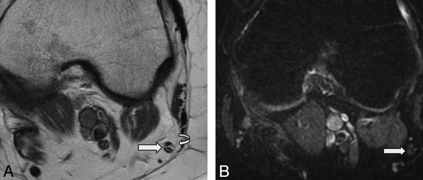
- Fig 1.
MRN appearance of a normal CPN. Axial T1-weighted (A) and axial T2 SPAIR (B) images at the level of popliteal fossa show an example of a normal CPN (straight arrows). Note the fascicular appearance and isointensity of the CPN on T1WI and T2WI with respect to the skeletal muscle. The nerve also appears normal in size and is surrounded by a rim of perineural fat (curved arrow) and thin epineurium.
- Fig 2.
MRN appearance of a normal sciatic nerve. Coronal T2 SPACE image demonstrates a normal sciatic nerve (arrows). Individual fascicles outlined by fat are clearly seen coursing in the craniocaudal direction. Note the iso- to minimal T2 hyperintensity of the normal nerve with respect to the skeletal muscle. The nerve also appears normal in size and is surrounded by a rim of perineural fat.
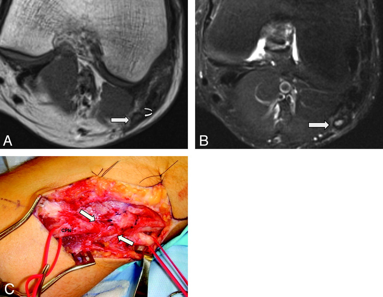
- Fig 3.
MRN appearance of abnormal CPN with axonotmesis. Axial T1WI (A) and axial T2 SPAIR (B) images of a 25-year-old man with clinical findings of left CPN related to a previous injury. The rim of perineural fat is disrupted with fibrosis on T1WI (curved arrow). Note the enlarged and T2 hyperintense CPN (white arrows) with lost fascicular appearance and thickened epineurium (a dark SI rim surrounding the hyperintense CPN). Regional denervation muscle edema and atrophy in the muscles of the extensor compartment were also seen (not shown). C, Intraoperative photograph confirms a thickened epineurium surrounding the CPN and perineural fibrosis (arrows).
- Fig 4.
MRN appearance of a Morton neuroma (interdigital neuroma or perineural fibrosis). A 42-year-old man presented with persistent neuropathic pain after resection of the common digital nerve in the third web space of the foot. A, Short-axis T1WI after IV injection of gadolinium shows focal enhancement in the third webspace, consistent with postoperative changes and a small residual neuroma (arrow). B, Intraoperative photograph confirms the residual neuroma in the third webspace (arrow). C, Short-axis T1WI of the same patient demonstrates an additional 6-mm lesion in the second webspace, representing another clinically unsuspected interdigital neuroma (arrow). D, Intraoperative photograph confirms the additional interdigital neuroma in the third webspace (arrow).
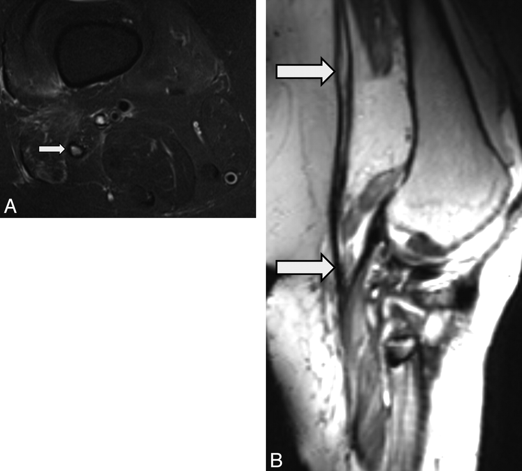
- Fig 5.
MRN appearance of an abnormal neurotomized CPN. A 42-year-old woman presented with progressively worsening foot drop following penetrating trauma, which occurred about 2 months earlier. A, Axial T2 SPAIR image at the level of the popliteal fossa shows a thickened perineurium and abnormal T2 hyperintensity of the peroneal nerve (arrow). Note the disrupted fascicular appearance of the nerve (arrow). Regional denervation muscle edema and atrophy in extensor compartment muscles were also seen (not shown). B, Oblique reconstruction from a 3D T2 SPACE sequence along the axis of the injured CPN demonstrates an enlarged hyperintense CPN with neurotmesis and fibrosis distally.
- Fig 6.
MRN appearance of an abnormal sciatic nerve with neurotmesis. A 50-year-old woman presented with foot drop following left hip-replacement surgery (metallic hardware) performed 6 months earlier. A, Coronal 3D T2 SPACE image shows focal discontinuity of the sciatic nerve (large arrow) with proximal atrophy (small arrow). Neurotmesis of the sciatic nerve was confirmed during surgery, and the nerve was repaired. B, Intraoperative photograph shows the sciatic nerve after repair with a cable sural nerve autograft (arrows), which bridges the torn ends of the sciatic nerve.
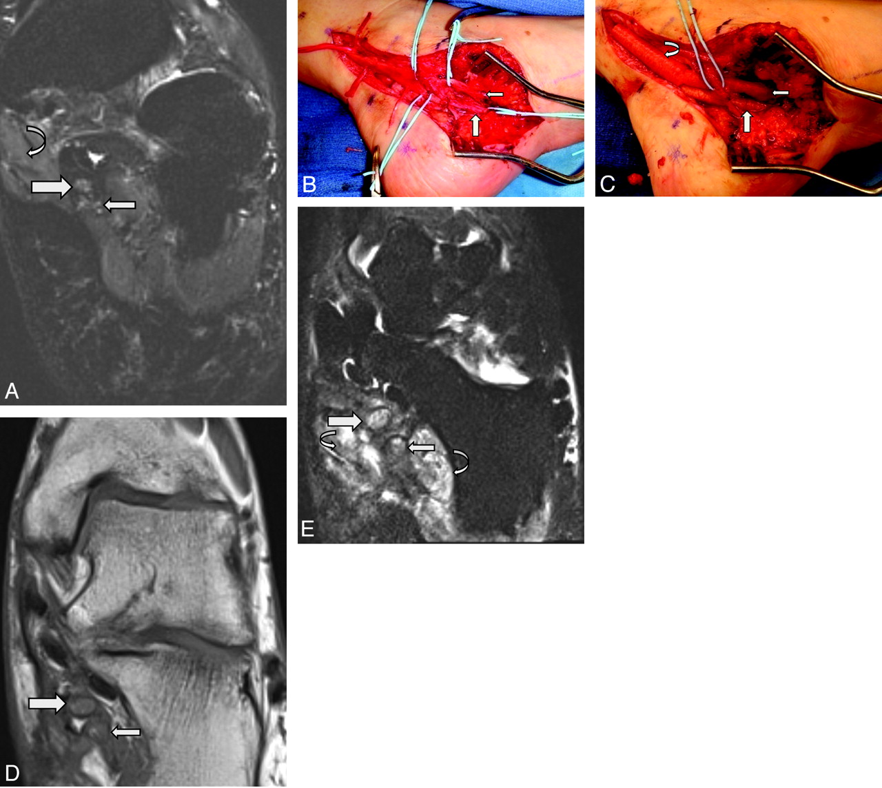
- Fig 7.
MRN appearance of abnormal MPN and LPN in a 32-year-old woman with persisting and worsening heel pain and toe weakness after a tarsal tunnel release surgery. A, Coronal oblique T2 SPAIR image obtained before redo surgery shows abnormally enlarged and T2 hyperintense MPN (larger arrow) and LPN (smaller arrow) in the tarsal tunnel. Note the mild denervation edema of the abductor hallucis (curved arrow). B, Intraoperative photograph during neurolysis confirms the MRN findings of abnormal MPN (larger arrow) and LPN (smaller arrow) in the distal tarsal tunnel. C, Another intraoperative photograph shows both the MPN (larger arrow) and LPN (smaller arrow) as well as the tibial nerve (curved arrow). All 3 nerves were placed in collagen-based nerve wraps. The patient, however, continued to have pain and was referred for MRN about 6 weeks following the redo tarsal release. D, Coronal oblique T1WI demonstrates markedly enlarged MPN (larger arrow) and LPN (smaller arrow). Both nerves are encased in exuberant fibrosis and show loss of the fascicular pattern as well. E, Coronal oblique T2 SPAIR image shows worsening neuropathy of MPN (larger arrow) and LPN (smaller arrow), indicated by further enlargement and increased T2 hyperintensity of both nerves. Worsening muscle denervation edema is also nicely depicted (curved arrows in E). In D and E, previously placed nerve wraps are seen as hypointense structures encircling both the medial and plantar nerves.
- Fig 8.
MRN appearance of an abnormal MPN and LPN in a 48-year-old woman who was referred for MRN due to persistent pain in the foot. She had undergone tarsal tunnel release, neurolysis, nerve tube, and wrap placement. A, Intraoperative photograph shows the MPN in a nerve wrap (larger arrow) and the LPN in a nerve tube (smaller arrow). B, Axial T2 SPAIR image from the MRN obtained 6 months after redo surgery shows abnormal T2 hyperintensity and enlargement of both the MPN (larger arrow) and LPN (smaller arrow). Note the circumscribed rim of T2 hypointensity around the MPN and LPN related to nerve wrap and nerve tube materials. C, Axial T1WI distally shows extensive fibrosis (arrow) below the level of abductor hallucis, not addressed during initial neurolysis, causing persistent distal entrapment of the MPN and LPN. The patient continued to have symptoms and another MRN was performed 11 months after redo surgery. D, Oblique coronal STIR image more distally shows worsening increasing size and hyperintensity of the MPN (larger arrow) and LPN (smaller arrow), compared with prior MRN. Persistent muscle denervation changes were again noted (not shown). E, Intraoperative photograph, about a year from the first redo tarsal tunnel surgery. The patient underwent another tarsal tunnel release surgery, which confirmed MRN findings of encasing perineural fibrosis. Notice the inflamed and hyperemic MPN (larger arrow) and LPN (smaller arrow) following neurolysis. The previously placed nerve wrap and nerve tube are resorbed.
- Fig 9.
MRN appearance of regeneration of a peripheral nerve. A 46-year-old woman with a prior median nerve injury underwent repair and nerve tube placement across median nerve branches. A, Axial STIR image shows fluid-filled nerve tubes (small arrows). B, Axial T1WI obtained after IV administration of gadolinium shows filling defects within the fluid-filled nerve tubes (small arrows). These appear as nonenhancing hypointense structures or filling defects within the tubes, which were confirmed as hypertrophied nerve sprouts that failed to unite at surgery.


Tables
Typical protocol for 3T MR imaging sequences used for MRN of the sciatic nervea
Sequence FOV (cm) In-Plane Resolution (mm) TR/TE (ms) Turbo Factor Coronal T1 TSE 30–40 4 780/10 3 Coronal T2 3D SPACE 30–40 1 1600/128 151 Coronal STIR 3D SPACE 30–40 1 1500/91 41 Axial T2 SPAIR TSE 35–40 LR × 20 AP 3 4000/75 17 Axial T1 TSE 35–40 LR × 20 AP 3 800/11 6 ↵a All sequences were run with a high-resolution matrix (256 × 392 or higher).


