Advertisement

Main menu

  • Home
  • Content
    • Current Issue
    • Accepted Manuscripts
    • Article Preview
    • Past Issue Archive
    • Video Articles
    • AJNR Case Collection
    • Case of the Week Archive
    • Case of the Month Archive
    • Classic Case Archive
  • Special Collections
    • AJNR Awards
    • Low-Field MRI
    • Alzheimer Disease
    • ASNR Foundation Special Collection
    • Photon-Counting CT
    • View All
  • Multimedia
    • AJNR Podcasts
    • AJNR SCANtastic
    • Trainee Corner
    • MRI Safety Corner
    • Imaging Protocols
  • For Authors
    • Submit a Manuscript
    • Submit a Video Article
    • Submit an eLetter to the Editor/Response
    • Manuscript Submission Guidelines
    • Statistical Tips
    • Fast Publishing of Accepted Manuscripts
    • Graphical Abstract Preparation
    • Imaging Protocol Submission
    • Author Policies
  • About Us
    • About AJNR
    • Editorial Board
    • Editorial Board Alumni
  • More
    • Become a Reviewer/Academy of Reviewers
    • Subscribers
    • Permissions
    • Alerts
    • Feedback
    • Advertisers
    • ASNR Home

User menu

  • Alerts
  • Log in

Search

  • Advanced search
American Journal of Neuroradiology

American Journal of Neuroradiology

ASHNR American Society of Functional Neuroradiology ASHNR American Society of Pediatric Neuroradiology ASSR
  • Alerts
  • Log in

Advanced Search

  • Home
  • Content
    • Current Issue
    • Accepted Manuscripts
    • Article Preview
    • Past Issue Archive
    • Video Articles
    • AJNR Case Collection
    • Case of the Week Archive
    • Case of the Month Archive
    • Classic Case Archive
  • Special Collections
    • AJNR Awards
    • Low-Field MRI
    • Alzheimer Disease
    • ASNR Foundation Special Collection
    • Photon-Counting CT
    • View All
  • Multimedia
    • AJNR Podcasts
    • AJNR SCANtastic
    • Trainee Corner
    • MRI Safety Corner
    • Imaging Protocols
  • For Authors
    • Submit a Manuscript
    • Submit a Video Article
    • Submit an eLetter to the Editor/Response
    • Manuscript Submission Guidelines
    • Statistical Tips
    • Fast Publishing of Accepted Manuscripts
    • Graphical Abstract Preparation
    • Imaging Protocol Submission
    • Author Policies
  • About Us
    • About AJNR
    • Editorial Board
    • Editorial Board Alumni
  • More
    • Become a Reviewer/Academy of Reviewers
    • Subscribers
    • Permissions
    • Alerts
    • Feedback
    • Advertisers
    • ASNR Home
  • Follow AJNR on Twitter
  • Visit AJNR on Facebook
  • Follow AJNR on Instagram
  • Join AJNR on LinkedIn
  • RSS Feeds

AJNR Awards, New Junior Editors, and more. Read the latest AJNR updates

Research ArticleBrainE

Clinical Stroke Penumbra: Use of National Institutes of Health Stroke Scale as a Surrogate for CT Perfusion in Patient Triage for Intra-Arterial Middle Cerebral Artery Stroke Therapy

J.L. Boxerman, M.V. Jayaraman, W.A. Mehan, J.M. Rogg and R.A. Haas
American Journal of Neuroradiology November 2012, 33 (10) 1893-1900; DOI: https://doi.org/10.3174/ajnr.A3102
J.L. Boxerman
aFrom the Department of Diagnostic Imaging (J.L.B., M.V.J., J.M.R., R.A.H.), Rhode Island Hospital and Alpert Medical School of Brown University, Providence, Rhode Island
  • Find this author on Google Scholar
  • Find this author on PubMed
  • Search for this author on this site
M.V. Jayaraman
aFrom the Department of Diagnostic Imaging (J.L.B., M.V.J., J.M.R., R.A.H.), Rhode Island Hospital and Alpert Medical School of Brown University, Providence, Rhode Island
  • Find this author on Google Scholar
  • Find this author on PubMed
  • Search for this author on this site
W.A. Mehan
bDepartment of Neuroradiology (W.A.M.), Massachusetts General Hospital, Boston, Massachusetts.
  • Find this author on Google Scholar
  • Find this author on PubMed
  • Search for this author on this site
J.M. Rogg
aFrom the Department of Diagnostic Imaging (J.L.B., M.V.J., J.M.R., R.A.H.), Rhode Island Hospital and Alpert Medical School of Brown University, Providence, Rhode Island
  • Find this author on Google Scholar
  • Find this author on PubMed
  • Search for this author on this site
R.A. Haas
aFrom the Department of Diagnostic Imaging (J.L.B., M.V.J., J.M.R., R.A.H.), Rhode Island Hospital and Alpert Medical School of Brown University, Providence, Rhode Island
  • Find this author on Google Scholar
  • Find this author on PubMed
  • Search for this author on this site
  • Article
  • Figures & Data
  • Info & Metrics
  • Responses
  • References
  • PDF
Loading

Article Figures & Data

Figures

  • Tables
  • Fig 1.
    • Download figure
    • Open in new tab
    • Download powerpoint
    Fig 1.

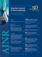
    Summary of the study design, highlighting independent and consensus analysis by neuroradiologists by using CTP (CTP group) and NIHSS with CTA (stroke scale group) to estimate infarct core and penumbra. Intragroup and intergroup consensus were determined (κ), as well as the significance of unblinding the stroke scale group to CTP (McNemar test).

  • Fig 2.
    • Download figure
    • Open in new tab
    • Download powerpoint
    Fig 2.

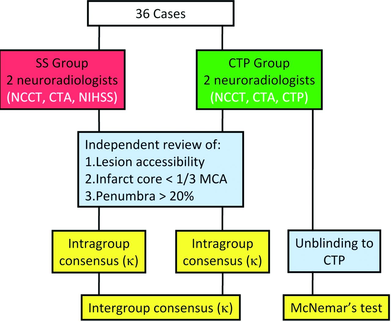
    A 76-year-old woman 5 hours 22 minutes after symptom onset (NIHSS score = 25). A, CTA demonstrates left M1 cutoff (white arrow). B, CTA source images demonstrate hypoattenuation in the left corona radiata, basal ganglia, and temporal lobe consistent with MCA infarct. C, Predominantly matched CBV and CBF defects corresponding to a portion of infarct core predicted on CTA source images, with peripheral prolonged TTP suggesting penumbra (Somatom 16). IAT recommendations for stroke scale (infarct core more than one-third of the MCA territory) and CTP (treatable penumbra) groups were discordant.

  • Fig 3.
    • Download figure
    • Open in new tab
    • Download powerpoint
    Fig 3.

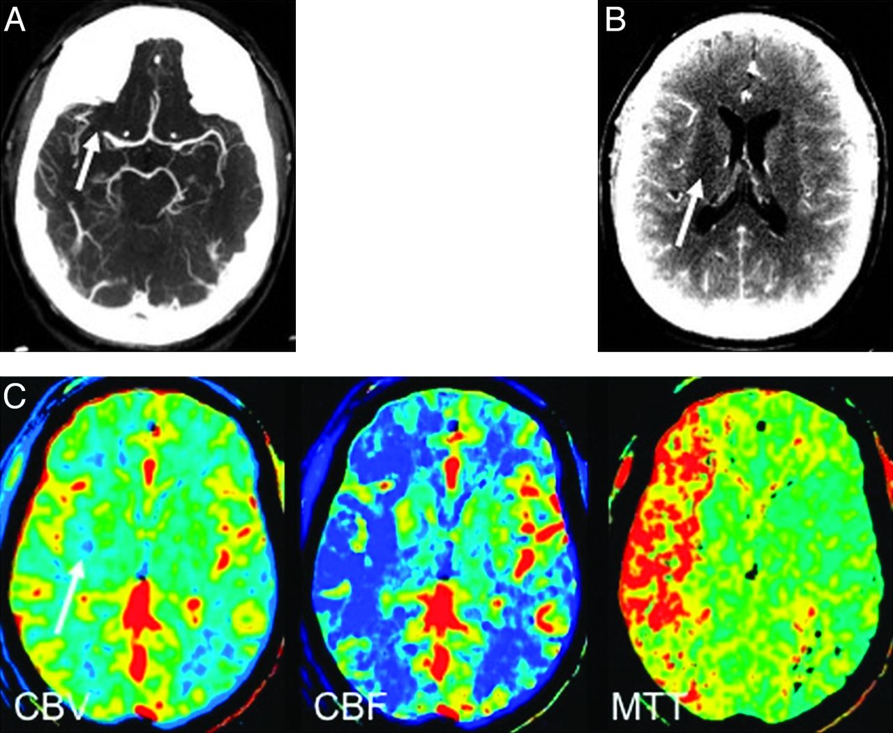
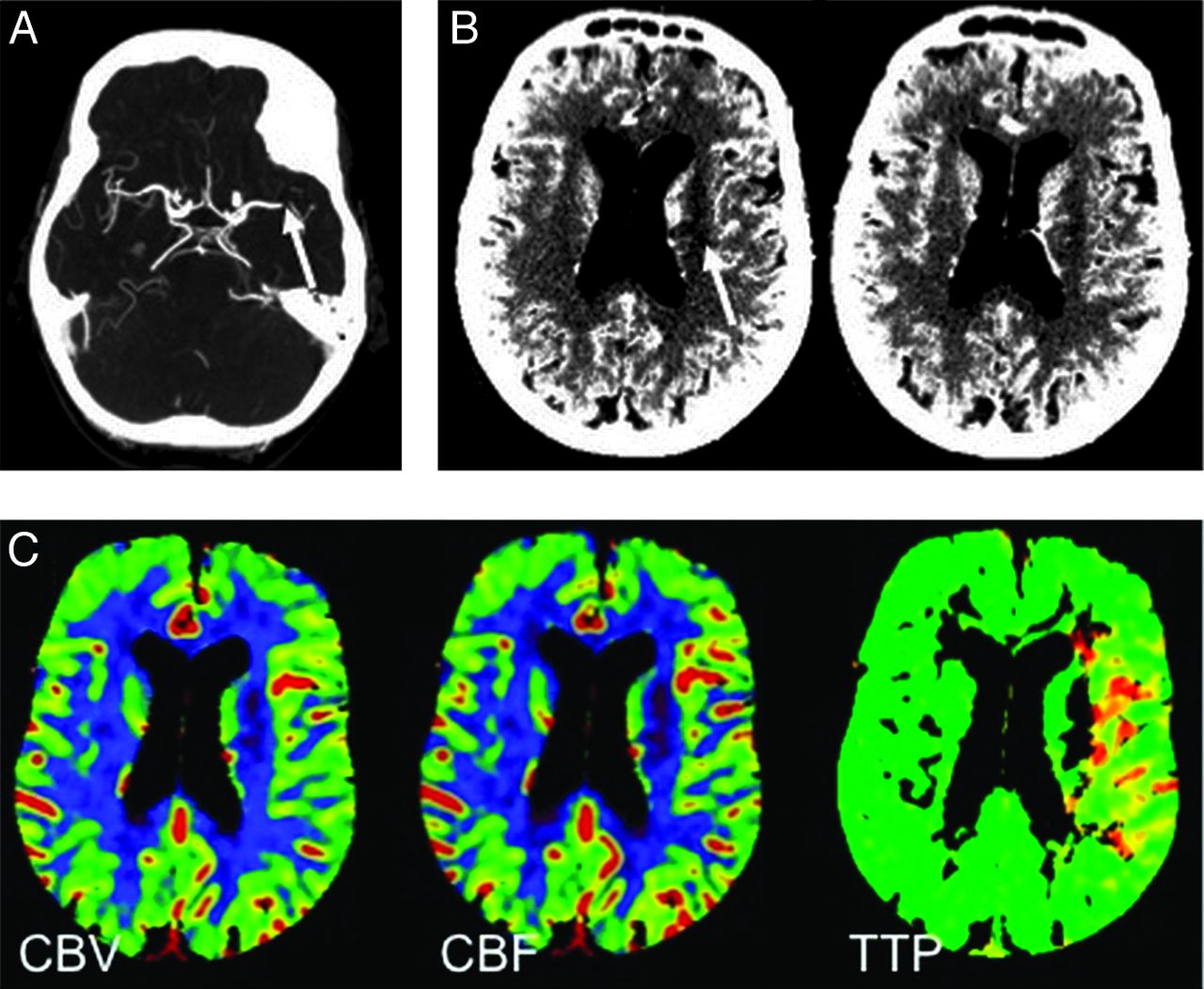
    A 47-year-old man 3 hours 37 minutes after symptom onset (NIHSS score = 17). A, CTA demonstrates right M1 cutoff (white arrow). B, CTA source image demonstrates equivocal hypoattenuation in the right corona radiata (white arrow), suggesting a small infarct. C, Large mismatched CBF and MTT compared with CBV (white arrow), consistent with treatable penumbra (LightSpeed Qx/I). On the basis of NIHSS score, locus of M1 cutoff and size of infarct core, the stroke scale group also recommended IAT.

  • Fig 4.
    • Download figure
    • Open in new tab
    • Download powerpoint
    Fig 4.

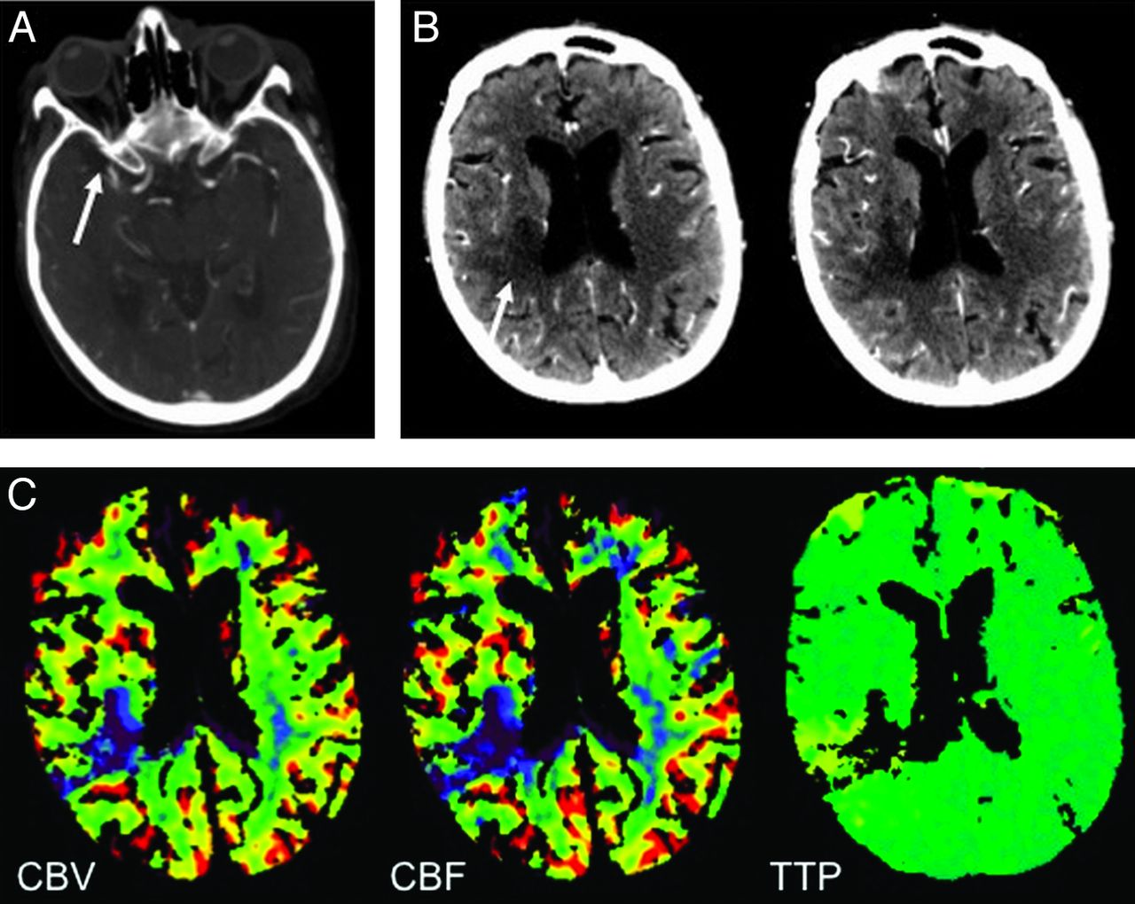
    An 88-year-old woman 3 hours 1 minute after symptom onset (NIHSS score = 17). A, CTA demonstrates right M1 cutoff (white arrow). B, CTA source images demonstrate hypoattenuation in the posterior right MCA territory (white arrow), consistent with a small infarct. C, Matched CBV and CBF defects corresponding to the infarct core on CTA source images, with matched TTP defect predicting the absence of treatable penumbra (Somatom 16). On the basis of the NIHSS score, locus of M1 cutoff, and size of infarct core, the stroke scale group predicted a treatable penumbra and recommended IAT. The stroke scale group revised its recommendation to “no treat” when unblinded to CTP.

  • Fig 5.
    • Download figure
    • Open in new tab
    • Download powerpoint
    Fig 5.

    A 92-year-old woman 4 hours 11 minutes after symptom onset (NIHSS score = 9). A, CTA demonstrates a focal defect in the distal left M1 (white arrow). B, CTA source images demonstrate hypoattenuation in the left corona radiata (white arrow), consistent with a small infarct. C, Essentially matched CBV and CBF defects corresponding to the infarct core on CTA source images, with a matched TTP defect including a small peripheral region of prolonged TTP predicting the absence of substantial penumbra (Somatom 16). On the basis of the NIHSS score, the locus of M1 cutoff, and size of infarct core, the stroke scale group predicted a treatable penumbra and recommended IAT. The stroke scale group revised its recommendation to “no treat” when unblinded to CTP.

  • Fig 6.
    • Download figure
    • Open in new tab
    • Download powerpoint
    Fig 6.

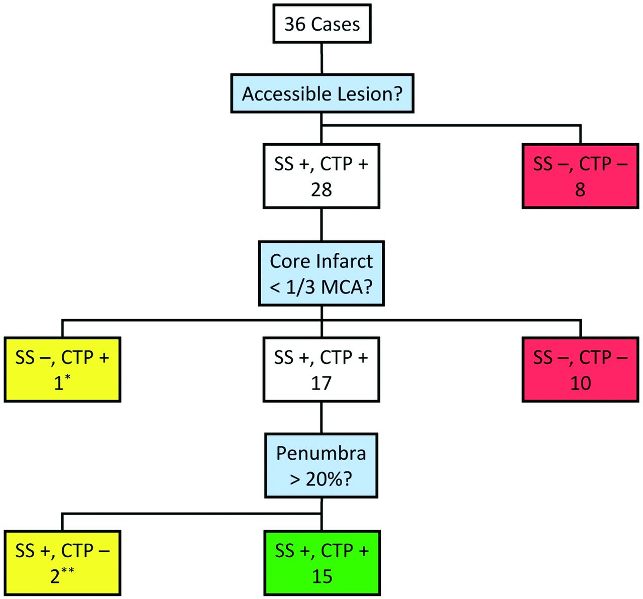
    Summary of treatment decisions for the stroke scale and CTP groups. Of the 28 patients with accessible lesions, 17 were concluded by both groups to have treatable infarct core (less than one-third of the MCA territory), 15 of whom had treatable penumbra (>20% infarct core). There was 1 discrepancy regarding infarct core (asterisk), treated by the CTP but not the stroke scale group, and 2 discrepancies regarding penumbra (double asterisks), both treated by the stroke scale but not the CTP group but reversed by the stroke scale group on unblinding.

Tables

  • Figures
  • Summary of intragroup and intergroup scores for the 3 IAT criteriaa

    Stroke Scale Group ConsensusCTP Group ConsensusIntergroup
    YesNoκ (SE)YesNoκ (SE)κ (SE)
    Lesion accessibility2881.02881.01.0
    Core infarct <1/3 MCA25110.94 (0.06)26100.66 (0.14)0.93 (0.07)
    Penumbra >20% core1840.64 (0.19)21150.83 (0.09)0.49 (0.22)
    Recommend IAT17190.83 (0.09)16200.78 (0.11)0.83 (0.09)
    • ↵a Stroke scale group used NCCT, CTA, and the NIHSS for IAT triage, and the CTP group used NCCT, CTA, and CTP.

PreviousNext
Back to top

In this issue

American Journal of Neuroradiology: 33 (10)
American Journal of Neuroradiology
Vol. 33, Issue 10
1 Nov 2012
  • Table of Contents
  • Index by author
Advertisement
Print
Download PDF
Email Article

Thank you for your interest in spreading the word on American Journal of Neuroradiology.

NOTE: We only request your email address so that the person you are recommending the page to knows that you wanted them to see it, and that it is not junk mail. We do not capture any email address.

Enter multiple addresses on separate lines or separate them with commas.
Clinical Stroke Penumbra: Use of National Institutes of Health Stroke Scale as a Surrogate for CT Perfusion in Patient Triage for Intra-Arterial Middle Cerebral Artery Stroke Therapy
(Your Name) has sent you a message from American Journal of Neuroradiology
(Your Name) thought you would like to see the American Journal of Neuroradiology web site.
CAPTCHA
This question is for testing whether or not you are a human visitor and to prevent automated spam submissions.
Cite this article
J.L. Boxerman, M.V. Jayaraman, W.A. Mehan, J.M. Rogg, R.A. Haas
Clinical Stroke Penumbra: Use of National Institutes of Health Stroke Scale as a Surrogate for CT Perfusion in Patient Triage for Intra-Arterial Middle Cerebral Artery Stroke Therapy
American Journal of Neuroradiology Nov 2012, 33 (10) 1893-1900; DOI: 10.3174/ajnr.A3102

Citation Manager Formats

  • BibTeX
  • Bookends
  • EasyBib
  • EndNote (tagged)
  • EndNote 8 (xml)
  • Medlars
  • Mendeley
  • Papers
  • RefWorks Tagged
  • Ref Manager
  • RIS
  • Zotero
0 Responses
Respond to this article
Share
Bookmark this article
Clinical Stroke Penumbra: Use of National Institutes of Health Stroke Scale as a Surrogate for CT Perfusion in Patient Triage for Intra-Arterial Middle Cerebral Artery Stroke Therapy
J.L. Boxerman, M.V. Jayaraman, W.A. Mehan, J.M. Rogg, R.A. Haas
American Journal of Neuroradiology Nov 2012, 33 (10) 1893-1900; DOI: 10.3174/ajnr.A3102
del.icio.us logo Twitter logo Facebook logo Mendeley logo
  • Tweet Widget
  • Facebook Like
  • Google Plus One
Purchase

Jump to section

  • Article
    • Abstract
    • ABBREVIATIONS:
    • Materials and Methods
    • Results
    • Discussion
    • Conclusions
    • Footnotes
    • References
  • Figures & Data
  • Info & Metrics
  • Responses
  • References
  • PDF

Related Articles

  • No related articles found.
  • PubMed
  • Google Scholar

Cited By...

  • Effect of Radiographic Contrast Media Shortage on Stroke Evaluation in the United States
  • Endovascular treatment for acute ischaemic stroke due to medium vessel occlusion: data from ANGEL-ACT registry
  • NCCT and CTA-based imaging protocol for endovascular treatment selection in late presenting or wake-up strokes
  • Initial hospital management of patients with emergent large vessel occlusion (ELVO): report of the standards and guidelines committee of the Society of NeuroInterventional Surgery
  • Future acute ischemic stroke trials should randomize on the angio table
  • Crossref (16)
  • Google Scholar

This article has been cited by the following articles in journals that are participating in Crossref Cited-by Linking.

  • Initial hospital management of patients with emergent large vessel occlusion (ELVO): report of the standards and guidelines committee of the Society of NeuroInterventional Surgery
    Ryan A McTaggart, Sameer A Ansari, Mayank Goyal, Todd A Abruzzo, Barb Albani, Adam J Arthur, Michael J Alexander, Felipe C Albuquerque, Blaise Baxter, Ketan R Bulsara, Michael Chen, Josser E Delgado Almandoz, Justin F Fraser, Donald Frei, Chirag D Gandhi, Don V Heck, Steven W Hetts, M Shazam Hussain, Michael Kelly, Richard Klucznik, Seon-Kyu Lee, Thabele Leslie-Mawzi, Philip M Meyers, Charles J Prestigiacomo, G Lee Pride, Athos Patsalides, Robert M Starke, Peter Sunenshine, Peter A Rasmussen, Mahesh V Jayaraman
    Journal of NeuroInterventional Surgery 2017 9 3
  • NCCT and CTA-based imaging protocol for endovascular treatment selection in late presenting or wake-up strokes
    Telma Santos, Andreia Carvalho, André Almeida Cunha, Marta Rodrigues, Tiago Gregório, Ludovina Paredes, Henrique Costa, José Mário Roriz, João Pinho, Miguel Veloso, Sérgio Castro, Pedro Barros, Manuel Ribeiro
    Journal of NeuroInterventional Surgery 2019 11 2
  • Association between initial NIHSS score and recanalization rate after endovascular thrombectomy
    Junya Aoki, Kentaro Suzuki, Takuya Kanamaru, Akihito Kutsuna, Takehiro Katano, Yohei Takayama, Yuji Nishi, Yuho Takeshi, Toru Nakagami, Shinichiro Numao, Arata Abe, Satoshi Suda, Yasuhiro Nishiyama, Kazumi Kimura
    Journal of the Neurological Sciences 2019 403
  • Neuroimaging Paradigms to Identify Patients for Reperfusion Therapy in Stroke of Unknown Onset
    Mark R. Etherton, Andrew D. Barreto, Lee H. Schwamm, Ona Wu
    Frontiers in Neurology 2018 9
  • Normalized wall index, intraplaque hemorrhage and ulceration of carotid plaques correlate with the severity of ischemic stroke
    Xin Cao, Qingluan Yang, Ye Tang, Lei Pan, Mao Lai, Zekuan Yu, Daoying Geng, Jun Zhang
    Atherosclerosis 2020 315
  • Characteristics and prognosis of acute basilar artery occlusion in minor to moderate stroke and severe stroke after endovascular treatment: A multicenter retrospective study
    Dayu Wu, Fei Guo, Diyan Liu, Rongguo Hu, Zhenkun Shen, Yuwei Yang, Yane Wang, Yumei Xu, Longxuan Li
    Clinical Neurology and Neurosurgery 2021 202
  • Endovascular treatment for acute ischaemic stroke due to medium vessel occlusion: data from ANGEL-ACT registry
    Dapeng Sun, Raynald ­, Xiaochuan Huo, Baixue Jia, Xu Tong, Anxin Wang, Gaoting Ma, Ning Ma, Feng Gao, Dapeng Mo, Zhongrong Miao
    Stroke and Vascular Neurology 2023 8 1
  • The Association Between National Institutes of Health Stroke Scale Score and Clinical Outcome in Patients with Large Core Infarctions Undergoing Endovascular Treatment
    Lingyu Zhang, Jinfu Ma, Mengmeng Wang, Lin Zhang, Wenzhe Sun, Honghong Ji, Chengsong Yue, Jiacheng Huang, Wenjie Zi, Fengli Li, Changwei Guo, Pengfei Wang
    Neurology and Therapy 2024 13 3
  • Interventional Management of Acute Ischemic Stroke: A Systematic Review
    J. Scott Pannell, David R. Santiago-Dieppa, Alexander A. Khalessi
    Current Treatment Options in Cardiovascular Medicine 2014 16 8
  • The value of whole-brain CT perfusion imaging combined with dynamic CT angiography in the evaluation of pial collateral circulation with middle cerebral artery occlusion
    Yi-Chuan Ma, Ai-Qi Chen, Fei Guo, Juan Yu, Min Xu, Dan-Dan Shan, Shun-Hua Zhang
    Technology and Health Care 2022 30 4

More in this TOC Section

  • Multimodal CT Provides Improved Performance for Lacunar Infarct Detection
  • Optimal MRI Sequence for Identifying Occlusion Location in Acute Stroke: Which Value of Time-Resolved Contrast-Enhanced MRA?
  • Evaluating the Effects of White Matter Multiple Sclerosis Lesions on the Volume Estimation of 6 Brain Tissue Segmentation Methods
Show more Brain

Similar Articles

Advertisement

Indexed Content

  • Current Issue
  • Accepted Manuscripts
  • Article Preview
  • Past Issues
  • Editorials
  • Editor's Choice
  • Fellows' Journal Club
  • Letters to the Editor
  • Video Articles

Cases

  • Case Collection
  • Archive - Case of the Week
  • Archive - Case of the Month
  • Archive - Classic Case

More from AJNR

  • Trainee Corner
  • Imaging Protocols
  • MRI Safety Corner
  • Book Reviews

Multimedia

  • AJNR Podcasts
  • AJNR Scantastics

Resources

  • Turnaround Time
  • Submit a Manuscript
  • Submit a Video Article
  • Submit an eLetter to the Editor/Response
  • Manuscript Submission Guidelines
  • Statistical Tips
  • Fast Publishing of Accepted Manuscripts
  • Graphical Abstract Preparation
  • Imaging Protocol Submission
  • Evidence-Based Medicine Level Guide
  • Publishing Checklists
  • Author Policies
  • Become a Reviewer/Academy of Reviewers
  • News and Updates

About Us

  • About AJNR
  • Editorial Board
  • Editorial Board Alumni
  • Alerts
  • Permissions
  • Not an AJNR Subscriber? Join Now
  • Advertise with Us
  • Librarian Resources
  • Feedback
  • Terms and Conditions
  • AJNR Editorial Board Alumni

American Society of Neuroradiology

  • Not an ASNR Member? Join Now

© 2025 by the American Society of Neuroradiology All rights, including for text and data mining, AI training, and similar technologies, are reserved.
Print ISSN: 0195-6108 Online ISSN: 1936-959X

Powered by HighWire