
Article Figures & Data
Figures
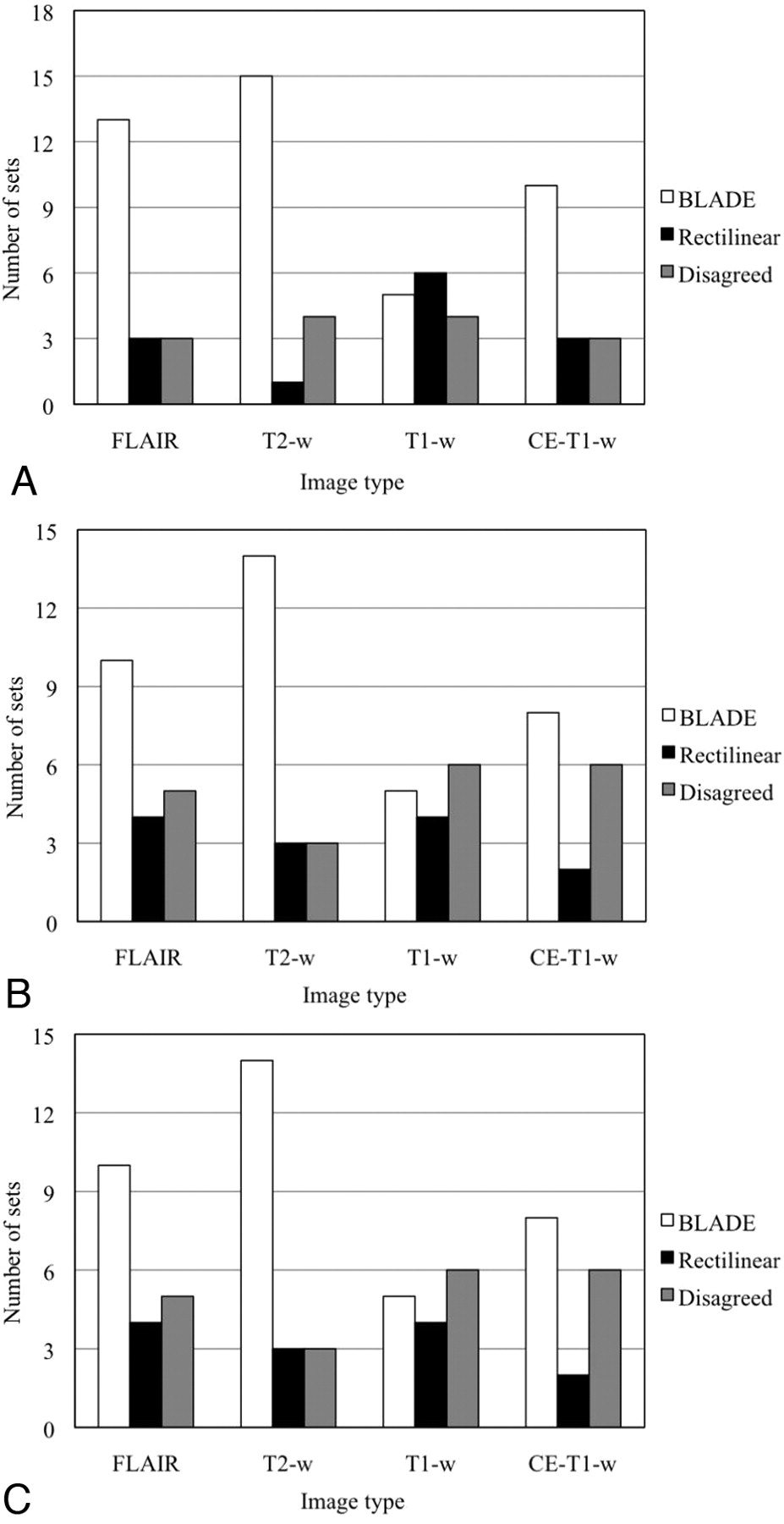
- Fig 1.
Number of image sets in which the evaluators preferred 1 kind of image over the other or disagreed in terms of (A) less motion artifact, (B) better lesion characterization, and (C) better overall image quality.
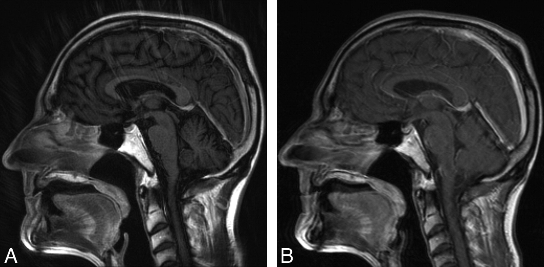
- Fig 2.
Sagittal CE-T1WI images of a patient obtained by using (A) BLADE and (B) rectilinear technique. Despite the presence of extensive radial artifact in the partially radial acquisition image, gray-white differentiation and correction of motion artifact remain superior.
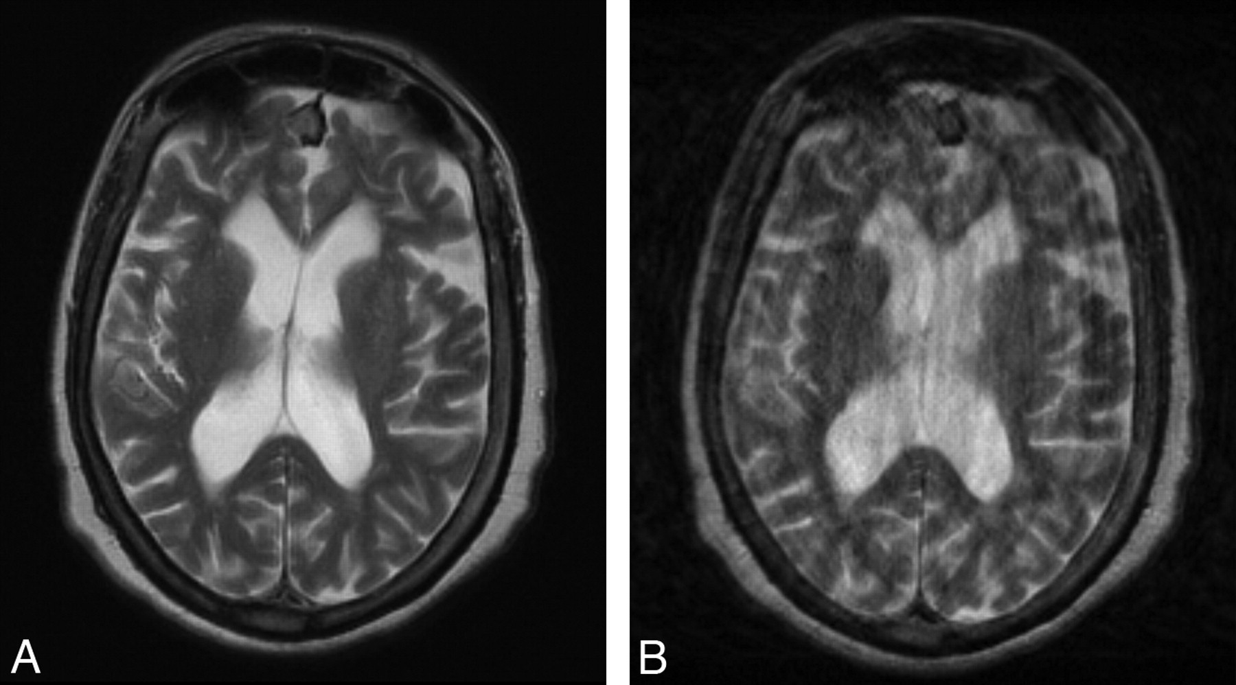
- Fig 3.
Axial T2WI images of a patient obtained by using (A) BLADE and (B) rectilinear techniques. The partially radial acquisition image is free of motion artifact, whereas extensive motion artifact degrades the rectilinear image.
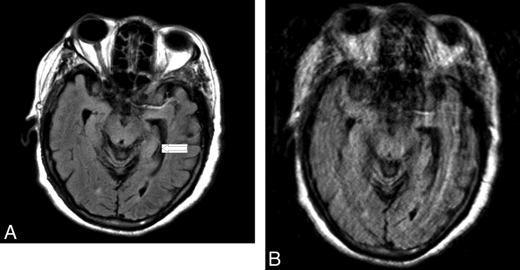
- Fig 4.
Axial FLAIR images of patient who presented with an acute left middle cerebral artery distribution stroke obtained by using (A) BLADE and (B) rectilinear techniques. The partially radial acquisition image is free of motion artifact, whereas extensive motion artifact degrades quality of the rectilinear image. The small, subtle ischemic lesion was also better characterized from the partially radial acquisition image (arrow).


Tables
- Table 1:
The imaging sequence, sequence parameters, and total scan time for acquisition of T2WI, FLAIR, T1WI, and CE-T1WI images by partially radial and rectilinear techniques
Image Type BLADE (Imaging sequence/TR/TE/α/TI/FOV/section thickness/base resolution/blade coverage/motion correction/turbo factor/echo-train length per section/averages/concatenation/imaging time) Rectilinear (Imaging sequence/TR/TE/α/TI/FOV/section thickness/base resolution/averages/concatenation/imaging time) T2WI TSE/4000 ms/107 ms/150°/-/230 × 230 mm/5 mm/256 × 256/91.7%/on/35/11/1/2/1 minute 38 seconds TSE/4320 ms/88 ms/150°/-/173 × 230 mm/5 mm/192 × 256/1/1/1 minute 13 seconds FLAIR FLAIR/9000 ms/107 ms/150°/150 ms/230 × 230 mm/5 mm/256 × 256/91.7%/on/35/11/1/2/3 minutes 38 seconds FLAIR/4320 ms/110 ms/150°/2500 ms/173 × 230 mm/5 mm/192 × 256/1/1/1 minute 21 seconds Axial T1WI FLAIR/2500 ms/59 ms/150°/860 ms/230 × 230 mm/5 mm/256 × 256/95.5%/off/19/21/1/3/2 minutes 47 seconds Spin-echo/740 ms/17 ms/90°/-/173 × 230 mm/5 mm/192 × 256/2/1/4 minutes 8 seconds Coronal T1WI FLAIR/2500 ms/59 ms/150°/860 ms/230 × 230 mm/5 mm/256 × 256/95.5%/off/19/21/1/5/4 minutes 37 seconds Spin-echo/740 ms/17 ms/90°/-/201 × 230 mm/5 mm/224 × 256/1/1/1 minute 23 seconds Sagittal T1WI FLAIR/2500 ms/59 ms/150°/860 ms/230 × 230 mm/5 mm/256 × 256/95.5%/off/19/21/1/3/2 minutes 52 seconds Spin-echo/525 ms/17 ms/90°/-/230 × 230 mm/5 mm/192 × 256/1/1/1 minute 52 seconds - Table 2:
The mean value and standard deviation of the motion artifact scores for each sequence and overall rating scores from the images obtained using rectilinear and partially radial acquisition techniques
Image Type Motion Score Mean Value ± Standard Deviation Rectilinear BLADE FLAIR 1.21 ± 0.63 0.82 ± 0.87 T2WI 1.18 ± 0.86 0.425 ± 0.65 T1WI 1.83 ± 1.21 1.57 ± 1.18 CE-T1WI 0.91 ± 0.64 0.97 ± 0.62 Overall 1.26 ± 0.89 0.90 ± 0.92 -
Motion artifact scoring scale: 0 indicates no visible motion artifact; 1, visible motion artifact with mild degradation of the image quality; 2, visible motion artifact with moderate degradation of the image quality; 3, visible motion artifact with severe degradation of the image quality; 4, visible motion artifact which renders the image of no diagnostic value.
-
- Table 3:
Total number of axial and non-axial T1WI and CE-T1WI with radial artifact for rectilinear and BLADE techniques and the number of images in which radial artifact compromised the diagnostic yield
Gridding and Section Orientation Total Radial Artifact Diagnostic Yield Absent Raters Disagreed Present Not Compromised Raters Disagreed Compromised Rectilinear Axial 10 9 1 0 10 0 0 Nonaxial 21 20 1 0 21 0 0 Total 31 29 2 0 31 0 0 BLADE Axial 10 1 4 5 10 0 0 Nonaxial 21 0 1 20 10 2 9 Total 31 1 5 25 20 2 9


