
Article Figures & Data
Figures
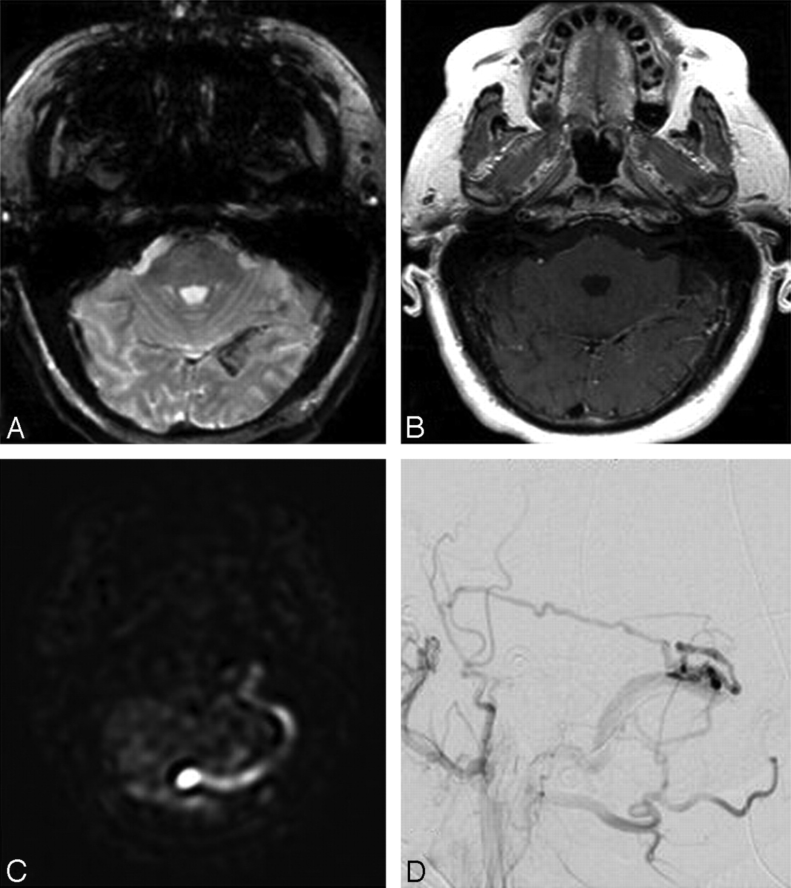
- Fig 1.
Patient 12. A 64-year-old man presenting with complex partial seizure, with electroencephalography localized to the left temporal lobe. A, Axial gradient recalled-echo image shows hemosiderin staining in the left temporo-occipital cortex and along the tentorium cerebelli. B, Axial T1-weighted postcontrast image shows mildly prominent serpiginous vasculature in this region. C, Axial ASL perfusion shows high-signal-intensity labeled spins within the venous confluence and left transverse and sigmoid sinuses, indicating the presence of venous shunting surgery. D, Lateral DSA left external carotid artery injection, shows a left transverse sinus DAVF with arterial feeding from the left middle meningeal artery and, to a lesser extent, from the left occipital artery. Venous drainage is to the left transverse and sigmoid sinuses and left internal jugular vein.
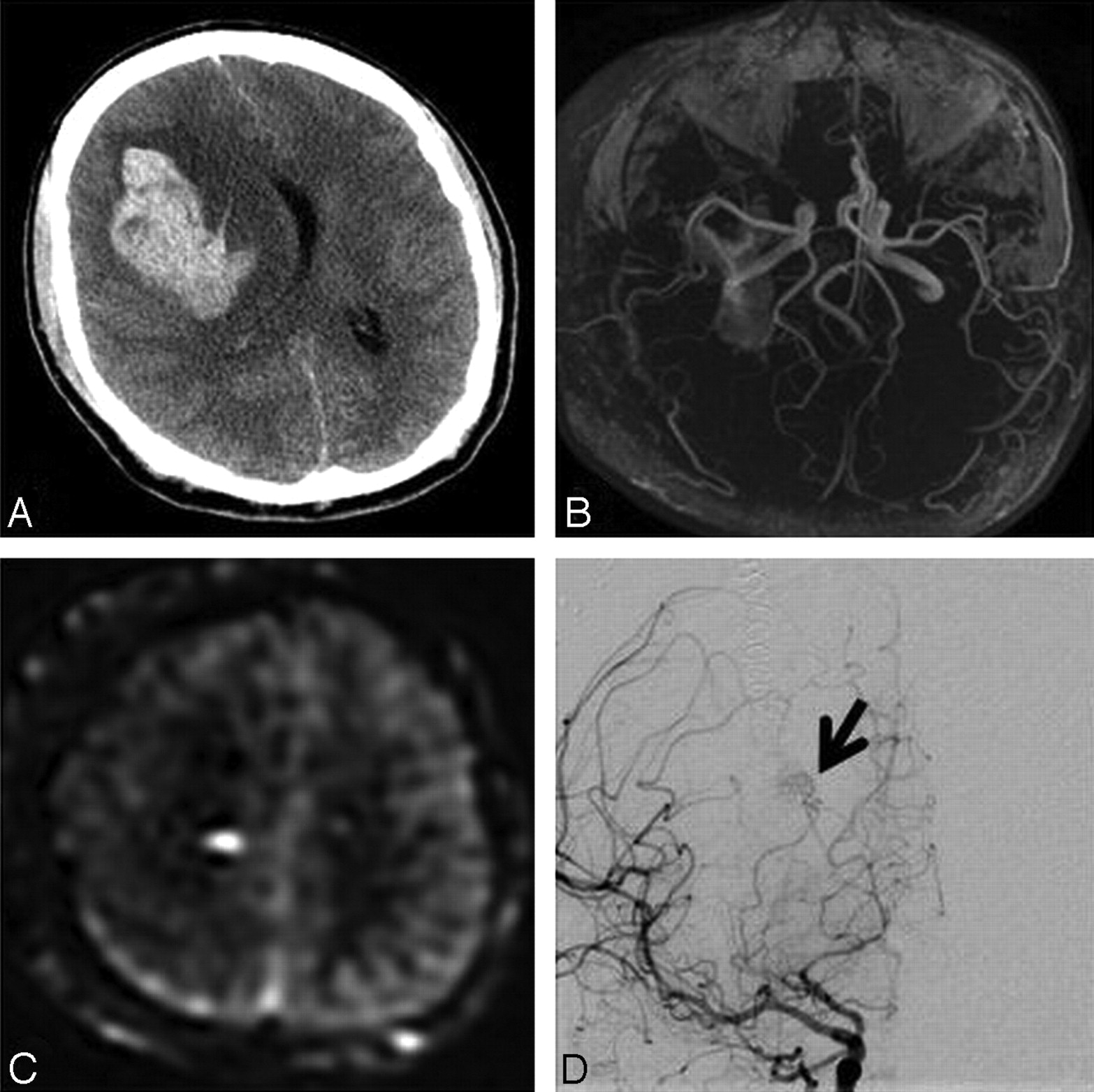
- Fig 2.
Patient 5. A 25-year-old man with sudden onset of headache while weightlifting, followed by confusion and loss of consciousness. A, Axial noncontrast CT scan shows a large right frontal hematoma with significant edema, mass effect, and brain herniation. The patient was immediately taken to the operating room for right frontal craniotomy decompression. B, Postdecompression time-of-flight MRA findings are normal, without MR imaging evidence of AVM or DAVF. C, Axial ASL image shows focal high signal intensity at the medial right frontal lobe (white arrow), concerning for the presence of a shunt lesion. Initial angiogram (not shown) obtained on the same day as the MR imaging study failed to demonstrate the presence of an AVM, likely due to edema and mass effect. D, Repeat angiogram after 4 weeks shows a small (<1 cm) right frontal lobe AVM (black arrow). The feeding vessel is the right anterior choroidal artery, with deep venous drainage into the right internal cerebral vein.
- Fig 3.
Patient 2. A 76-year-old man presenting with worsening headache and vomiting. A, Axial gradient recalled-echo MR image shows a left cerebellar hemorrhage. B, MRA does not reveal an obvious vascular lesion. C, ASL image shows high signal intensity within the venous confluence (white arrow) and left internal jugular vein (white arrowhead), suspicious for a shunt lesion. D, Frontal DSA confirms a small AVM, fed by a small branch of the left vertebral artery; venous drainage is to the venous confluence, left transverse sinus, and left internal jugular vein.
- Fig 4.
Histograms of pooled scores of the 3 readers regarding the likelihood of DAVF/AVM (1 = very unlikely, 2 = unlikely, 3 = neutral, 4 = likely, 5 = very likely) before and after reviewing the ASL images, for cases with both negative and positive findings on DSA. Note that the distribution before reviewing ASL was skewed toward the correct diagnosis, but this effect was magnified following ASL review. In particular, the number of cases deemed “neutral” (score of 3) markedly decreased following review of ASL, suggesting that the readers had higher confidence in their decisions post-ASL.
- Fig 5.
ROC curves for the pooled and individual readers pre- and postreview of the ASL images. The increase in the AUC was significant (P = .02). The post-ASL curve is a more well-rounded predictor and indicates that the readers' sensitivity for identifying a vascular malformation has increased after reviewing the ASL images. Reader 1 is a neurologist; readers 2 and 3 are neuroradiologists.
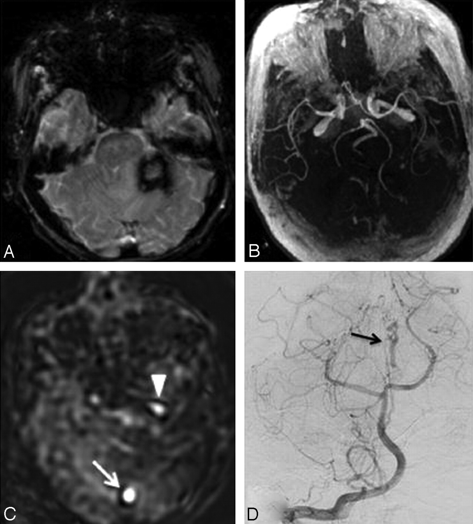
- Fig 6.
Patient 7. A 38-year-old man who presented to the medical center with new-onset headache. A and B, Time-of-flight MRA image (A) and axial T1-weighted postcontrast image (B) show no evidence of DAVF, AVM, or aneurysm. C, ASL shows focal high signal intensity at the left petrous apex, concerning for a focal lesion (aneurysm or shunt lesion). The initial angiogram failed to demonstrate a vascular lesion (not shown). D, Frontal projection from the repeat angiogram (left vertebral artery injection) shows a very slow-flow left petrous apex DAVF (arrows), fed by a muscular branch of the left vertebral artery and drained via perimedullary veins. A small DAVF was confirmed at surgical resection.


Tables
Readersa κ 95% CI 1 and 2 0.45 0.23–.61 1 and 3 0.42 0.18–.62 2 and 3 0.72 0.47–.89 Signs ICH 0.30 −0.11–.74 Edema 0.54 0.28–.74 Abnormal MRA 0.43 0.17–.68 Serpiginous vessels 0.58 0.32–.80 Venous enhancement 0.29 0.02–.54 Venous ASL signal 0.64 0.40–.85 -
↵a Reader 1 is a neurologist; readers 2 and 3 are neuroradiologists.
-
- Table 2:
Univariate and multivariate logistic regression analysis of imaging features on DSA result
Imaging Features Odds Ratio Standard Error Z P > z 95% CI Univariate analysis ICH 0.17 0.19 −1.62 .105 0.02–1.45 Edema 0.30 0.15 −2.48 .013 0.12–0.78 Abnormal MRAa 3.45 1.68 2.55 .011 1.33–8.94 Serpiginous vesselsa 17.85 11.18 4.60 <.001 5.23–60.94 Venous enhancementa 2.73 1.38 1.99 .046 1.02–7.35 Venous ASL signala 19.60 11.83 4.93 <.001 6.00–63.97 Multivariate analysis ICH 1.18 1.84 0.11 .915 0.06–24.93 Edemaa 0.12 0.11 −2.34 .019 0.02–0.71 Abnormal MRA 0.82 0.88 −0.18 .856 0.10–6.72 Serpiginous vesselsa 2.53 13.35 2.37 .018 1.55–101.19 Venous enhancementa 0.79 0.77 −0.24 .811 0.12–5.31 Venous ASL signala 17.30 14.57 3.39 .001 3.32–90.14 -
↵a Significant at P < .05.
-
Multivariate post-ASL model is an improvement over the pre-ASL model (P = .0001).
-
In this issue


Jump to section
Related Articles
Cited By...
- Arterial Spin-Labeling MR Imaging in the Detection of Intracranial Arteriovenous Malformations in Patients with Hereditary Hemorrhagic Telangiectasia
- Noninvasive Follow-up Imaging of Ruptured Pediatric Brain AVMs Using Arterial Spin-Labeling
- Follow-Up MRI for Small Brain AVMs Treated by Radiosurgery: Is Gadolinium Really Necessary?
- Arterial Spin-Labeling Improves Detection of Intracranial Dural Arteriovenous Fistulas with MRI
- Standard and Guidelines: Intracranial Dural Arteriovenous Shunts
- Intracranial Arteriovenous Shunting: Detection with Arterial Spin-Labeling and Susceptibility-Weighted Imaging Combined