
Article Figures & Data
Figures
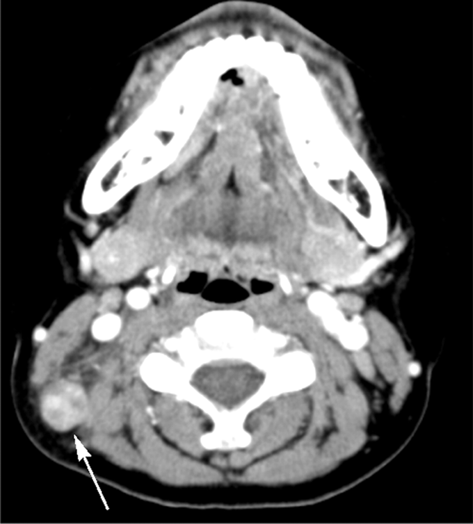
- Fig 1.
KD in an 8-year-old girl. Transverse CT image shows a lymph node (arrow) containing multiple necrotic foci in the peripheral portion of the node. The necrotic foci have indistinct margins, and the extent of nodal necrosis accounts for <30% (mild degree) of the lymph node. The obliteration of the fat plane surrounding the node is seen (arrowhead). The CTN and CTN/M were calculated as 79 HU and 1.0, respectively.
- Fig 2.
KD in a 13-year-old boy. Transverse CT image shows a lymph node (arrow) containing multiple necrotic foci in the peripheral portion of the node. The necrotic foci have indistinct margins, and the extent of nodal necrosis accounts for approximately 30%–70% (moderate degree) of the lymph node. The CTN and CTN/M were calculated as 65 HU and 0.9, respectively.
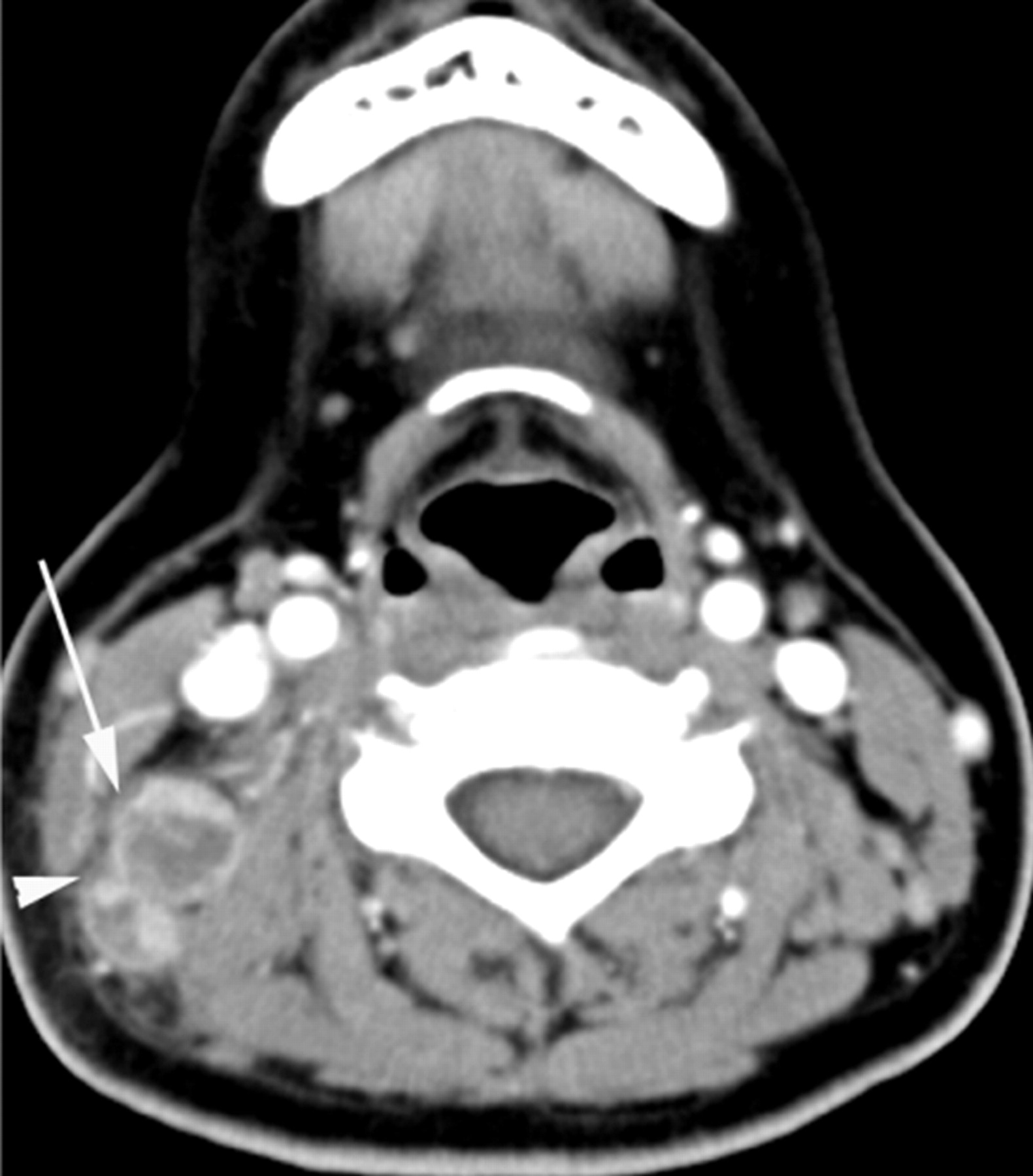
- Fig 3.
KD in a 31-year-old woman. Transverse CT scan shows 2 lymph nodes with necrotic foci. The larger one (arrow) has a single necrotic focus with a relatively indistinct margin, and the extent accounts for >70% (severe degree) of the lymph node. Perinodal infiltration is seen (arrowhead). The CTN and CTN/M were 43 HU and 0.7, respectively.
- Fig 4.
TL in a 38-year-old woman. Transverse CT scan demonstrates a necrotic lymph node. The lymph node (arrow) has a single necrotic focus with a well-defined margin, and the extent of nodal necrosis accounts for >70% (severe degree) of the lymph node. The CTN and CTN/M were 29 HU and 0.4, respectively.
- Fig 5.
TL in a 35-year-old woman. Transverse image shows a lymph node (arrow) having multiple necrotic foci with relatively well-defined margins and a moderate extent of nodal necrosis. Note calcification (arrowhead) within the node. CTN and CTN/M were 38 HU and 0.6, respectively.
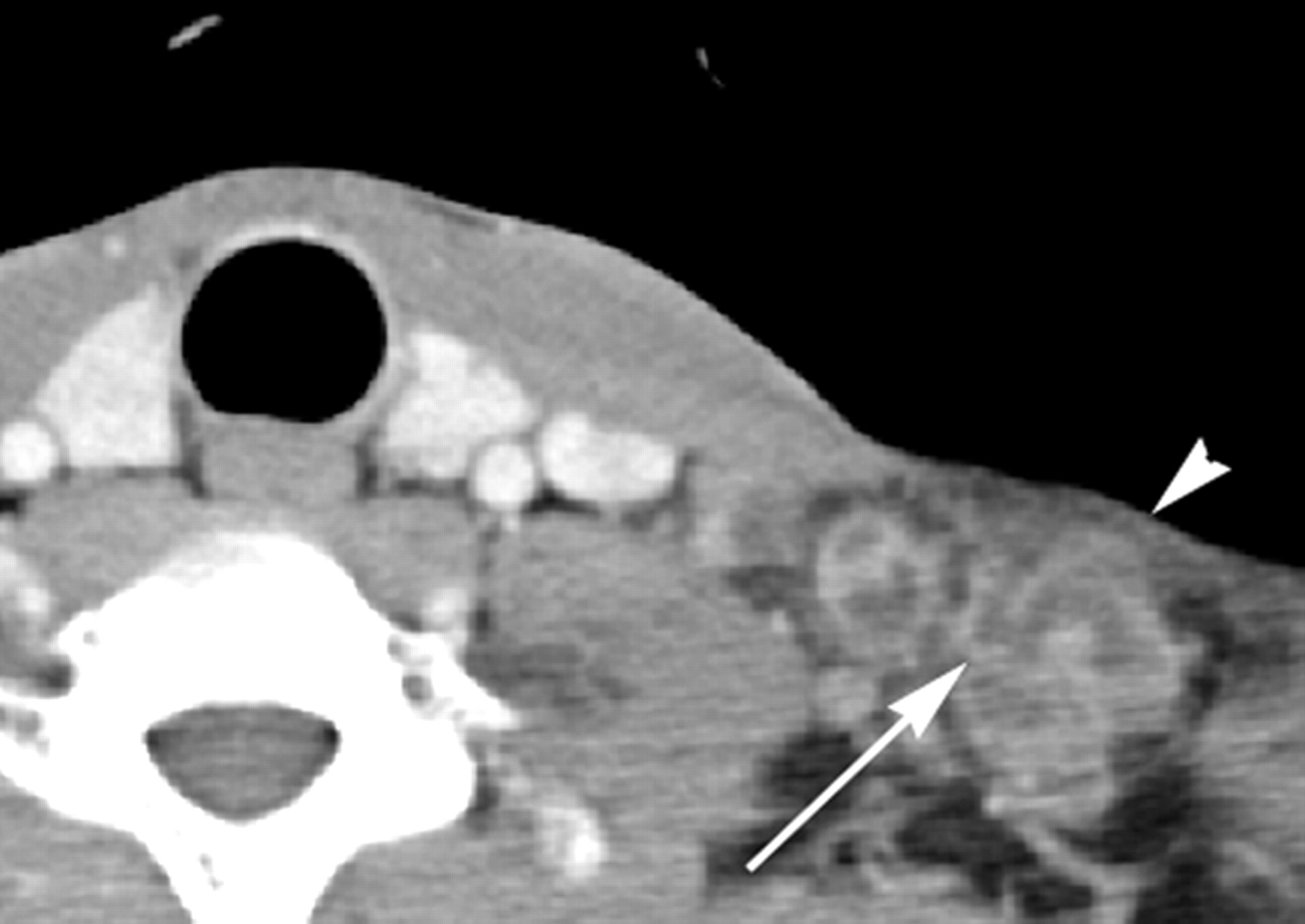
- Fig 6.
TL in a 37-year-old man. Transverse CT scan shows several lymph nodes with necrotic foci. The larger one (arrow) has multiple necrotic foci (not shown in this figure) with well-defined margins and a severe extent of nodal necrosis. Note the obliteration of perinodal fat around the lymph node (arrowhead). The CTN and CTN/M were 22 HU and 0.4, respectively.
- Fig 7.
Graph of CTN of TL and KD. The mean CTN of KD (70.3 ± 18.01) is significantly higher than that of TL (37.4 ± 20.00). When 44.5 was used as the cutoff value for the CTN, a sensitivity of 89.5% and a specificity of 86.0% were achieved for differentiating KD from TL.
- Fig 8.
Graph of CTN/M of TL and KD. The mean CTN/M of KD (1.1 ± 0.27) is significantly higher than that of TL (0.6 ± 0.31). When 0.7 was used as the cutoff value for the CTN, a sensitivity of 94.7% and a specificity of 76.7% were achieved for differentiating KD from TL.


Tables
Characteristics KD (n = 24) TL (n = 45) Age (yr) Range 7–49 16–85 Mean 25.1 39.9 Sex 14 women, 10 men 36 women, 9 men 16-Channel CT 15 (62.5%) 28 (62.2%) 64-Channel CT 9 (37.5%) 17 (37.8%) Time interval between CT and excisional biopsy (day) Range 0–44 0–27 Mean 8.2 7.4 Size (cm) Range 1.0–3.4 1.1–5.2 Mean 2.0 2.5 Level of analyzed lymph nodes I 2 (8.3%) 1 (2.2%) II 16 (66.7%) 20 (44.4%) III 1 (4.2%) 1 (2.2%) IV 1 (4.2%) 7 (15.6%) V 4 (16.6%) 16 (35.6%) -
↵a Unless otherwise indicated, data are expressed as the number of patients, with percentages in parentheses.
-
KD (n = 24) TL (n = 45) P Value Extent of necrosis .000 Mild or moderate 21 (87.5%) 17 (37.8%) Severe 3 (12.5%) 28 (62.2%) No. of necroses .003 Single 2 (8.4%) 20 (44.4%) Multiple 22 (91.6%) 25 (55.6%) Location of necrosis .339b Central 6 (25.0%) 7 (15.6%) Peripheral 18 (75.0%) 38 (84.4%) Margin of necrosis .000 Indistinct 19 (79.2%) 9 (20.0%) Relatively WD or WD 5 (20.8%) 36 (80.0%) Perinodal infiltration .192b Absent 2 (9%) 11 (24%) Present 22 (91%) 34 (76%) Calcification .012b Absent 24 (100%) 34 (76%) Present 0 (0%) 11 (24%) CTNc 22 (92%) 19 (42%) .000 CTN/Md 23 (96%) 11 (24%) .000 -
Note:—WD indicates well-defined; mild, <30% of an affected lymph node; moderate, 30%–70% of an affected lymph node; severe, >70% of an affected lymph node.
-
↵a Unless otherwise indicated, data are expressed as the number of patients, with percentages in parentheses.
-
↵b Calculated with the Fisher exact test. The other categoric variables were compared using the χ2 test.
-
↵c CTN of >44.5 HU is used as the threshold for differentiating KD from TL.
-
↵d CTN/M of >0.7 is used as the threshold for differentiating KD from TL.
-
In this issue


Jump to section
Related Articles
Cited By...
- No citing articles found.