Advertisement

Main menu

  • Home
  • Content
    • Current Issue
    • Accepted Manuscripts
    • Article Preview
    • Past Issue Archive
    • Video Articles
    • AJNR Case Collection
    • Case of the Week Archive
    • Case of the Month Archive
    • Classic Case Archive
  • Special Collections
    • AJNR Awards
    • Low-Field MRI
    • Alzheimer Disease
    • ASNR Foundation Special Collection
    • Photon-Counting CT
    • View All
  • Multimedia
    • AJNR Podcasts
    • AJNR SCANtastic
    • Trainee Corner
    • MRI Safety Corner
    • Imaging Protocols
  • For Authors
    • Submit a Manuscript
    • Submit a Video Article
    • Submit an eLetter to the Editor/Response
    • Manuscript Submission Guidelines
    • Statistical Tips
    • Fast Publishing of Accepted Manuscripts
    • Graphical Abstract Preparation
    • Imaging Protocol Submission
    • Author Policies
  • About Us
    • About AJNR
    • Editorial Board
    • Editorial Board Alumni
  • More
    • Become a Reviewer/Academy of Reviewers
    • Subscribers
    • Permissions
    • Alerts
    • Feedback
    • Advertisers
    • ASNR Home

User menu

  • Alerts
  • Log in

Search

  • Advanced search
American Journal of Neuroradiology

American Journal of Neuroradiology

ASHNR American Society of Functional Neuroradiology ASHNR American Society of Pediatric Neuroradiology ASSR
  • Alerts
  • Log in

Advanced Search

  • Home
  • Content
    • Current Issue
    • Accepted Manuscripts
    • Article Preview
    • Past Issue Archive
    • Video Articles
    • AJNR Case Collection
    • Case of the Week Archive
    • Case of the Month Archive
    • Classic Case Archive
  • Special Collections
    • AJNR Awards
    • Low-Field MRI
    • Alzheimer Disease
    • ASNR Foundation Special Collection
    • Photon-Counting CT
    • View All
  • Multimedia
    • AJNR Podcasts
    • AJNR SCANtastic
    • Trainee Corner
    • MRI Safety Corner
    • Imaging Protocols
  • For Authors
    • Submit a Manuscript
    • Submit a Video Article
    • Submit an eLetter to the Editor/Response
    • Manuscript Submission Guidelines
    • Statistical Tips
    • Fast Publishing of Accepted Manuscripts
    • Graphical Abstract Preparation
    • Imaging Protocol Submission
    • Author Policies
  • About Us
    • About AJNR
    • Editorial Board
    • Editorial Board Alumni
  • More
    • Become a Reviewer/Academy of Reviewers
    • Subscribers
    • Permissions
    • Alerts
    • Feedback
    • Advertisers
    • ASNR Home
  • Follow AJNR on Twitter
  • Visit AJNR on Facebook
  • Follow AJNR on Instagram
  • Join AJNR on LinkedIn
  • RSS Feeds

AJNR Awards, New Junior Editors, and more. Read the latest AJNR updates

Research ArticleHead and Neck

CT Analysis Demonstrates That Cochlear Height Does Not Change with Age

M.C. Mori and K.W. Chang
American Journal of Neuroradiology January 2012, 33 (1) 119-123; DOI: https://doi.org/10.3174/ajnr.A2713
M.C. Mori
  • Find this author on Google Scholar
  • Find this author on PubMed
  • Search for this author on this site
K.W. Chang
  • Find this author on Google Scholar
  • Find this author on PubMed
  • Search for this author on this site
  • Article
  • Figures & Data
  • Supplemental
  • Info & Metrics
  • Responses
  • References
  • PDF
Loading

Article Figures & Data

Figures

  • Tables
  • Fig 1.
    • Download figure
    • Open in new tab
    • Download powerpoint
    Fig 1.

    Cochlear height measured from the midpoint of the basal turn to the midpoint of the apical turn on a coronal section.

  • Fig 2.
    • Download figure
    • Open in new tab
    • Download powerpoint
    Fig 2.

    CH versus age categorized by patient hearing category. The upper dashed line (4.48 mm) is 2 SDs below the male mean CH. The lower dashed line (4.25 mm) is 2 SDs below female mean CH.\.

  • Fig 3.
    • Download figure
    • Open in new tab
    • Download powerpoint
    Fig 3.

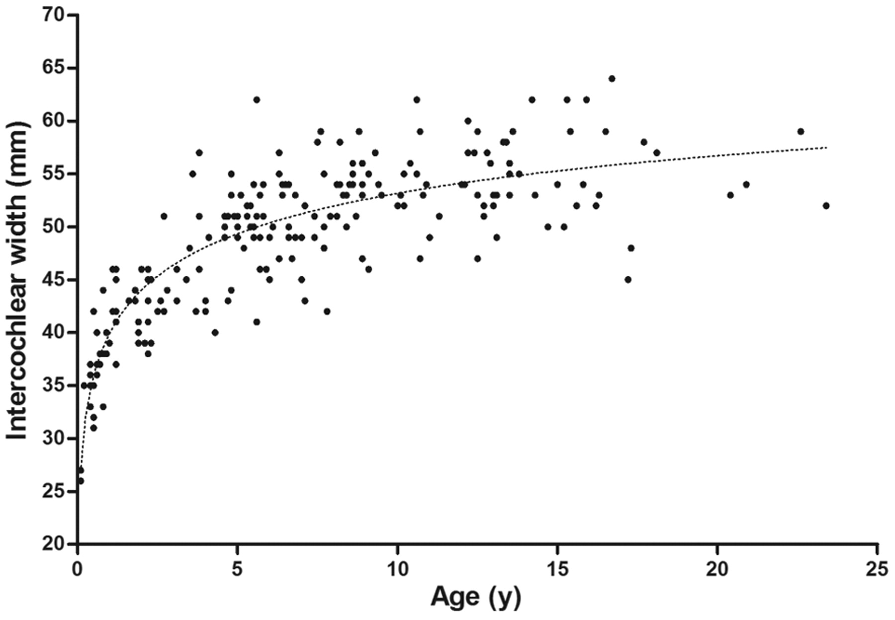
    ICW versus age for all patients.

  • Fig 4.
    • Download figure
    • Open in new tab
    • Download powerpoint
    Fig 4.

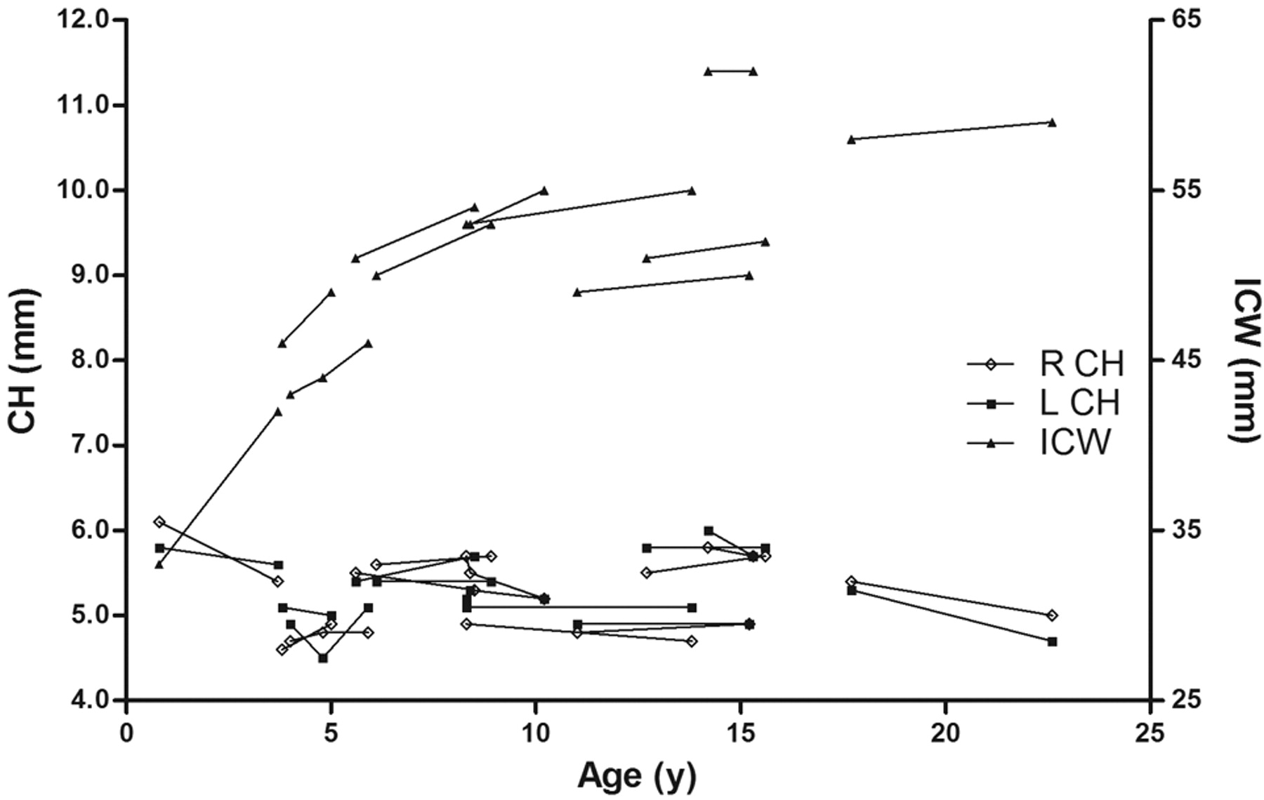
    CH versus age and ICW versus age for patients with multiple imaging studies. Each set of data points connected by a line represents a single patient's left CH, right CH, or ICW.

Tables

  • Figures
    • View popup
    Table 1:

    Results of CT studies of CHa

    Hearing CategoryNo. of EarsMin.Max.MeanSDPValue
    SNHL (all)1622.56.15.230.46.002b
        Males904.06.15.290.37.004b
        Females722.56.15.160.55.33
    CHL (all)1014.26.15.320.37.09
        Males614.26.15.380.39.17
        Females404.76.15.240.33.60
    Mixed HL (all)253.45.54.670.69<.00001b
        Males113.45.54.530.69<.00001b
        Females143.55.44.790.70.008b
    Normal (all)744.56.35.420.381
        Males524.56.35.480.381
        Females224.66.05.280.351
    Unknown (all)604.65.95.290.30.04
        Males324.65.95.320.35.06
        Females284.65.65.260.26.82
    Total (all)4222.56.35.260.45
        Males2463.46.35.320.43.009c
        Females1762.56.15.180.47
    • Note:—Min. indicates minimum; Max, maximum.

    • ↵a Two-sample t test P values are for comparisons among SNHL, CHL, mixed HL, or unknown-hearing ears and normal-hearing ears divided by all, male, or female.

    • ↵b Statistically significant differences (P < .01).

    • ↵c For the comparison of male and female CHs, multivariate linear regression controlling for age and ICW was used.

    • View popup
    Table 2:

    Patients with cochlear hypoplasiaa

    PatientAge (yr) SexL CH (mm)R CH (mm)TYPE HLDiagnosisCT Findings
    16.3 Female2.82.5SNHL, bilaterallyBilateral vestibulocochlear dysplasia, L Mondini malformationBilateral vestibulocochlear dysplasia
    R common chamber malformation, absent vestibular aqueduct, aplastic modiolus
    L Mondini malformation (hypoplastic cochlea with partition defect)
    21.9 Male3.43.6Mixed HLBOR syndromeBilateral dilated vestibular aqueduct with small modiolus, trumpet-shaped IAC, small mass in R middle ear (possible congenital cholesteatoma)
    30.8 Female3.73.5Mixed HLBOR syndromeBilateral tympanostomy tubes, hypoplasia of modiolus, vestibular ectasia, dilated vestibular aqueducts, Mondini deformities (cochlear ectasia with partition defects), question of ossicular fusion
    R middle ear and mastoid air cell opacification; absent vs hypoplastic stapes
    L hypoplastic mastoid with soft tissue thickening at L mesotympanum
    46.7 Female3.64.3Mixed HL, bilaterallyCHARGE syndromeBilateral hypoplastic SCCs, prominent EAC, vestibular dysplasia, cochlear ectasia with partition defects, hypoplastic stapes
    R sclerosis of ossicles, poorly visualized oval window, L poorly defined modiolus
    50.1 Male4.23.8Mixed HL, bilaterallyCHARGE syndromeBilateral absent SCCs, middle ear/mastoid air cell congestion or inflammation, normal middle ear morphology
    66.6 Male4.04.5SNHL, bilaterallyEVA syndromeBilateral enlarged vestibular aqueducts, R cochlear implant, L hypoplastic modiolus
    77.1 Male4.24.3CHL, bilaterallyBilateral class II microtia and EAC atresiaR malformed IAC, partially formed middle ear cavity, poorly defined ossicles, narrow EAC, normal SCCs/vestibule
    L absent lateral SCC, vestibular dysplasia, ossicular dysplasia, absent membranous EAC, normal cochlea
    • Note:—R indicates right; L, left; IAC, internal auditory canal; SCC, semicircular canals; BOR, branchio-oto-renal; EVA, enlarged vestibular aqueduct; EAC, external auditory canal..

    • ↵a CHs >2 SDs below the mean CH.

PreviousNext
Back to top

In this issue

American Journal of Neuroradiology: 33 (1)
American Journal of Neuroradiology
Vol. 33, Issue 1
1 Jan 2012
  • Table of Contents
  • Index by author
Advertisement
Print
Download PDF
Email Article

Thank you for your interest in spreading the word on American Journal of Neuroradiology.

NOTE: We only request your email address so that the person you are recommending the page to knows that you wanted them to see it, and that it is not junk mail. We do not capture any email address.

Enter multiple addresses on separate lines or separate them with commas.
CT Analysis Demonstrates That Cochlear Height Does Not Change with Age
(Your Name) has sent you a message from American Journal of Neuroradiology
(Your Name) thought you would like to see the American Journal of Neuroradiology web site.
CAPTCHA
This question is for testing whether or not you are a human visitor and to prevent automated spam submissions.
Cite this article
M.C. Mori, K.W. Chang
CT Analysis Demonstrates That Cochlear Height Does Not Change with Age
American Journal of Neuroradiology Jan 2012, 33 (1) 119-123; DOI: 10.3174/ajnr.A2713

Citation Manager Formats

  • BibTeX
  • Bookends
  • EasyBib
  • EndNote (tagged)
  • EndNote 8 (xml)
  • Medlars
  • Mendeley
  • Papers
  • RefWorks Tagged
  • Ref Manager
  • RIS
  • Zotero
0 Responses
Respond to this article
Share
Bookmark this article
CT Analysis Demonstrates That Cochlear Height Does Not Change with Age
M.C. Mori, K.W. Chang
American Journal of Neuroradiology Jan 2012, 33 (1) 119-123; DOI: 10.3174/ajnr.A2713
del.icio.us logo Twitter logo Facebook logo Mendeley logo
  • Tweet Widget
  • Facebook Like
  • Google Plus One
Purchase

Jump to section

  • Article
    • Abstract
    • ABBREVIATIONS:
    • Materials and Methods
    • Results
    • Discussion
    • Conclusions
    • Footnotes
    • References
  • Figures & Data
  • Supplemental
  • Info & Metrics
  • Responses
  • References
  • PDF

Related Articles

  • PubMed
  • Google Scholar

Cited By...

  • No citing articles found.
  • Crossref (21)
  • Google Scholar

This article has been cited by the following articles in journals that are participating in Crossref Cited-by Linking.

  • 3D printed biomimetic cochleae and machine learning co-modelling provides clinical informatics for cochlear implant patients
    Iek Man Lei, Chen Jiang, Chon Lok Lei, Simone Rosalie de Rijk, Yu Chuen Tam, Chloe Swords, Michael P. F. Sutcliffe, George G. Malliaras, Manohar Bance, Yan Yan Shery Huang
    Nature Communications 2021 12 1
  • Automated measurement of hydrops ratio from MRI in patients with Ménière’s disease using CNN-based segmentation
    Young Sang Cho, Kyeongwon Cho, Chae Jung Park, Myung Jin Chung, Jong Hyuk Kim, Kyunga Kim, Yi-Kyung Kim, Hyung-Jin Kim, Jae-Wook Ko, Baek Hwan Cho, Won-Ho Chung
    Scientific Reports 2020 10 1
  • Age as a Factor of Growth in Mastoid Thickness and Skull Width
    Fida Abdulaziz Almuhawas, Anandhan E. Dhanasingh, Dijana Mitrovic, Yassin Abdelsamad, Farid Alzhrani, Abdulrahman Hagr, Abdulrahman Al Sanosi
    Otology & Neurotology 2020 41 5
  • The Role of Electrode Placement in Bilateral Simultaneously Cochlear‐Implanted Adult Patients
    Daniele De Seta, Yann Nguyen, Damien Bonnard, Evelyne Ferrary, Benoit Godey, David Bakhos, Michel Mondain, Olivier Deguine, Olivier Sterkers, Daniele Bernardeschi, Isabelle Mosnier
    Otolaryngology–Head and Neck Surgery 2016 155 3
  • Establishing Normal Diameter Range of the Cochlear and Facial Nerves with 3D-CISS at 3T
    Rei NAKAMICHI, Masahiro YAMAZAKI, Mitsuru IKEDA, Haruo ISODA, Hisashi KAWAI, Michihiko SONE, Tsutomu NAKASHIMA, Shinji NAGANAWA
    Magnetic Resonance in Medical Sciences 2013 12 4
  • CT-scan contouring technique allows for direct and reliable measurements of the cochlear duct length: implication in cochlear implantation with straight electrode-arrays
    Thi Hau Vu, Chiara Perazzini, Mathilde Puechmaille, Aurélie Bachy, Aurélien Mulliez, Louis Boyer, Thierry Mom, Jean Gabrillargues
    European Archives of Oto-Rhino-Laryngology 2019 276 8
  • The developing temporal bone: computed tomography measurements and assessment of suture closure from birth to 18 years of age
    P. Paetz, G. F. Goetz, H. Lanfermann, A. M. Giesemann
    Surgical and Radiologic Anatomy 2017 39 6
  • Age-related changes in the number of cresyl-violet-stained, parvalbumin and NMDAR 2B expressing neurons in the human spiral ganglion
    Charanjeet Kaur, Shubhi Saini, Indra Pal, Punit Kumar, Hem Chandra Sati, Tony George Jacob, Daya Nand Bhardwaj, Tara Sankar Roy
    Hearing Research 2020 388
  • CT Scan Imaging of the Human Fetal Labyrinth
    Mehdi Mejdoubi, Fabrice Dedouit, Fatima-Zohra Mokrane, Norbert Telmon
    Otology & Neurotology 2016 37 5
  • Diagnostic Value of 3D Segmentation in Understanding the Anatomy of Human Inner Ear Including Malformation Types
    Saad Alenzi, Anandhan Dhanasingh, Hani Alanazi, Abdulrahman Alsanosi, Abdulrahman Hagr
    Ear, Nose & Throat Journal 2021 100 5_suppl

More in this TOC Section

  • Parathyroid Lesions: Characterization with Dual-Phase Arterial and Venous Enhanced CT of the Neck
  • Correlation of Apparent Diffusion Coefficient at 3T with Prognostic Parameters of Retinoblastoma
  • Efficacy of Diffusion-Weighted Imaging for the Differentiation between Lymphomas and Carcinomas of the Nasopharynx and Oropharynx: Correlations of Apparent Diffusion Coefficients and Histologic Features
Show more HEAD AND NECK

Similar Articles

Advertisement

Indexed Content

  • Current Issue
  • Accepted Manuscripts
  • Article Preview
  • Past Issues
  • Editorials
  • Editor's Choice
  • Fellows' Journal Club
  • Letters to the Editor
  • Video Articles

Cases

  • Case Collection
  • Archive - Case of the Week
  • Archive - Case of the Month
  • Archive - Classic Case

More from AJNR

  • Trainee Corner
  • Imaging Protocols
  • MRI Safety Corner
  • Book Reviews

Multimedia

  • AJNR Podcasts
  • AJNR Scantastics

Resources

  • Turnaround Time
  • Submit a Manuscript
  • Submit a Video Article
  • Submit an eLetter to the Editor/Response
  • Manuscript Submission Guidelines
  • Statistical Tips
  • Fast Publishing of Accepted Manuscripts
  • Graphical Abstract Preparation
  • Imaging Protocol Submission
  • Evidence-Based Medicine Level Guide
  • Publishing Checklists
  • Author Policies
  • Become a Reviewer/Academy of Reviewers
  • News and Updates

About Us

  • About AJNR
  • Editorial Board
  • Editorial Board Alumni
  • Alerts
  • Permissions
  • Not an AJNR Subscriber? Join Now
  • Advertise with Us
  • Librarian Resources
  • Feedback
  • Terms and Conditions
  • AJNR Editorial Board Alumni

American Society of Neuroradiology

  • Not an ASNR Member? Join Now

© 2025 by the American Society of Neuroradiology All rights, including for text and data mining, AI training, and similar technologies, are reserved.
Print ISSN: 0195-6108 Online ISSN: 1936-959X

Powered by HighWire