
Article Figures & Data
Figures
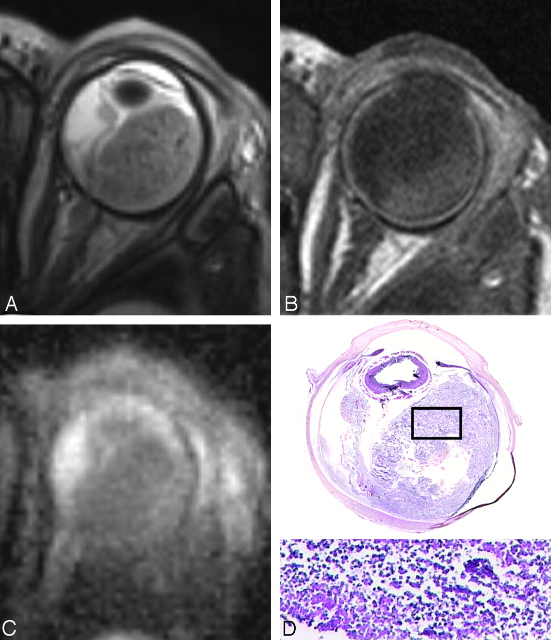
- Fig 1.
A 34-month-old girl with sporadic unilateral retinoblastoma of the right eye. A and B, Axial T2-weighted (A) and contrast-enhanced T1-weighted (B) MR images show an anteriorly located retinoblastoma with inhomogeneous SI. C, Axial DWI (b-value, 1000 s/mm2) shows a markedly increased SI anteriorly in the mass on DWI (arrowhead) compared with the posterior part of the mass (arrow). D, On the ADC map, a reciprocal pattern is recognized with the lowest ADC values anterior in the tumor (mean ADC value, 1.09 × 10⁻³ mm2/s). Axial ADC map shows linear blurring artifacts caused by the long echo train in the HASTE sequence. E, Histopathologic specimen shows poorly differentiated vital tumor tissue anteriorly (upper inset) and necrosis with some foci of vital tumor tissue posteriorly (lower inset) in the eye (H&E, original magnification ×3.5).
- Fig 2.
A and B, A 27-month-old boy with sporadic unilateral retinoblastoma of the left eye. Axial T2-weighted (A) and contrast-enhanced T1-weighted (B) MR images show a diffuse infiltrating retinoblastoma along the totally detached retina. C and D, On the axial DWI (b-value, 1000 s/mm2) (C), the tumor shows diffuse hyperintense SI with a corresponding low ADC value (mean ADC value, 1.09 × 10⁻³ mm2/s) (D). The mean ADC value of subretinal fluid is slightly lower compared with the remaining vitreous (1.99 versus 2.64 × 10⁻³ mm2/s, respectively). E, Histopathologic specimen shows a totally detached retina, diffusely infiltrated by retinoblastoma (H&E, original magnification 3.5). Slightly more tumor tissue is present in the medial part of the eye on histopathology compared with the MR imaging because of a minor angulation within the cutting plane compared with the axial plane of the MR image. F, Inset: photomicrograph of the specimen shows poorly differentiated vital retinoblastoma, with densely packed cells (H&E, original magnification ×20).
- Fig 3.
A 30-month-old girl with enucleation of the right eye because of sporadic bilateral retinoblastoma. A−C, Axial T2-weighted MR image (A), contrast-enhanced T1-weighted MR image (B), and ADC map (C) show multifocal retinoblastoma with a large subretinal mass (mean ADC value of the tumor mass, 1.03 × 10⁻³ mm2/s; of the subretinal fluid, 2.35 × 10⁻³ mm2/s) and multiple small nodules along the detached retina, which can also be encountered on the ADC map (too small for ADC measurement). D, Photomicrograph of a histopathologic specimen shows a multifocal poorly differentiated vital retinoblastoma, with multiple small noduli along the detached retina (H&E, original magnification ×3.5).
- Fig 4.
A 9-month-old boy with sporadic unilateral retinoblastoma of the left eye. A and B, Axial T2-weighted (A) and contrast-enhanced T1-weighted (B) MR images show a relatively homogeneous tumor mass combined with retinal detachment, compatible with retinoblastoma. C and D, On axial DWI (b-value, 1000 s/mm2) (C), the tumor shows diffuse hyperintense SI with corresponding low ADC values (mean ADC value, 0.82 × 10⁻³ mm2/s) (D). E, Histopathologic specimen shows a compact subretinal tumor mass located posteriorly in the eye combined with retinal detachment (H&E, original magnification × 3.5). F, Inset: photomicrograph of the specimen shows poorly differentiated vital retinoblastoma, with densely packed cells (H&E, original magnification ×20).
- Fig 5.
A 1-month-old girl with sporadic unilateral retinoblastoma of the left eye, who presented with orbital cellulitis and uveitis secondary to necrotic multifocal retinoblastoma. A, Total retinal detachment with a large subretinal mass temporal in the eye with low SI on a T2-weighted MR image, in combination with lens luxation and inflammatory changes in the preseptal soft tissues. B, The contrast-enhanced T1-weighted MR image shows absence of enhancement of the retinoblastoma; high SI posterior and temporal in the eye is also present on the precontrast T1-weighted image (not shown) and is suspicious for (hemorrhagic) necrosis. Note strong enhancement of the choroid with absence of iris enhancement. C, ADC map shows intermediate SI throughout the tumor (mean ADC values, tumor 1.73 × 10⁻³ mm2/s). D, Histopathologic specimen shows multifocal retinoblastoma with a large friable subretinal mass (H&E, original magnification × 3.5). Inset: photomicrograph of the specimen shows details of necrotic tumor cells with loss of cell cohesion (H&E, original magnification, ×3.5).
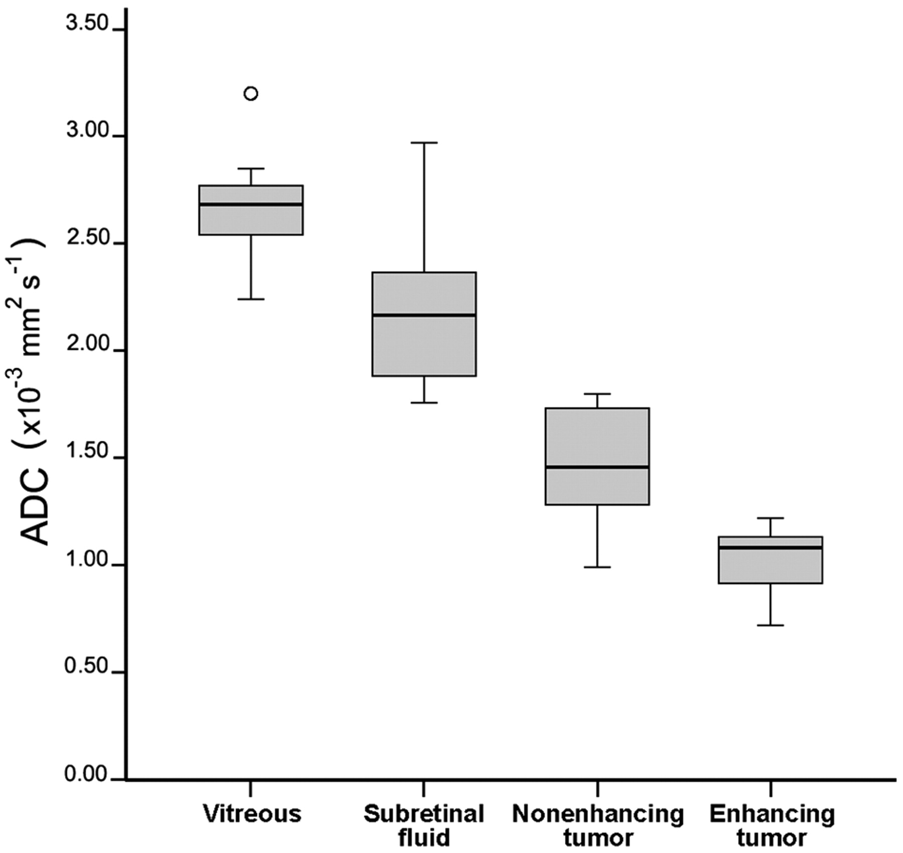
- Fig 6.
Box-and-whisker plots show the results for ADCs of the vitreous, subretinal fluid, and nonenhancing and enhancing tumor parts. There is hardly any overlap in the range of ADC values between enhancing and nonenhancing tumor parts of the affected eyes or between nonenhancing tumor parts and vitreous or subretinal fluid. The center line indicates the median; the bottom and top of the box, 25th and 75th percentiles respectively; ο, outlier.


Tables
Mean of ADC values for retinoblastoma, subretinal fluid, and vitreous
Parameter ADC Value Range Retinoblastoma Enhancing tumor part 1.03 ± 0.15 0.72–1.22 Nonenhancing tumor part 1.47 ± 0.27 0.99–1.80 Subretinal fluid 2.20 ± 0.37 1.76–2.97 Hemorrhage 2.08 ± 0.22 Effusion 2.24 ± 0.19 Vitreous 2.67 ± 0.28 2.24–3.20 With tumor seeding 2.58 ± 0.21 Without tumor seeding 2.75 ± 0.33


