Advertisement

Main menu

  • Home
  • Content
    • Current Issue
    • Accepted Manuscripts
    • Article Preview
    • Past Issue Archive
    • Video Articles
    • AJNR Case Collection
    • Case of the Week Archive
    • Case of the Month Archive
    • Classic Case Archive
  • Special Collections
    • AJNR Awards
    • Low-Field MRI
    • Alzheimer Disease
    • ASNR Foundation Special Collection
    • Photon-Counting CT
    • View All
  • Multimedia
    • AJNR Podcasts
    • AJNR SCANtastic
    • Trainee Corner
    • MRI Safety Corner
    • Imaging Protocols
  • For Authors
    • Submit a Manuscript
    • Submit a Video Article
    • Submit an eLetter to the Editor/Response
    • Manuscript Submission Guidelines
    • Statistical Tips
    • Fast Publishing of Accepted Manuscripts
    • Graphical Abstract Preparation
    • Imaging Protocol Submission
    • Author Policies
  • About Us
    • About AJNR
    • Editorial Board
    • Editorial Board Alumni
  • More
    • Become a Reviewer/Academy of Reviewers
    • Subscribers
    • Permissions
    • Alerts
    • Feedback
    • Advertisers
    • ASNR Home

User menu

  • Alerts
  • Log in

Search

  • Advanced search
American Journal of Neuroradiology

American Journal of Neuroradiology

ASHNR American Society of Functional Neuroradiology ASHNR American Society of Pediatric Neuroradiology ASSR
  • Alerts
  • Log in

Advanced Search

  • Home
  • Content
    • Current Issue
    • Accepted Manuscripts
    • Article Preview
    • Past Issue Archive
    • Video Articles
    • AJNR Case Collection
    • Case of the Week Archive
    • Case of the Month Archive
    • Classic Case Archive
  • Special Collections
    • AJNR Awards
    • Low-Field MRI
    • Alzheimer Disease
    • ASNR Foundation Special Collection
    • Photon-Counting CT
    • View All
  • Multimedia
    • AJNR Podcasts
    • AJNR SCANtastic
    • Trainee Corner
    • MRI Safety Corner
    • Imaging Protocols
  • For Authors
    • Submit a Manuscript
    • Submit a Video Article
    • Submit an eLetter to the Editor/Response
    • Manuscript Submission Guidelines
    • Statistical Tips
    • Fast Publishing of Accepted Manuscripts
    • Graphical Abstract Preparation
    • Imaging Protocol Submission
    • Author Policies
  • About Us
    • About AJNR
    • Editorial Board
    • Editorial Board Alumni
  • More
    • Become a Reviewer/Academy of Reviewers
    • Subscribers
    • Permissions
    • Alerts
    • Feedback
    • Advertisers
    • ASNR Home
  • Follow AJNR on Twitter
  • Visit AJNR on Facebook
  • Follow AJNR on Instagram
  • Join AJNR on LinkedIn
  • RSS Feeds

AJNR Awards, New Junior Editors, and more. Read the latest AJNR updates

LetterLetter

MR Imaging of Fetal Goiter

J. H. Harreld, R.K. Kilani, C.D. Lascola and S.K. Bartz
American Journal of Neuroradiology September 2011, 32 (8) E160; DOI: https://doi.org/10.3174/ajnr.A2646
J. H. Harreld
  • Find this author on Google Scholar
  • Find this author on PubMed
  • Search for this author on this site
R.K. Kilani
  • Find this author on Google Scholar
  • Find this author on PubMed
  • Search for this author on this site
C.D. Lascola
  • Find this author on Google Scholar
  • Find this author on PubMed
  • Search for this author on this site
S.K. Bartz
  • Find this author on Google Scholar
  • Find this author on PubMed
  • Search for this author on this site
  • Article
  • Figures & Data
  • Info & Metrics
  • Responses
  • References
  • PDF
Loading

Fetal goiter is a rare potential cause of airway obstruction and neonatal hypoxic-ischemic brain injury or death.1 MR imaging evaluation of the enlarged gland and airway can provide information crucial to perinatal management. Because the neuroradiologist may be required to apply expertise in MR imaging and in head and neck anatomy to these cases, we describe imaging findings in fetal goiter and discuss fetal MR imaging protocol optimization.

Screening sonography of the 33-week fetus of a woman with Graves disease on propylthiouracil (PTU) therapy revealed a homogeneous 5.3-cm bilobed anterior neck mass suspicious for goiter and mild polyhydramnios. The distal femoral ossification center was not seen, and fetal heart rate was normal. The PTU dose was reduced, and subsequent fetal MR imaging (Fig 1) at 34.5 weeks' gestation demonstrated a T1 hyperintense and T2 low-intensity bilobed anterior neck mass consistent with goiter, slightly decreased in size. The airway was poorly visualized at the level of the oropharynx but was otherwise well-defined to the level of the carina, with ∼1 cm left-to-right midline tracheal shift. Due to potential airway compromise, the ex utero intrapartum treatment procedure was performed during cesarean delivery at 36 weeks to secure the neonatal airway. American Pediatric Gross Assessment Records (APGARs) were 3 at 1 minute and 8 at 5 minutes. Sonography on day 2 of life demonstrated decreasing enlargement of the thyroid with mild hyperemia. By day 3 of life, the patient was extubated and did well on room air, with occasional stridor at 1-month follow-up. Results of thyroid function and hormonal studies remained normal during the first 2 months of life.

Fig 1.
  • Download figure
  • Open in new tab
  • Download powerpoint
Fig 1.

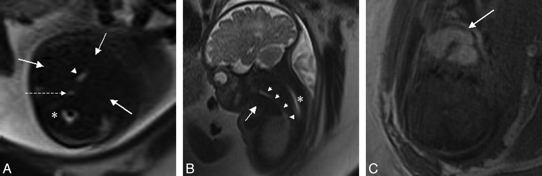
MR imaging of fetal goiter at 34.5 weeks' gestation. A and B, The fluid-filled airway (arrowheads) is well-evaluated on axial (A) and sagittal (B) T2-weighted images to the level of the carina, with poor visualization at the level of the oropharynx. The fluid-filled esophagus (dashed arrow) and spinal canal (asterisk) are also seen. Goitrous fetal thyroid (solid arrows) is isointense to muscle on T2-weighted images, limiting delineation. C, Hyperintensity on T1-weighted images better defines the enlarged fetal thyroid.

Fetal goiter occurs in ∼8% of fetuses of hyperthyroid mothers on antithyroid therapy and manifests as a symmetric homogeneous solid bilobed anterior neck mass on sonography after the fetal thyroid begins to function late in the first trimester.2,3 Depending on the mother's antibody and medication status, the fetus may be hyperthyroid, euthyroid, or hypothyroid. Fetal tachycardia suggests a hyperthyroid state, and hypoplasia of the distal femoral ossification center may indicate hypothyroidism.2

Although neonatal thyroid status generally normalizes within 1 month of birth,3 fetal goiter may result in significant complications at delivery due to airway obstruction, including hypoxic-ischemic brain injury and death.1 Fixed hyperextension of the neck due to mass effect may necessitate cesarean delivery; polyhydramnios due to poor fetal swallowing indicates potential airway obstruction and should prompt MR imaging.2 Although MR imaging may be performed at any time after the first trimester, MR imaging performed closer to delivery will best predict peripartum anatomy. The rapidity of the T2-weighted single-shot fast spin-echo (SS-FSE) acquisition enables imaging of the moving fetus without significant motion degradation and provides excellent delineation of the fluid-filled fetal airway. Although thyroid T2 signal intensity varies with functional status, hypointense if protein and iodine concentration are high and hyperintense if they are low, T1 hyperintensity is more consistent.4 T1-weighted images to better define the thyroid are, therefore, recommended to complement visualization of the airway on standard T2-weighted SS-FSE fetal MR images.

References

  1. 1.↵
    1. Ogamo M,
    2. Sugiyama T,
    3. Maeda Y,
    4. et al
    . The ex utero intrapartum treatment (EXIT) procedure in giant fetal neck masses. Fetal Diagn Ther 2005;20:214–18
    CrossRefPubMed
  2. 2.↵
    1. Polak M,
    2. Le Gac I,
    3. Vuillard E,
    4. et al
    . Fetal and neonatal thyroid function in relation to maternal Graves' disease. Best Pract Res Clin Endocrinol Metab 2004;18:289–302
    CrossRefPubMed
  3. 3.↵
    1. Rosenfeld H,
    2. Ornoy A,
    3. Shechtman S,
    4. et al
    . Pregnancy outcome, thyroid dysfunction and fetal goitre after in utero exposure to propylthiouracil: a controlled cohort study. Br J Clin Pharmacol 2009;68:609–17
    CrossRefPubMed
  4. 4.↵
    1. Kondoh M,
    2. Miyazaki O,
    3. Imanishi Y,
    4. et al
    . Neonatal goiter with congenital thyroid dysfunction in two infants diagnosed by MRI. Pediatr Radiol 2004;34:570–73
    PubMed
  • © 2011 by American Journal of Neuroradiology
PreviousNext
Back to top

In this issue

American Journal of Neuroradiology: 32 (8)
American Journal of Neuroradiology
Vol. 32, Issue 8
1 Sep 2011
  • Table of Contents
  • Index by author
Advertisement
Print
Download PDF
Email Article

Thank you for your interest in spreading the word on American Journal of Neuroradiology.

NOTE: We only request your email address so that the person you are recommending the page to knows that you wanted them to see it, and that it is not junk mail. We do not capture any email address.

Enter multiple addresses on separate lines or separate them with commas.
MR Imaging of Fetal Goiter
(Your Name) has sent you a message from American Journal of Neuroradiology
(Your Name) thought you would like to see the American Journal of Neuroradiology web site.
CAPTCHA
This question is for testing whether or not you are a human visitor and to prevent automated spam submissions.
Cite this article
J. H. Harreld, R.K. Kilani, C.D. Lascola, S.K. Bartz
MR Imaging of Fetal Goiter
American Journal of Neuroradiology Sep 2011, 32 (8) E160; DOI: 10.3174/ajnr.A2646

Citation Manager Formats

  • BibTeX
  • Bookends
  • EasyBib
  • EndNote (tagged)
  • EndNote 8 (xml)
  • Medlars
  • Mendeley
  • Papers
  • RefWorks Tagged
  • Ref Manager
  • RIS
  • Zotero
0 Responses
Respond to this article
Share
Bookmark this article
MR Imaging of Fetal Goiter
J. H. Harreld, R.K. Kilani, C.D. Lascola, S.K. Bartz
American Journal of Neuroradiology Sep 2011, 32 (8) E160; DOI: 10.3174/ajnr.A2646
del.icio.us logo Twitter logo Facebook logo Mendeley logo
  • Tweet Widget
  • Facebook Like
  • Google Plus One
Purchase

Jump to section

  • Article
    • References
  • Figures & Data
  • Info & Metrics
  • Responses
  • References
  • PDF

Related Articles

  • No related articles found.
  • PubMed
  • Google Scholar

Cited By...

  • No citing articles found.
  • Crossref
  • Google Scholar

This article has not yet been cited by articles in journals that are participating in Crossref Cited-by Linking.

More in this TOC Section

  • Brain AVM’s Nidus: What if We Hadn’t Understood Anything?
  • Letter to the Editor regarding “Automated Volumetric Software in Dementia: Help or Hindrance to the Neuroradiologist?”
  • Reply:
Show more LETTERS

Similar Articles

Advertisement

Indexed Content

  • Current Issue
  • Accepted Manuscripts
  • Article Preview
  • Past Issues
  • Editorials
  • Editor's Choice
  • Fellows' Journal Club
  • Letters to the Editor
  • Video Articles

Cases

  • Case Collection
  • Archive - Case of the Week
  • Archive - Case of the Month
  • Archive - Classic Case

More from AJNR

  • Trainee Corner
  • Imaging Protocols
  • MRI Safety Corner
  • Book Reviews

Multimedia

  • AJNR Podcasts
  • AJNR Scantastics

Resources

  • Turnaround Time
  • Submit a Manuscript
  • Submit a Video Article
  • Submit an eLetter to the Editor/Response
  • Manuscript Submission Guidelines
  • Statistical Tips
  • Fast Publishing of Accepted Manuscripts
  • Graphical Abstract Preparation
  • Imaging Protocol Submission
  • Evidence-Based Medicine Level Guide
  • Publishing Checklists
  • Author Policies
  • Become a Reviewer/Academy of Reviewers
  • News and Updates

About Us

  • About AJNR
  • Editorial Board
  • Editorial Board Alumni
  • Alerts
  • Permissions
  • Not an AJNR Subscriber? Join Now
  • Advertise with Us
  • Librarian Resources
  • Feedback
  • Terms and Conditions
  • AJNR Editorial Board Alumni

American Society of Neuroradiology

  • Not an ASNR Member? Join Now

© 2025 by the American Society of Neuroradiology All rights, including for text and data mining, AI training, and similar technologies, are reserved.
Print ISSN: 0195-6108 Online ISSN: 1936-959X

Powered by HighWire