
Article Figures & Data
Figures
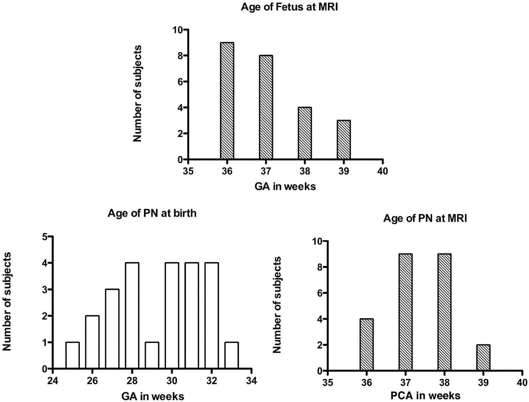
- Fig 1.
Age distribution of fetus and PN. The GA and PCA are expressed in weeks.
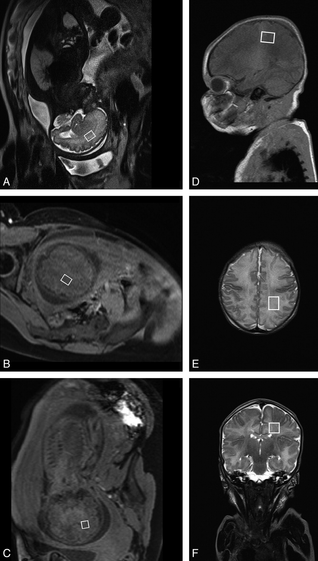
- Fig 2.
Localization of the voxel for 1H-MR spectroscopy. A–C, fetus; D–F, PN.
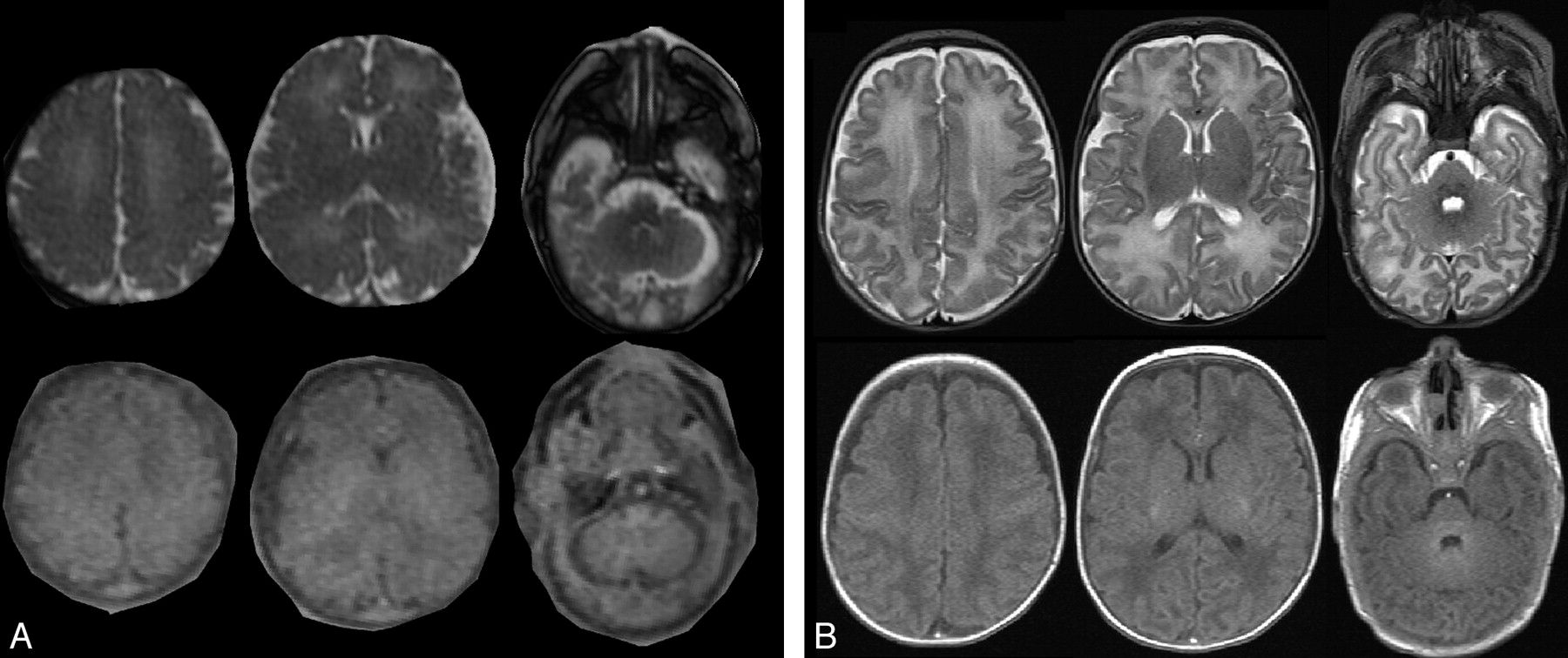
- Fig 3.
Typical axial T1- and T2-weighted MR imaging from an in utero fetus and a PN. A, Brain MR imaging of a fetus explored at 37 weeks GA. Axial T2-weighted images (HASTE sequence; top row). Axial T1-weighted images (bottom row). B, Brain MR imaging of a PN born at 27 weeks GA and explored at 38 weeks PCA. Top row, Axial T2-weighted images. Bottom row, Axial T1-weighted images. Images were obtained at the level of the centrum semiovale, basal ganglia, and posterior fossa (from left to right).
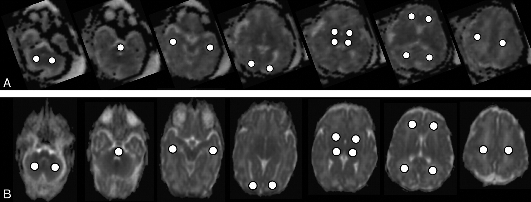
- Fig 4.
Typical cerebral ADC maps from a fetus and a preterm neonate. Row A, ADC maps of a fetus (GA = 36 weeks). Row B, ADC maps of a preterm neonate (PCA = 36 weeks). The location of the ROIs within all analyzed brain areas is displayed within the images (for each row from left to right: cerebellum, pons, temporal white matter; occipital white matter, thalamus and basal ganglia; frontal and parietal white matter, centrum semiovale).
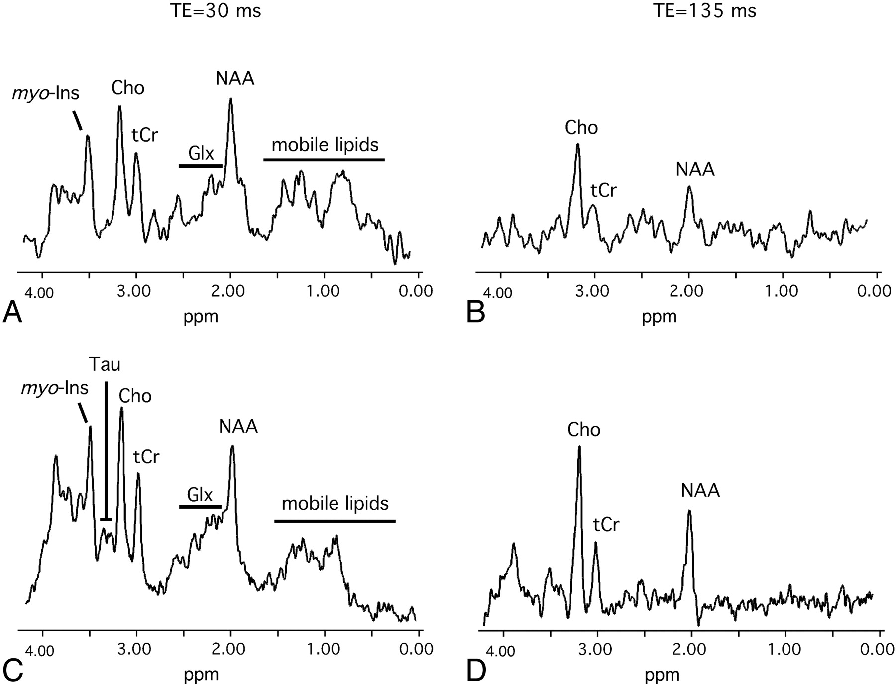
- Fig 5.
Typical brain 1H-MR spectra from a fetus and a PN obtained in the deep white matter. A and B, MR spectra from a fetus in utero near term (GA = 36 weeks). C and D, MR spectra from a preterm neonate at term equivalent (PCA = 36 weeks).


Tables
Cerebral Structure Fetuses (n = 24) ADC (× 10−6 mm2/s) PNs (n = 24) ADC (× 10−6 mm2/s) Unpaired t Test, P Value Cerebellum 1.20 ± 0.02 1.19 ± 0.02 .6839 Pons 1.07 ± 0.02 0.98 ± 0.02 .0008 Thalamus 1.14 ± 0.02 1.10 ± 0.01 .1251 Basal ganglia 1.26 ± 0.02 1.21 ± 0.01 .0869 Temporal white matter 1.53 ± 0.03 1.57 ± 0.02 .2257 Occipital white matter 1.50 ± 0.03 1.56 ± 0.02 .1236 Frontal white matter 1.75 ± 0.02 1.77 ± 0.03 .4981 Parietal white matter 1.66 ± 0.03 1.77 ± 0.03 .0172 Centrum semiovale 1.44 ± 0.03 1.44 ± 0.03 .8654 -
Note:—Data are mean ± SEM.
-
Metabolite Ratio Fetuses (n = 20 or 19a) PNs (n = 20) Unpaired t Test, P Value TE = 30 ms NAA/H2O 2.19 ± 0.19 2.76 ± 0.16 .0279 tCr/H2O 2.18 ± 0.20 2.80 ± 0.16 .0184 Cho/H2O 2.98 ± 0.21 3.53 ± 0.22 .0818 Glx/H2O 5.36 ± 0.35 6.38 ± 0.29 .0324 Tau/H2O 0.56 ± 0.09 0.63 ± 0.06 .6525 mIns/H2O 1.64 ± 0.17 2.17 ± 0.17 .0332 Lipids/H2O 9.98 ± 2.44 7.28 ± 0.65 .2986 TE = 135 ms NAA/H2O 2.00 ± 0.22 2.07 ± 0.12 .7685 tCr/H2O 1.33 ± 0.21 1.30 ± 0.07 .8697 Cho/H2O 3.50 ± 0.20 3.47 ± 0.16 .8891 Lac/H2O 0.00 ± 0.00 0.00 ± 0.00 -
Note:—Data are mean ± SEM.
-
a n = 20 for a TE of 30 ms and 19 for TE of 135 ms.
-
Metabolite Ratio Fetuses (n = 20 or 19a) PNs (n = 20) Unpaired t Test, P Value TE = 30 ms NAA/Cho 0.73 ± 0.05 0.77 ± 0.03 .3926 NAA/Cr 1.02 ± 0.06 0.98 ± 0.03 .5588 Cho/tCr 1.39 ± 0.06 1.25 ± 0.03 .0318 mIns/tCr 0.75 ± 0.06 0.76 ± 0.04 .8682 mIns/Cho 0.55 ± 0.05 0.76 ± 0.04 .0013 mIns/NAA 0.75 ± 0.06 0.77 ± 0.03 .7542 Glx/tCr 2.67 ± 0.24 2.36 ± 0.13 .2594 Glx/Cho 1.93 ± 0.16 1.90 ± 0.11 .9145 Glx/NAA 2.80 ± 0.34 2.40 ± 0.14 .2868 TE = 135 ms NAA/Cho 0.59 ± 0.08 0.60 ± 0.03 .8463 NAA/Cr 1.74 ± 0.32 1.64 ± 1.08 .8352 Cho/tCr 3.06 ± 0.34 2.73 ± 0.12 .4597 -
Note:—Data are mean ± SEM.
-
a n = 20 for a TE of 30 ms and 19 for TE of 135 ms.
-


