
Article Figures & Data
Figures
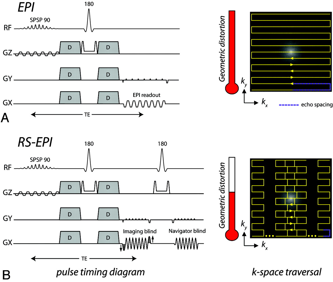
- Fig 1.
Pulse-timing diagram and k-space traversal of the (A) EPI and (B) RS-EPI trajectories. In RS-EPI, each imaging blind is accompanied by a navigator blind acquired at the center of k-space (not shown in the k-space traversal schematic) to perform a phase correction that will correct for phase differences between blinds. The purple dotted line denotes the “echo-spacing”—that is, the time between consecutive echoes in an EPI echo-train. The amount of geometric distortion in an image is proportional to this. The distortion meter shows the resulting distortion reduction (drawn to scale) with the use of RS-EPI, assuming a blind width of 64 and a matrix size of 192 × 192.
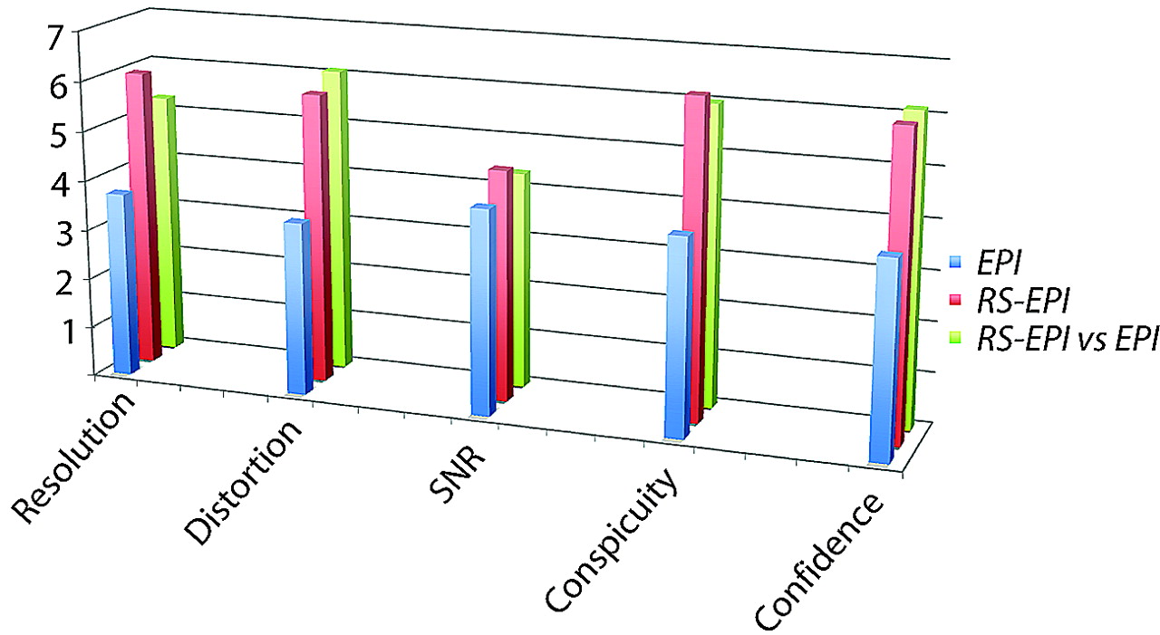
- Fig 2.
Comparison between the routine ASSET-accelerated EPI sequence and our implementation of RS-EPI in terms of 5 categories averaged over 35 patients.
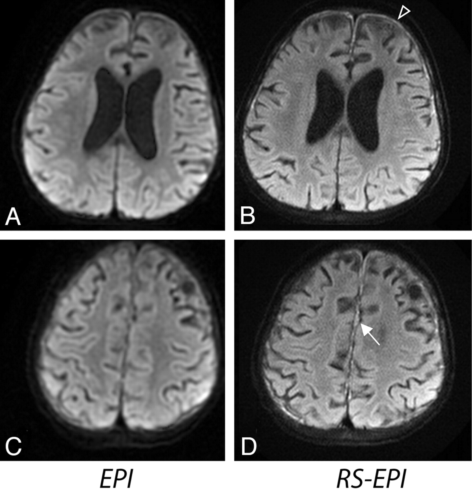
- Fig 3.
An 18-month-old boy presenting with subdural empyema. A and B, Abscess depicted with greater diagnostic confidence on RS-EPI. C and D, Pus present along the falx on RS-EPI (D), not well seen on EPI (C).
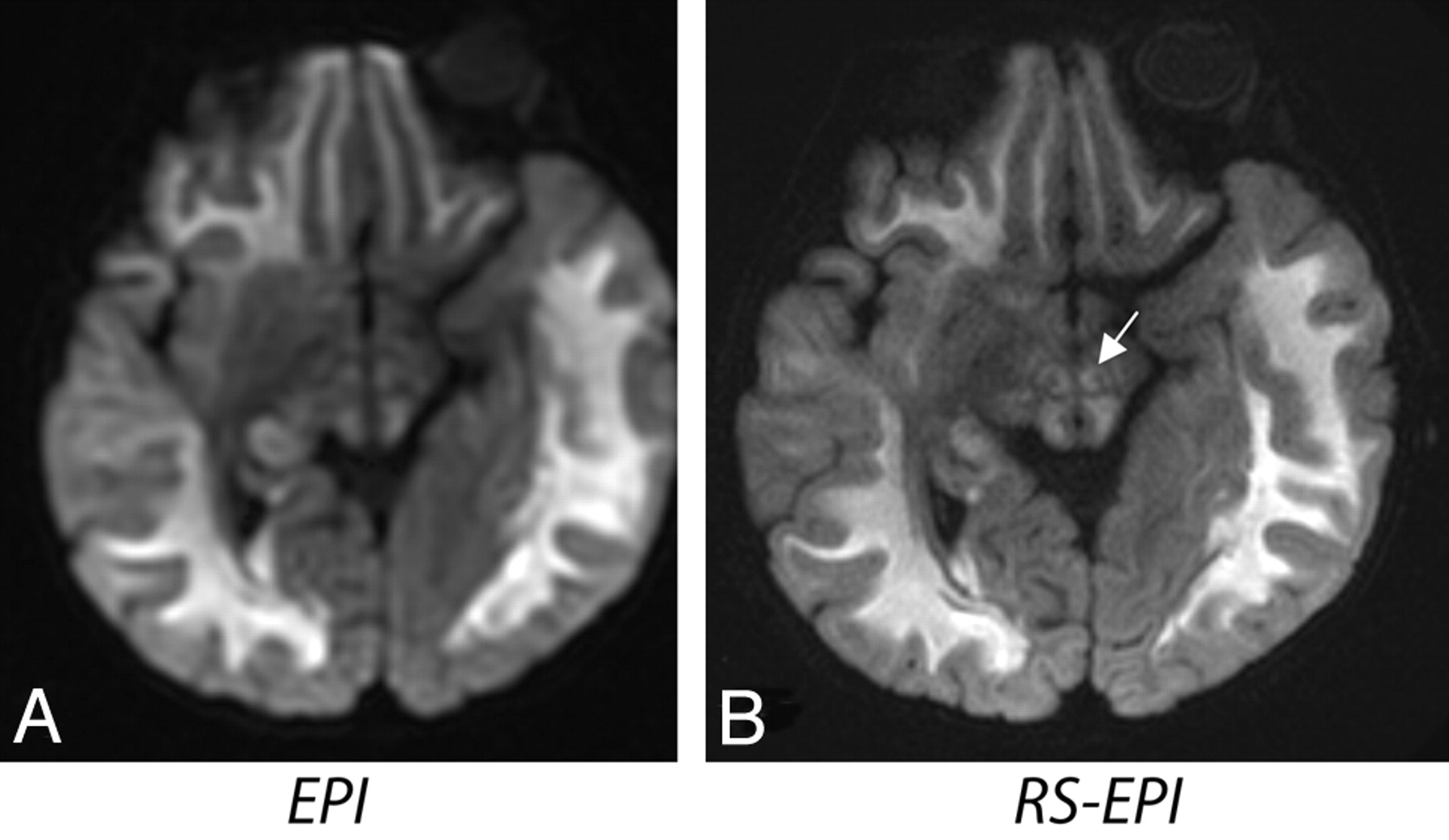
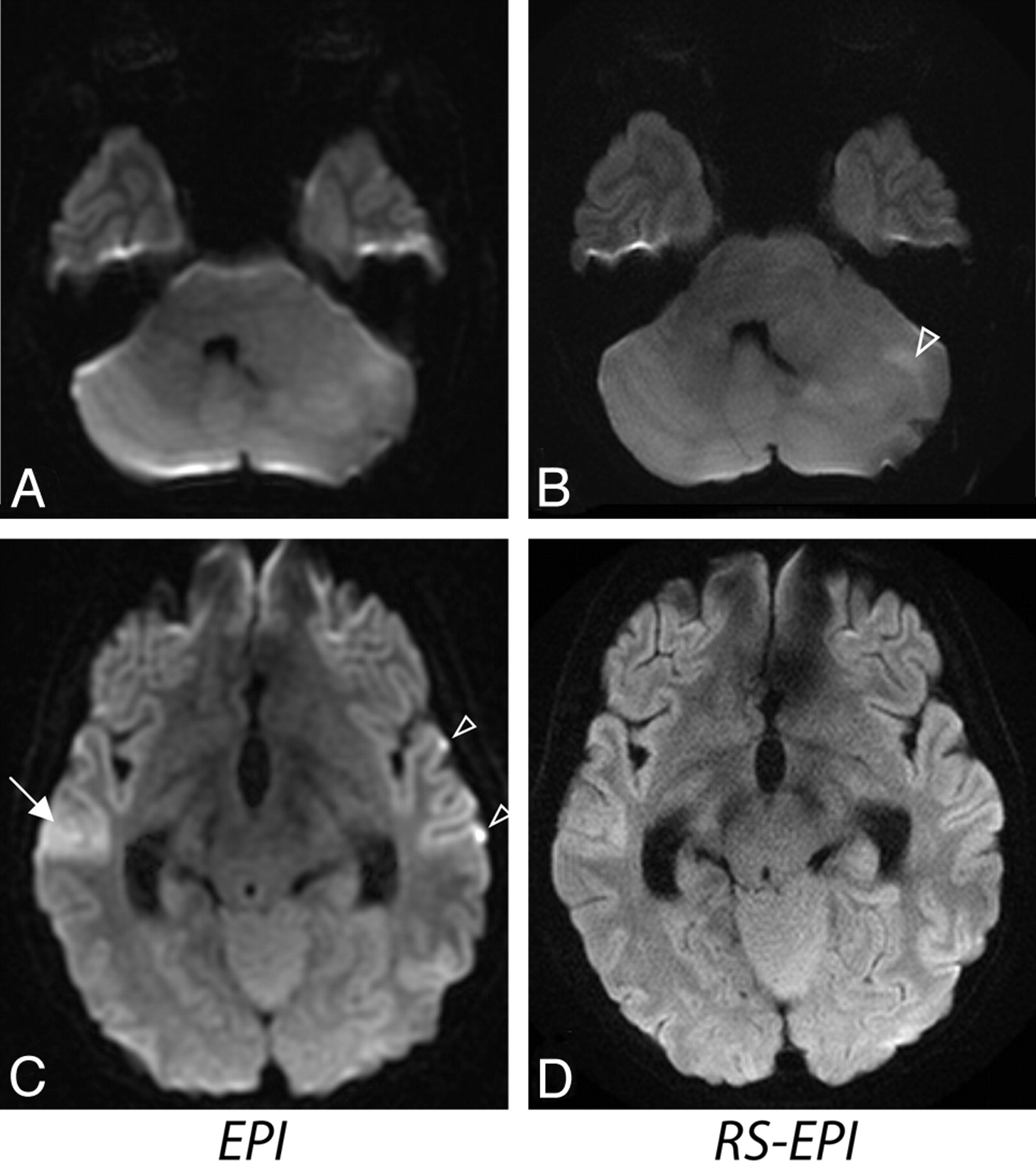
- Fig 4.
A 10-month-old girl with Leigh disease. RS-EPI shows exquisite resolution at the level of the brain stem, specifically in the midbrain. The red nucleus is also better defined on RS-EPI (white arrow).
- Fig 5.
A 10-month-old boy presenting with cystic encephalomalacia. RS-EPI demonstrates cystic encephalomalacic changes with higher resolution.
- Fig 6.
Two patients with Moyamoya disease. A and B, An 8-year-old girl presenting with possible infarct or blood product at the surgical site on EPI (solid white arrow). The absence of these distortion artifacts on RS-EPI makes this confidently negative. The open white arrows indicate undesirable brightening of the flocculus due to susceptibility artifacts from the brain/bone interface. C and D, A 3-year-old boy presenting with possible postoperative blood-product artifacts on EPI (white arrow), but the lesion appears more suspicious for an ischemic lesion on RS-EPI. On closer inspection, the lesion demonstrates subtle cortical T2 high intensity on the T2 FSE sequence, confirming the suspicion that this represents a true ischemic focus, rather than distortion related to postoperative changes. These 2 cases demonstrate that RS-EPI is both sensitive and specific.
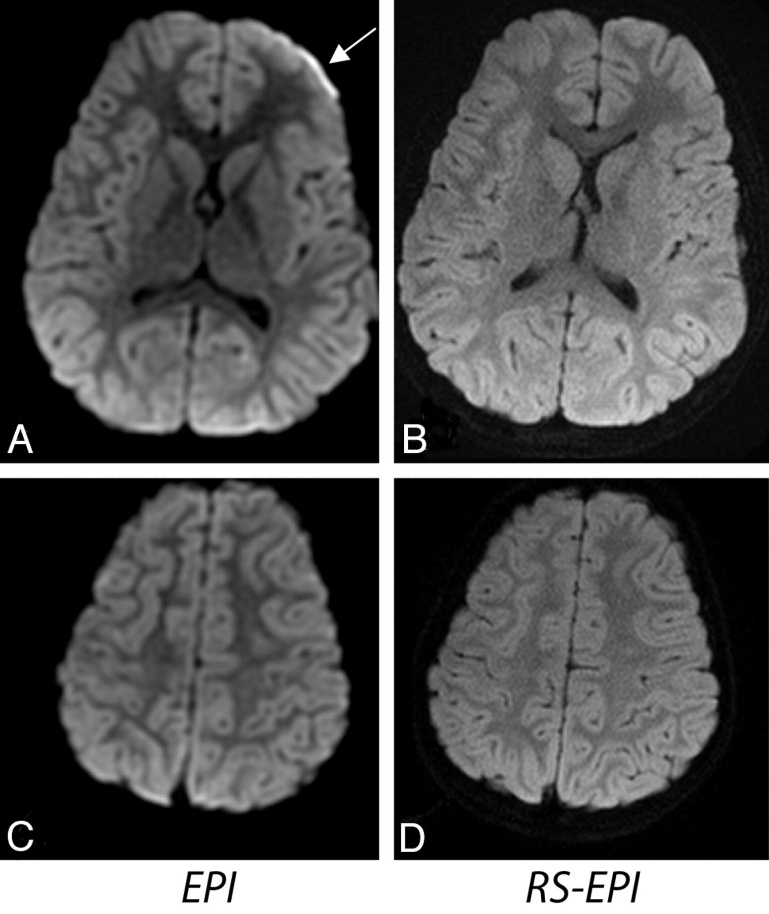
- Fig 7.
Two sections from a patient with diffuse infiltrating anaplastic astrocytoma (a 14-year-old girl). A and B, DWI shows areas of increased cellularity (open white arrow). C and D, Areas of increased signal intensity on EPI (white arrow) suggest the possibility of cortical and subcortical tumor involvement, which may be seen with gliomatosis or diffuse infiltrating glioma. Reduction of distortion in RS-EPI shows a normal cortical ribbon. Also, the open arrows show areas of increased distortion on EPI compared with RS-EPI.
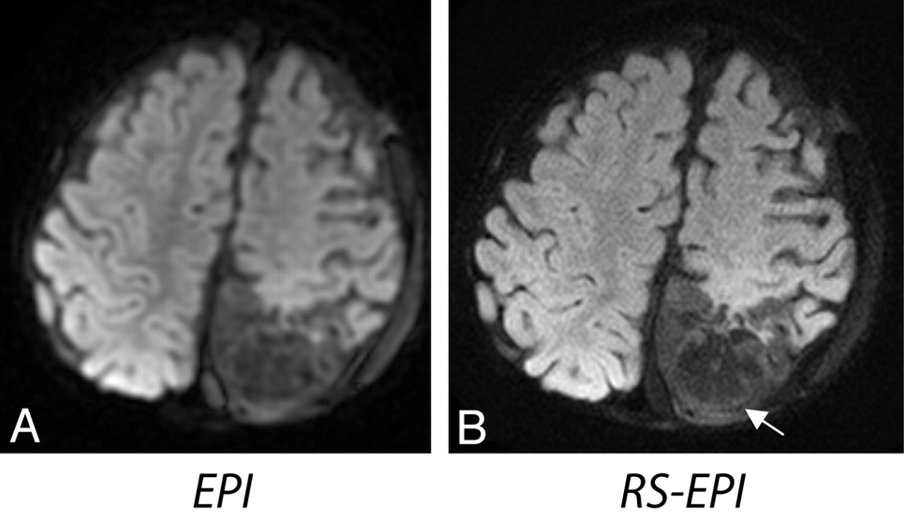
- Fig 8.
Two sections from an 8-year-old female patient. A, Distortion artifacts on EPI could be confused with subdural hemorrhage or empyema. This artifact disappears on RS-EPI (B). A,C, Elevated contrast on EPI, not appearent on RS-EPI (B,D), could be confused with diffuse cortical ischemic injury, encephalitis, or even seizure-related changes.
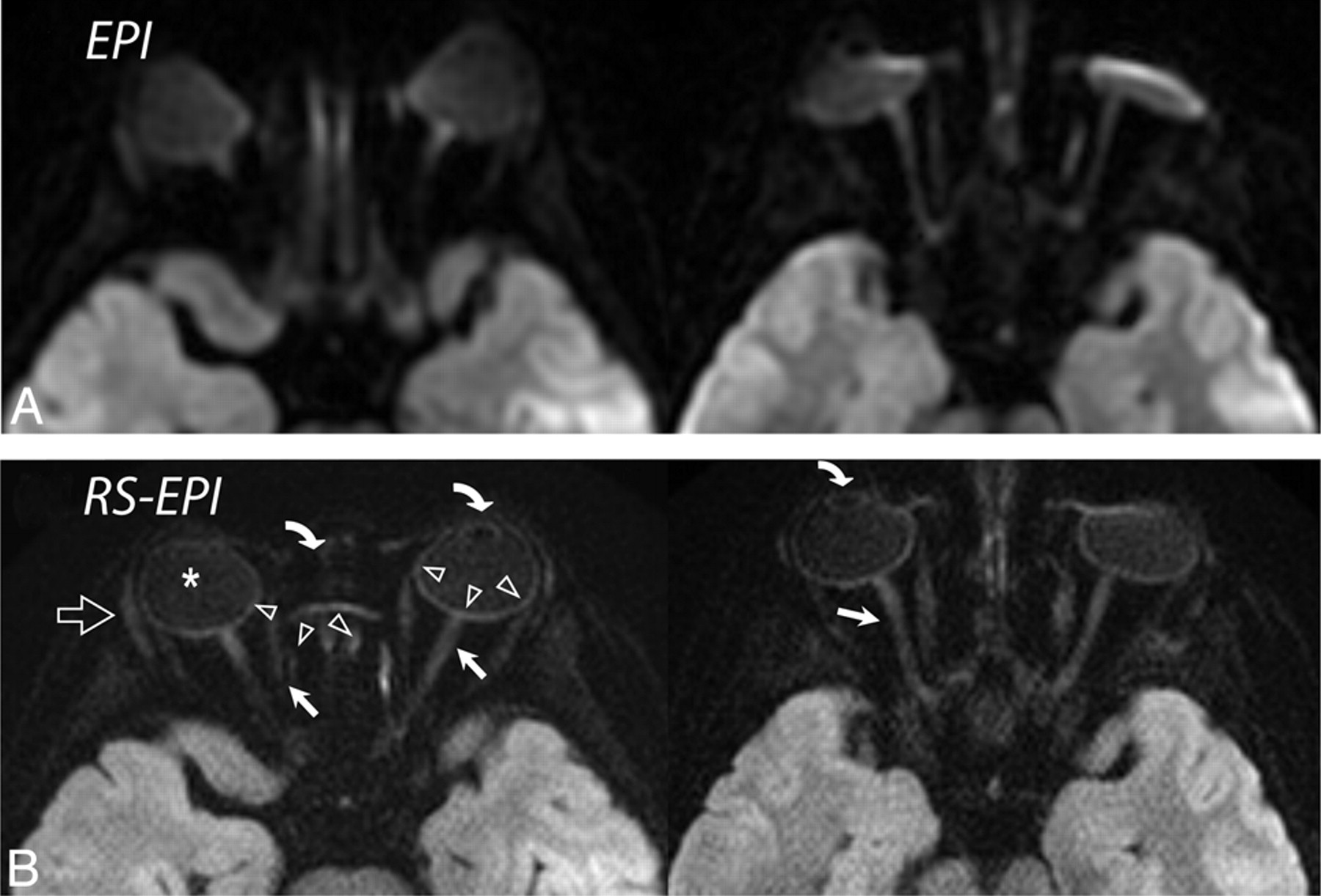
- Fig 9.
A, ASSET-accelerated (×2) diffusion-weighted single-shot EPI in 2 different subjects (a 3-year-old girl and a 10-year-old boy) and their corresponding diffusion-weighted RS-EPI scans. B, ASSET EPI scans have a better SNR, but orbital anatomy, such as the lens (curved arrows), optic nerve (arrows), sclera (arrowheads), vitreous humor (asterisk), and lacrimal glands (open arrow), are clearly better depicted on RS-EPI than on conventional ASSET-accelerated EPI scans.


