
Article Figures & Data
Figures
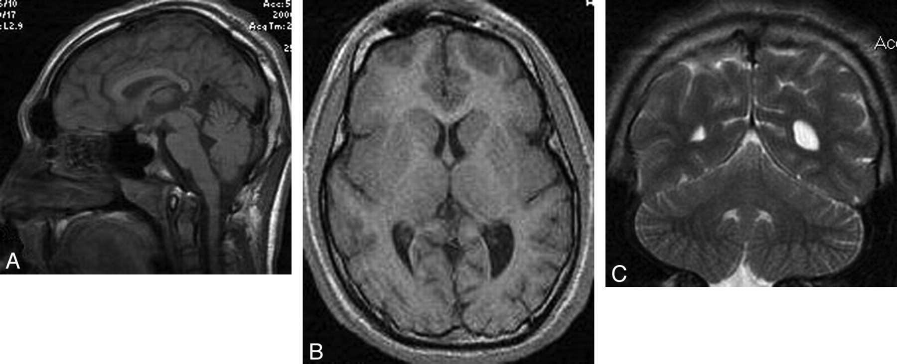
- Fig 1.
A 14-year-old boy with microcephaly and developmental delay. A, Sagittal T1-weighted image shows extreme microcephaly. The corpus callosum is fully formed but appears diffusely thin. B, Axial T1-weighted image shows a mildly simplified gyral pattern with normal cortical thickness. C, Coronal T2-weighted image shows a disproportionally large cerebellum.
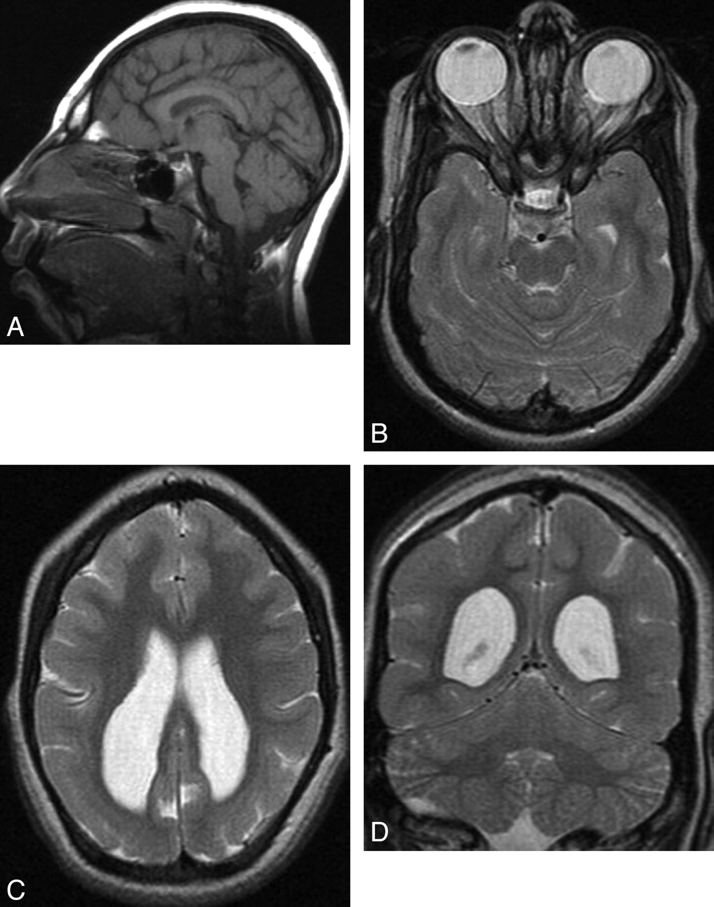
- Fig 2.
A 10-year-old girl with microcephaly. A, Sagittal T1-weighted image shows extreme microcephaly. The corpus callosum is fully formed but diffusely thin. B and C, Axial T2-weighted images show a moderately simplified gyral pattern with too few sulci but normal cortical thickness (measured at <3 mm). Symmetric posterior horn−dominant lateral ventricular enlargement is shown. White matter volume is severely reduced. D, Coronal T2-weighted image shows a disproportionally large cerebellum.
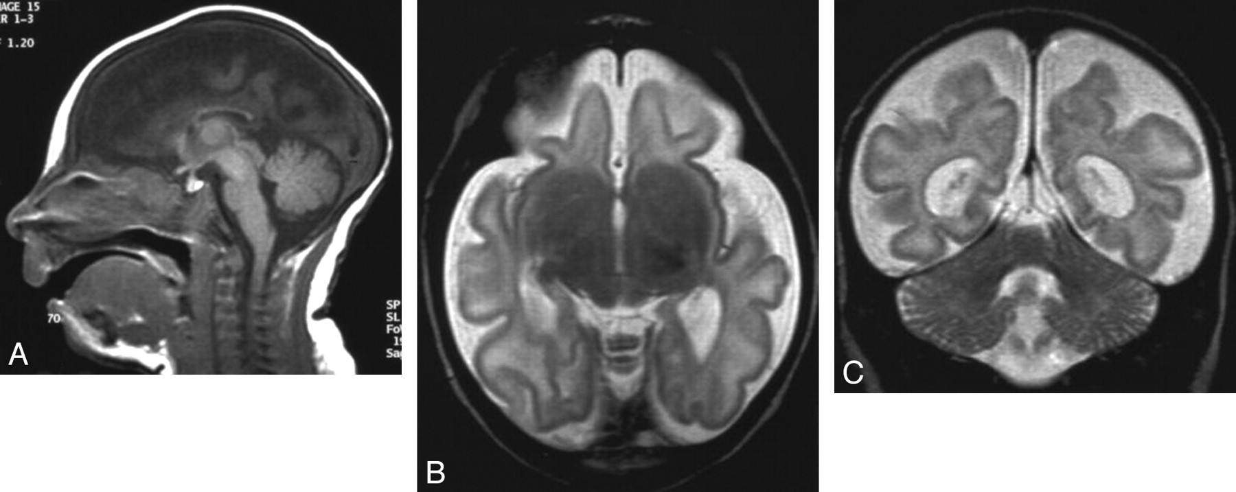
- Fig 3.
A 1-month-old girl with microcephaly, global developmental delay, and seizures. A, Sagittal T1-weighted image shows extreme microcephaly. The corpus callosum is partially formed, the rostrum is absent, and other parts (genu, body, and splenium) are diffusely thin. B, Axial T2-weighted image shows a severely simplified gyral pattern; sulci are both too few and too shallow. Although myelination is shown in the posterior limb of the internal capsule, the abnormal high intensity of the white matter is observed diffusely and the volume of the white matter is severely diminished. C, Coronal T2-weighted image shows a disproportionally large cerebellum.
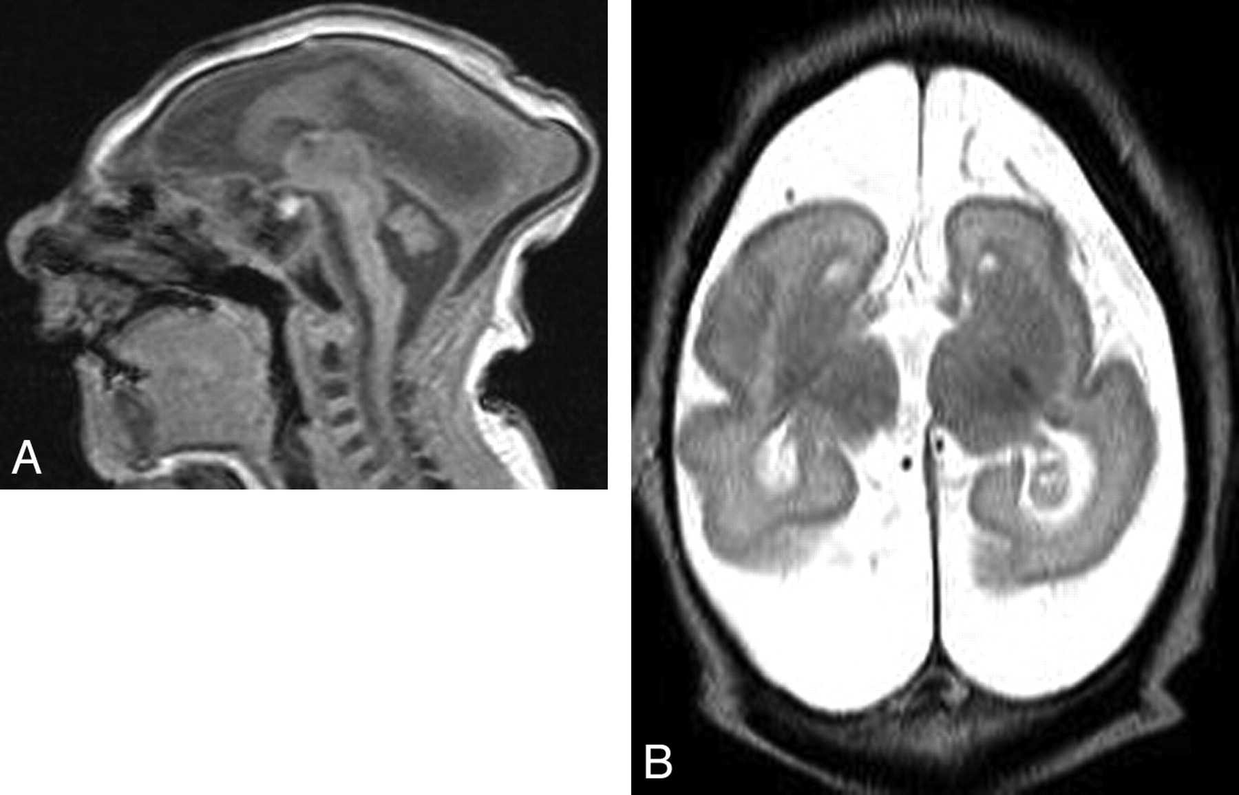
- Fig 4.
A 2-week-old boy with profound microcephaly. A, Sagittal T1-weighted image shows extreme microcephaly with absence of the corpus callosum. All structures seem proportionately affected. B, Axial T2-weighted image shows a severely simplified gyral pattern (almost no sulci). The volume of white matter is also extremely reduced proportional to the simplification of the gyral pattern, and the pericerebral spaces are enlarged.


Tables
Age distribution of patients with microcephaly
Age Male Female Unknown Total 0–1 mo 11 10 21 1–2 mo 7 6 13 3–6 mo 4 10 14 7–12 mo 6 8 14 13–24 mo 9 5 1 15 2–3 yr 7 4 11 3–6 yr 7 5 12 6–10 yr 3 7 1 11 >10 yr 2 2 4 unknown 4
In this issue


Jump to section
Related Articles
Cited By...
- A kinase-independent function of cyclin-dependent kinase 6 promotes outer radial glia expansion and neocortical folding
- Delineating FOXG1 syndrome: From congenital microcephaly to hyperkinetic encephalopathy
- Nonmicrocephalic Infants with Congenital Zika Syndrome Suspected Only after Neuroimaging Evaluation Compared with Those with Microcephaly at Birth and Postnatally: How Large Is the Zika Virus "Iceberg"?
- Cerebral cortex expansion and folding: what have we learned?